Upper GI · Oncology · 52 slides
Carcinoma stomach.
From the first vague symptom to the right operation: the anatomy, pathology and decisions behind the care of a patient with gastric cancer.
About the downloads
PowerPoint without speaker notes. SHA-256 checksums verify the downloaded files.
Contents · 7 parts
Introduction
Carcinoma Stomach
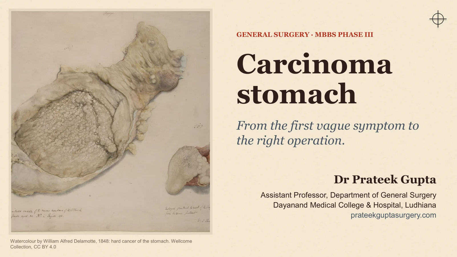
Watercolour by William Alfred Delamotte, 1848: hard cancer of the stomach. Wellcome Collection, CC BY 4.0
GENERAL SURGERY · MBBS PHASE III
Carcinoma stomach
From the first vague symptom to the right operation.
Dr Prateek Gupta
Assistant Professor, Department of General Surgery Dayanand Medical College & Hospital, Ludhiana prateekguptasurgery.com
Three months of “gas”, and now he vomits
A PATIENT IN THE SURGICAL OPD
A 58-year-old farmer has had three months of upper abdominal discomfort that antacids from the chemist did not settle.
History. Early fullness after small meals, loss of appetite and about 7 kg weight loss. For two weeks he has vomited large amounts of food eaten the day before.
General examination. Pale and thin. No jaundice. No palpable left supraclavicular node.
Abdomen. A hard, nodular epigastric lump above the umbilicus that moves with respiration. Visible gastric peristalsis and a succussion splash after a 4-hour fast.
Keep these questions in mind
1. What is the most likely diagnosis?
2. Why is he vomiting?
3. How will you confirm it, and how will you stage it?
4. Can he be cured, and with which operation?
5. What will you do if it has already spread?
We return to him at the end of the class.
Illustrative case: not a real patient.
CARCINOMA STOMACH
2
What you should be able to do by the end
THIS CLASS IN FIVE PARTS
I
Where it starts
Relate gastric anatomy and lymph drainage to surgery; list risk factors, precursor lesions and the H. pylori pathway.
II
Pathology and spread
Classify early and advanced cancer (Japanese, Borrmann, Lauren) and explain each route of spread.
III
The patient
Elicit the symptoms and signs, including the named signs of spread, and build a differential diagnosis.
IV
Confirm and stage
Plan endoscopy, biopsy and staging (CT, endoscopic ultrasound, laparoscopy) and apply TNM 8th edition.
V
Treat and follow up
Choose between endoscopic resection, gastrectomy with D2 lymphadenectomy, chemotherapy and palliation.
CARCINOMA STOMACH
3
Anatomy and risk
Where it starts
I
Anatomy decides the operation. Epidemiology and aetiology decide who gets the disease.
Surgical anatomy, blood supply and lymph node stations
Who gets gastric cancer, and where in the world
Proximal and distal cancer: two different diseases
Risk factors, H. pylori and the Correa cascade
Hereditary cancer, polyps and other precursor lesions
CARCINOMA STOMACH
4
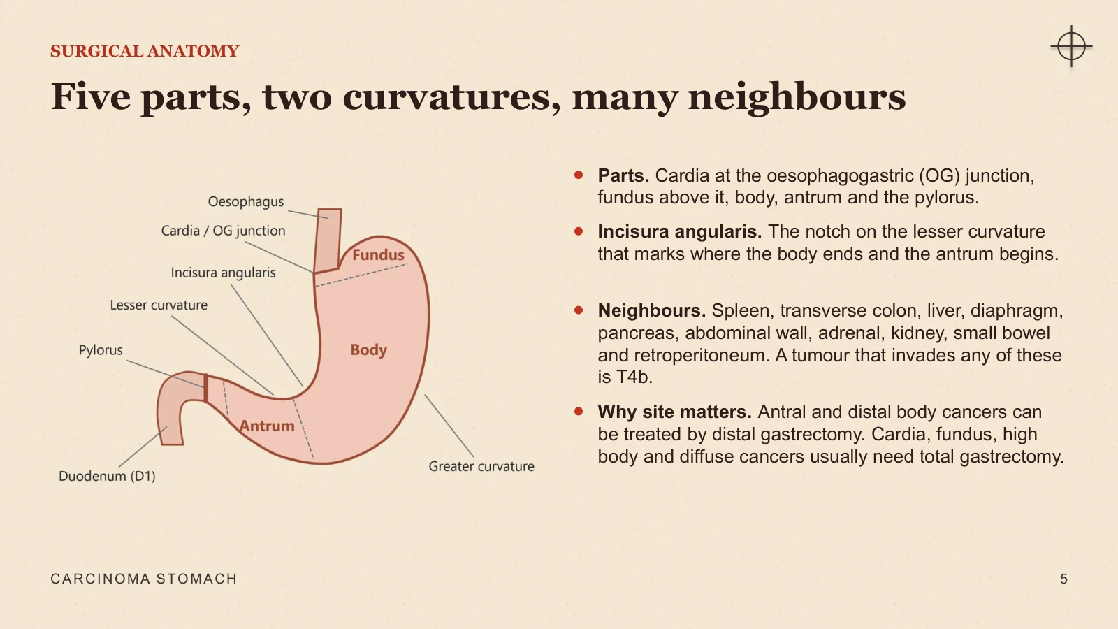
Five parts, two curvatures, many neighbours
SURGICAL ANATOMY
Parts. Cardia at the oesophagogastric (OG) junction, fundus above it, body, antrum and the pylorus.
Incisura angularis. The notch on the lesser curvature that marks where the body ends and the antrum begins.
Neighbours. Spleen, transverse colon, liver, diaphragm, pancreas, abdominal wall, adrenal, kidney, small bowel and retroperitoneum. A tumour that invades any of these is T4b.
Why site matters. Antral and distal body cancers can be treated by distal gastrectomy. Cardia, fundus, high body and diffuse cancers usually need total gastrectomy.
CARCINOMA STOMACH
5
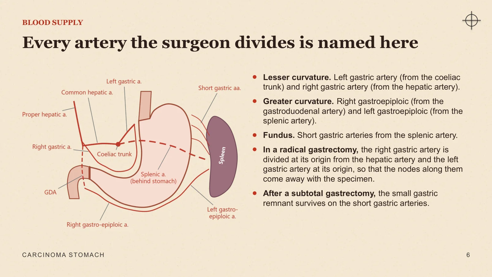
Every artery the surgeon divides is named here
BLOOD SUPPLY
Lesser curvature. Left gastric artery (from the coeliac trunk) and right gastric artery (from the hepatic artery).
Greater curvature. Right gastroepiploic (from the gastroduodenal artery) and left gastroepiploic (from the splenic artery).
Fundus. Short gastric arteries from the splenic artery.
In a radical gastrectomy, the right gastric artery is divided at its origin from the hepatic artery and the left gastric artery at its origin, so that the nodes along them come away with the specimen.
After a subtotal gastrectomy, the small gastric remnant survives on the short gastric arteries.
CARCINOMA STOMACH
6
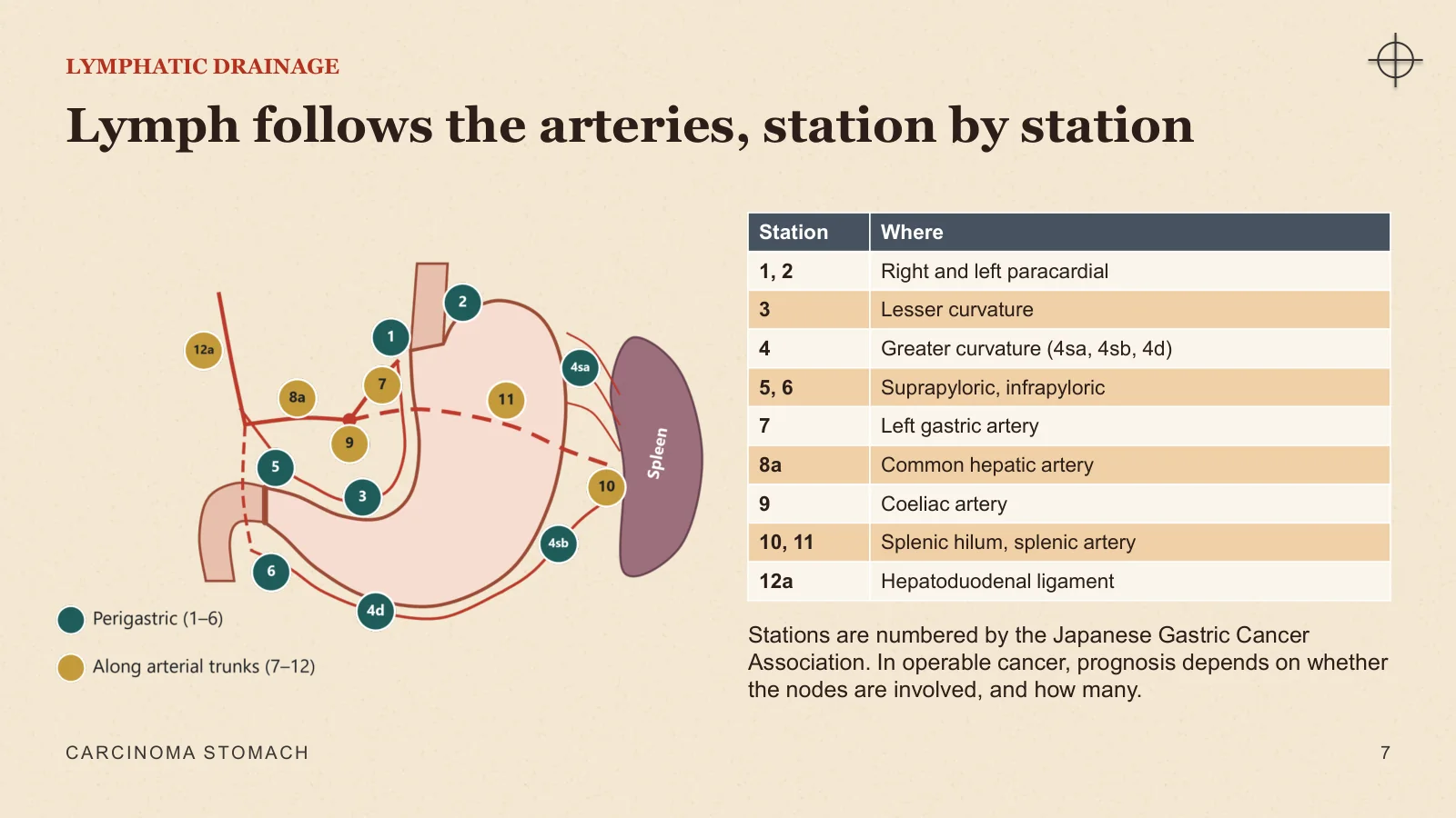
Lymph follows the arteries, station by station
LYMPHATIC DRAINAGE
Stations are numbered by the Japanese Gastric Cancer Association. In operable cancer, prognosis depends on whether the nodes are involved, and how many.
CARCINOMA STOMACH
7
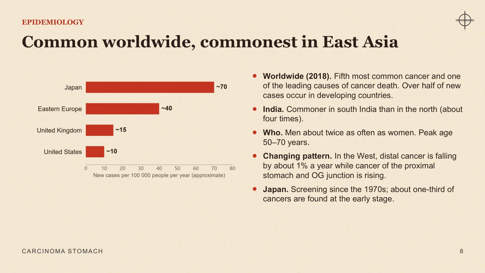
Common worldwide, commonest in East Asia
EPIDEMIOLOGY
Worldwide (2018). Fifth most common cancer and one of the leading causes of cancer death. Over half of new cases occur in developing countries.
India. Commoner in south India than in the north (about four times).
Who. Men about twice as often as women. Peak age 50–70 years.
Changing pattern. In the West, distal cancer is falling by about 1% a year while cancer of the proximal stomach and OG junction is rising.
Japan. Screening since the 1970s; about one-third of cancers are found at the early stage.
CARCINOMA STOMACH
8
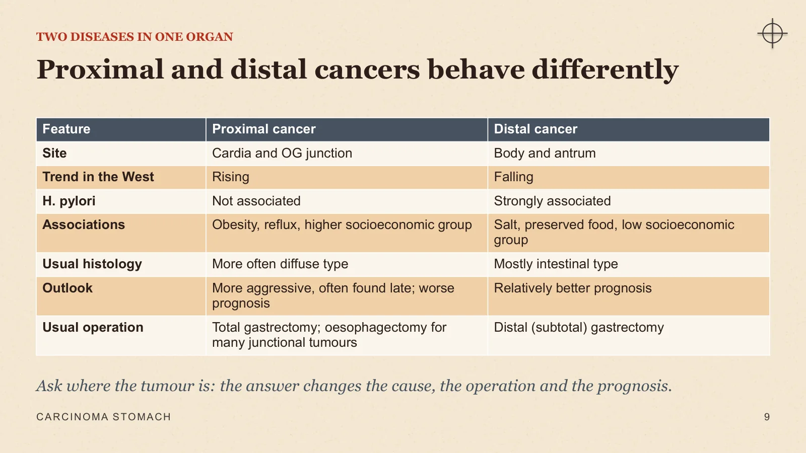
Proximal and distal cancers behave differently
TWO DISEASES IN ONE ORGAN
Ask where the tumour is: the answer changes the cause, the operation and the prognosis.
CARCINOMA STOMACH
9
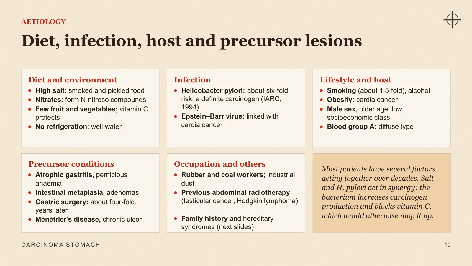
Diet, infection, host and precursor lesions
AETIOLOGY
Diet and environment
High salt: smoked and pickled food
Nitrates: form N-nitroso compounds
Few fruit and vegetables; vitamin C protects
No refrigeration; well water
Infection
Helicobacter pylori: about six-fold risk; a definite carcinogen (IARC, 1994)
Epstein–Barr virus: linked with cardia cancer
Lifestyle and host
Smoking (about 1.5-fold), alcohol
Obesity: cardia cancer
Male sex, older age, low socioeconomic class
Blood group A: diffuse type
Precursor conditions
Atrophic gastritis, pernicious anaemia
Intestinal metaplasia, adenomas
Gastric surgery: about four-fold, years later
Ménétrier's disease, chronic ulcer
Occupation and others
Rubber and coal workers; industrial dust
Previous abdominal radiotherapy (testicular cancer, Hodgkin lymphoma)
Family history and hereditary syndromes (next slides)
Most patients have several factors acting together over decades. Salt and H. pylori act in synergy: the bacterium increases carcinogen production and blocks vitamin C, which would otherwise mop it up.
CARCINOMA STOMACH
10
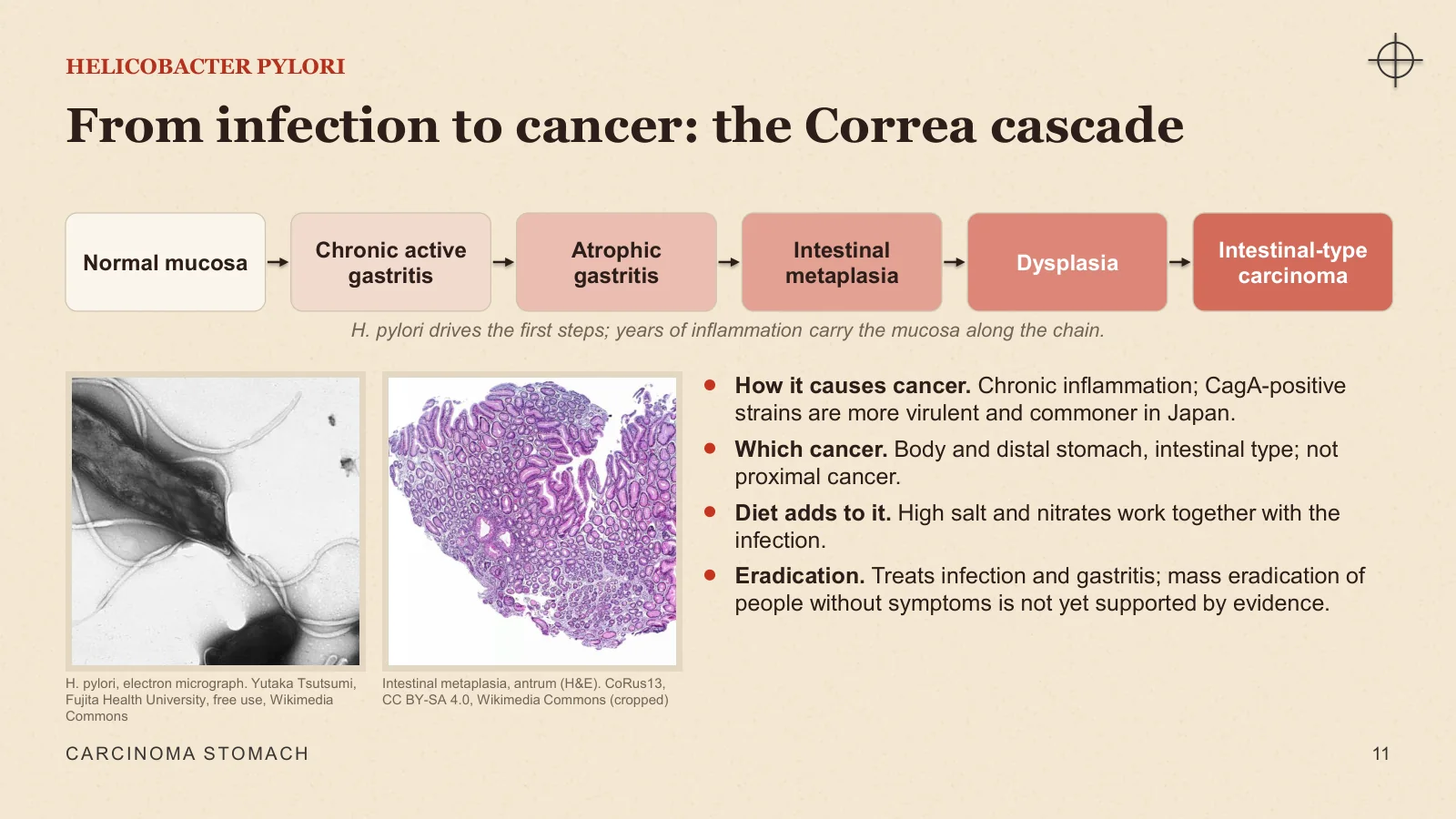
From infection to cancer: the Correa cascade
HELICOBACTER PYLORI
Normal mucosa
Chronic active gastritis
Atrophic gastritis
Intestinal metaplasia
Dysplasia
Intestinal-type carcinoma
H. pylori drives the first steps; years of inflammation carry the mucosa along the chain.
H. pylori, electron micrograph. Yutaka Tsutsumi, Fujita Health University, free use, Wikimedia Commons
Intestinal metaplasia, antrum (H&E). CoRus13, CC BY-SA 4.0, Wikimedia Commons (cropped)
How it causes cancer. Chronic inflammation; CagA-positive strains are more virulent and commoner in Japan.
Which cancer. Body and distal stomach, intestinal type; not proximal cancer.
Diet adds to it. High salt and nitrates work together with the infection.
Eradication. Treats infection and gastritis; mass eradication of people without symptoms is not yet supported by evidence.
CARCINOMA STOMACH
11
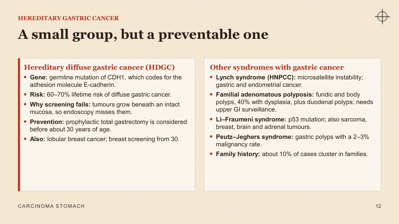
A small group, but a preventable one
HEREDITARY GASTRIC CANCER
Hereditary diffuse gastric cancer (HDGC)
Gene: germline mutation of CDH1, which codes for the adhesion molecule E-cadherin.
Risk: 60–70% lifetime risk of diffuse gastric cancer.
Why screening fails: tumours grow beneath an intact mucosa, so endoscopy misses them.
Prevention: prophylactic total gastrectomy is considered before about 30 years of age.
Also: lobular breast cancer; breast screening from 30.
Other syndromes with gastric cancer
Lynch syndrome (HNPCC): microsatellite instability; gastric and endometrial cancer.
Familial adenomatous polyposis: fundic and body polyps, 40% with dysplasia, plus duodenal polyps; needs upper GI surveillance.
Li–Fraumeni syndrome: p53 mutation; also sarcoma, breast, brain and adrenal tumours.
Peutz–Jeghers syndrome: gastric polyps with a 2–3% malignancy rate.
Family history: about 10% of cases cluster in families.
CARCINOMA STOMACH
12
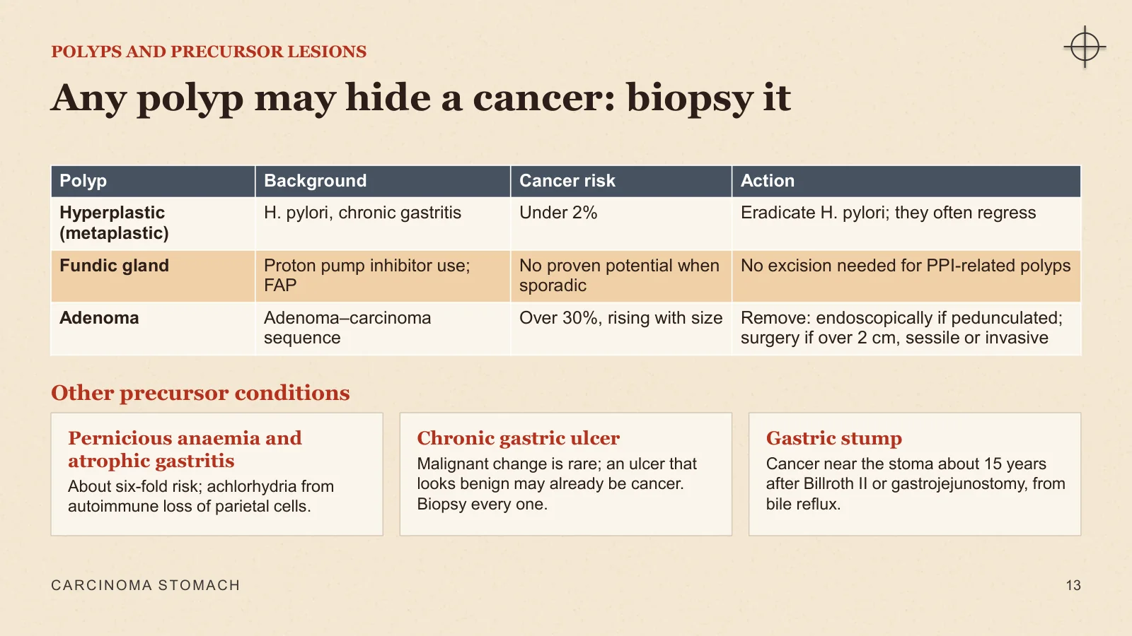
Any polyp may hide a cancer: biopsy it
POLYPS AND PRECURSOR LESIONS
Other precursor conditions
Pernicious anaemia and atrophic gastritis
About six-fold risk; achlorhydria from autoimmune loss of parietal cells.
Chronic gastric ulcer
Malignant change is rare; an ulcer that looks benign may already be cancer. Biopsy every one.
Gastric stump
Cancer near the stoma about 15 years after Billroth II or gastrojejunostomy, from bile reflux.
CARCINOMA STOMACH
13
Pathology and spread
Pathology and spread
II
How deep it has grown and how it looks decide what can be done.
Where in the stomach, and early versus advanced
Early gastric cancer: the Japanese classification
Advanced cancer: Borrmann types and linitis plastica
Lauren types, microscopy and molecular subtypes
Four routes of spread and the named signs they produce
CARCINOMA STOMACH
14
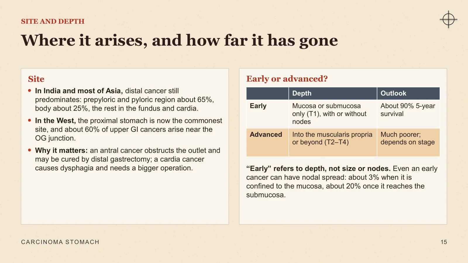
Where it arises, and how far it has gone
SITE AND DEPTH
Site
In India and most of Asia, distal cancer still predominates: prepyloric and pyloric region about 65%, body about 25%, the rest in the fundus and cardia.
In the West, the proximal stomach is now the commonest site, and about 60% of upper GI cancers arise near the OG junction.
Why it matters: an antral cancer obstructs the outlet and may be cured by distal gastrectomy; a cardia cancer causes dysphagia and needs a bigger operation.
Early or advanced?
“Early” refers to depth, not size or nodes. Even an early cancer can have nodal spread: about 3% when it is confined to the mucosa, about 20% once it reaches the submucosa.
CARCINOMA STOMACH
15
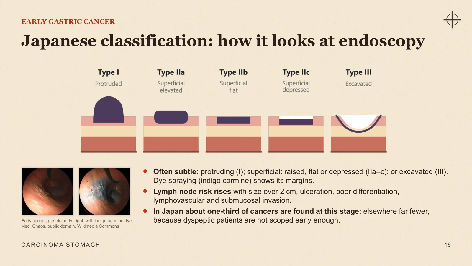
Japanese classification: how it looks at endoscopy
EARLY GASTRIC CANCER
Early cancer, gastric body; right: with indigo carmine dye. Med_Chaos, public domain, Wikimedia Commons
Often subtle: protruding (I); superficial: raised, flat or depressed (IIa–c); or excavated (III). Dye spraying (indigo carmine) shows its margins.
Lymph node risk rises with size over 2 cm, ulceration, poor differentiation, lymphovascular and submucosal invasion.
In Japan about one-third of cancers are found at this stage; elsewhere far fewer, because dyspeptic patients are not scoped early enough.
CARCINOMA STOMACH
16
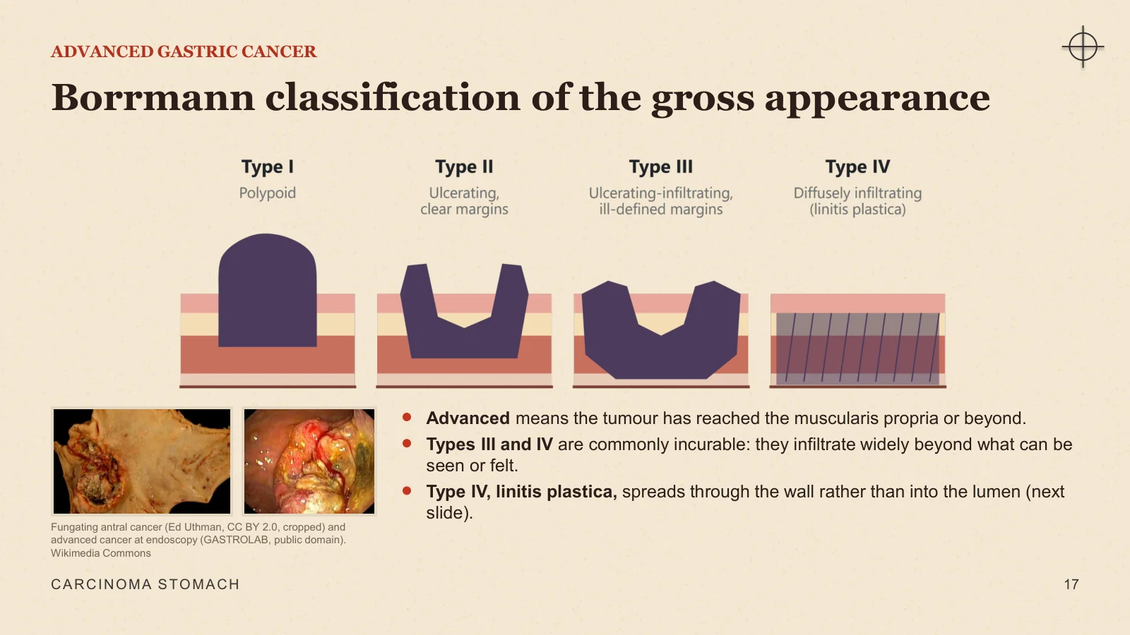
Borrmann classification of the gross appearance
ADVANCED GASTRIC CANCER
Fungating antral cancer (Ed Uthman, CC BY 2.0, cropped) and advanced cancer at endoscopy (GASTROLAB, public domain). Wikimedia Commons
Advanced means the tumour has reached the muscularis propria or beyond.
Types III and IV are commonly incurable: they infiltrate widely beyond what can be seen or felt.
Type IV, linitis plastica, spreads through the wall rather than into the lumen (next slide).
CARCINOMA STOMACH
17
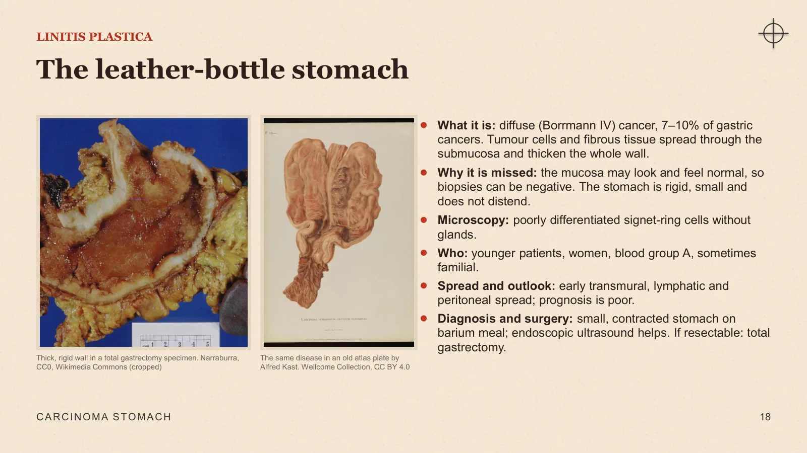
The leather-bottle stomach
LINITIS PLASTICA
Thick, rigid wall in a total gastrectomy specimen. Narraburra, CC0, Wikimedia Commons (cropped)
The same disease in an old atlas plate by Alfred Kast. Wellcome Collection, CC BY 4.0
What it is: diffuse (Borrmann IV) cancer, 7–10% of gastric cancers. Tumour cells and fibrous tissue spread through the submucosa and thicken the whole wall.
Why it is missed: the mucosa may look and feel normal, so biopsies can be negative. The stomach is rigid, small and does not distend.
Microscopy: poorly differentiated signet-ring cells without glands.
Who: younger patients, women, blood group A, sometimes familial.
Spread and outlook: early transmural, lymphatic and peritoneal spread; prognosis is poor.
Diagnosis and surgery: small, contracted stomach on barium meal; endoscopic ultrasound helps. If resectable: total gastrectomy.
CARCINOMA STOMACH
18
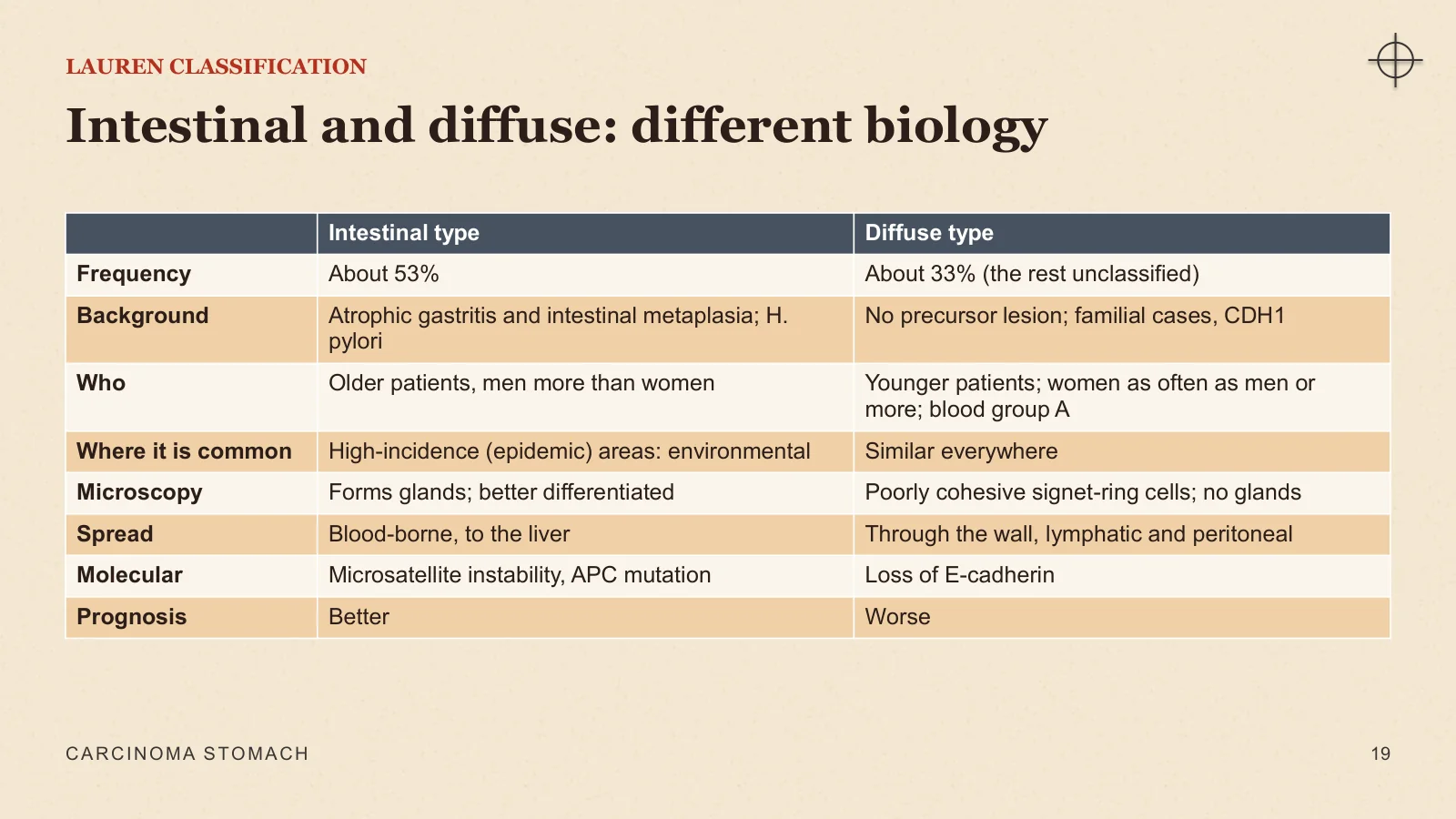
Intestinal and diffuse: different biology
LAUREN CLASSIFICATION
CARCINOMA STOMACH
19
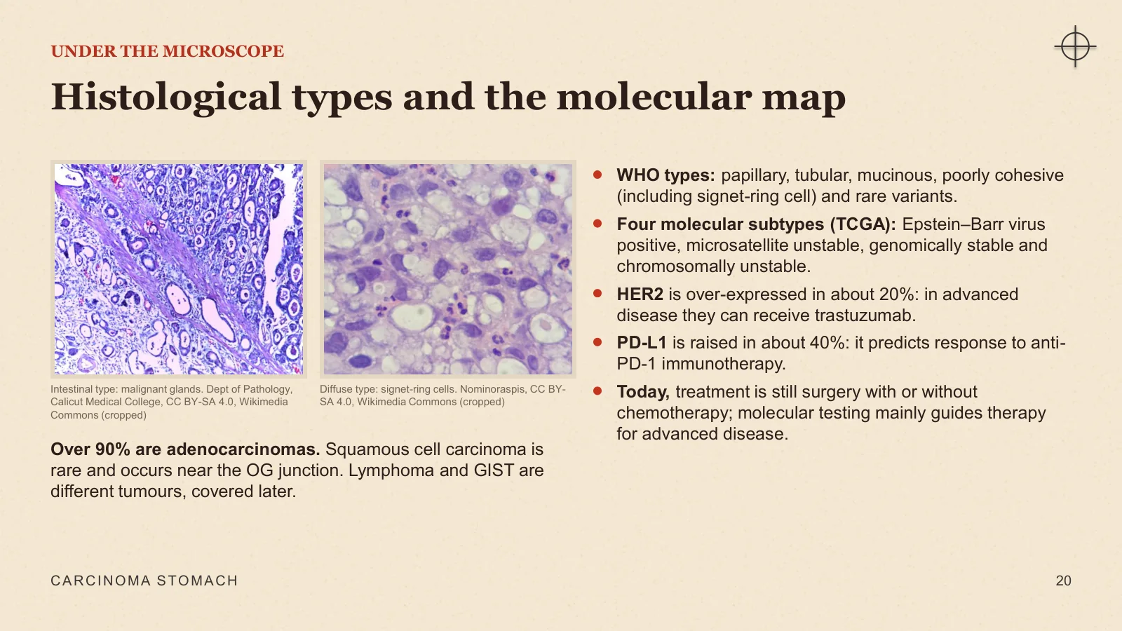
Histological types and the molecular map
UNDER THE MICROSCOPE
Intestinal type: malignant glands. Dept of Pathology, Calicut Medical College, CC BY-SA 4.0, Wikimedia Commons (cropped)
Diffuse type: signet-ring cells. Nominoraspis, CC BY-SA 4.0, Wikimedia Commons (cropped)
Over 90% are adenocarcinomas. Squamous cell carcinoma is rare and occurs near the OG junction. Lymphoma and GIST are different tumours, covered later.
WHO types: papillary, tubular, mucinous, poorly cohesive (including signet-ring cell) and rare variants.
Four molecular subtypes (TCGA): Epstein–Barr virus positive, microsatellite unstable, genomically stable and chromosomally unstable.
HER2 is over-expressed in about 20%: in advanced disease they can receive trastuzumab.
PD-L1 is raised in about 40%: it predicts response to anti-PD-1 immunotherapy.
Today, treatment is still surgery with or without chemotherapy; molecular testing mainly guides therapy for advanced disease.
CARCINOMA STOMACH
20
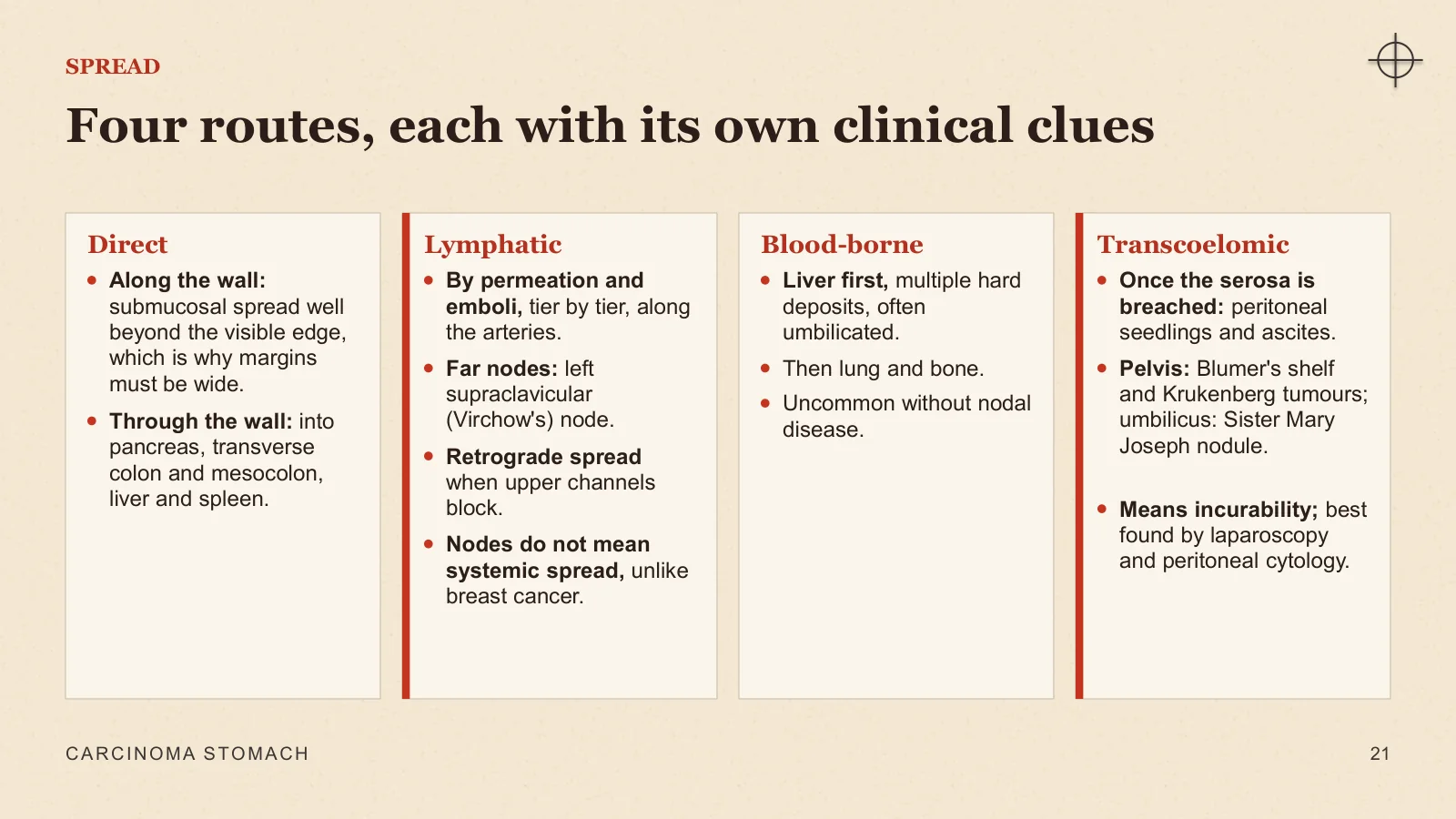
Four routes, each with its own clinical clues
SPREAD
Direct
Along the wall: submucosal spread well beyond the visible edge, which is why margins must be wide.
Through the wall: into pancreas, transverse colon and mesocolon, liver and spleen.
Lymphatic
By permeation and emboli, tier by tier, along the arteries.
Far nodes: left supraclavicular (Virchow's) node.
Retrograde spread when upper channels block.
Nodes do not mean systemic spread, unlike breast cancer.
Blood-borne
Liver first, multiple hard deposits, often umbilicated.
Then lung and bone.
Uncommon without nodal disease.
Transcoelomic
Once the serosa is breached: peritoneal seedlings and ascites.
Pelvis: Blumer's shelf and Krukenberg tumours; umbilicus: Sister Mary Joseph nodule.
Means incurability; best found by laparoscopy and peritoneal cytology.
CARCINOMA STOMACH
21
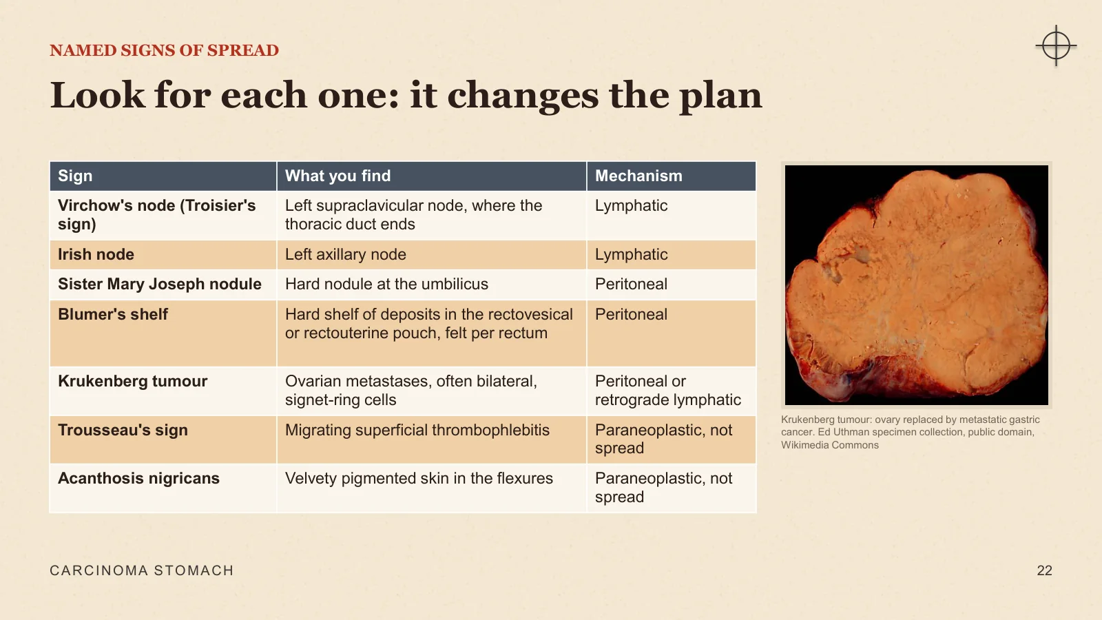
Look for each one: it changes the plan
NAMED SIGNS OF SPREAD
Krukenberg tumour: ovary replaced by metastatic gastric cancer. Ed Uthman specimen collection, public domain, Wikimedia Commons
CARCINOMA STOMACH
22
Clinical presentation
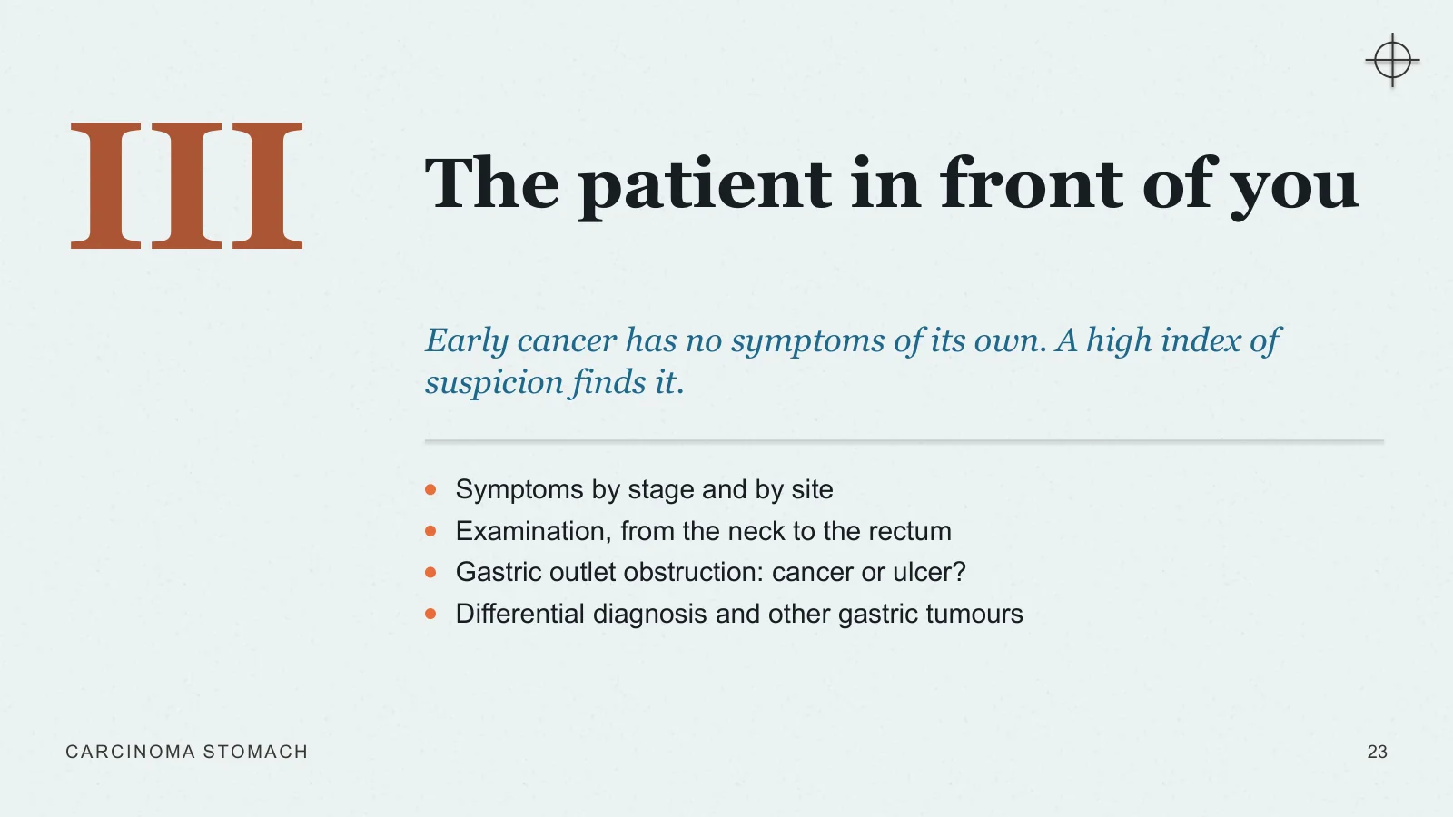
The patient in front of you
III
Early cancer has no symptoms of its own. A high index of suspicion finds it.
Symptoms by stage and by site
Examination, from the neck to the rectum
Gastric outlet obstruction: cancer or ulcer?
Differential diagnosis and other gastric tumours
CARCINOMA STOMACH
23
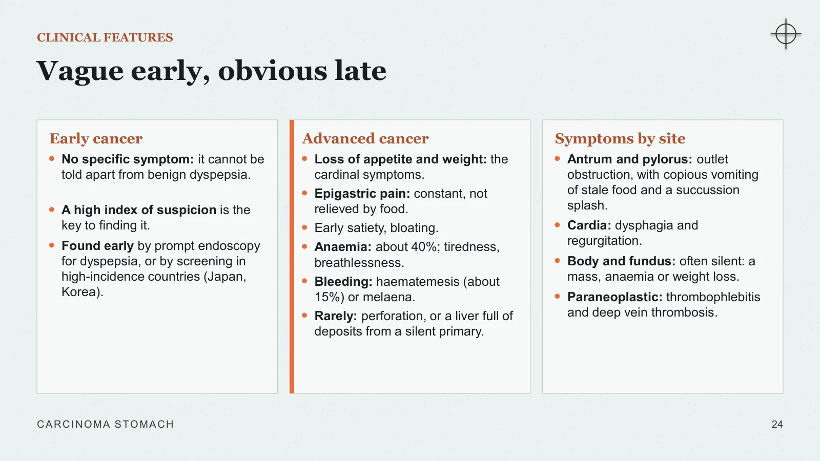
Vague early, obvious late
CLINICAL FEATURES
Early cancer
No specific symptom: it cannot be told apart from benign dyspepsia.
A high index of suspicion is the key to finding it.
Found early by prompt endoscopy for dyspepsia, or by screening in high-incidence countries (Japan, Korea).
Advanced cancer
Loss of appetite and weight: the cardinal symptoms.
Epigastric pain: constant, not relieved by food.
Early satiety, bloating.
Anaemia: about 40%; tiredness, breathlessness.
Bleeding: haematemesis (about 15%) or melaena.
Rarely: perforation, or a liver full of deposits from a silent primary.
Symptoms by site
Antrum and pylorus: outlet obstruction, with copious vomiting of stale food and a succussion splash.
Cardia: dysphagia and regurgitation.
Body and fundus: often silent: a mass, anaemia or weight loss.
Paraneoplastic: thrombophlebitis and deep vein thrombosis.
CARCINOMA STOMACH
24
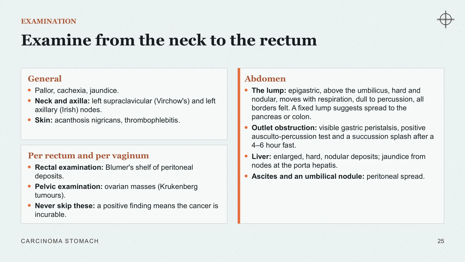
Examine from the neck to the rectum
EXAMINATION
General
Pallor, cachexia, jaundice.
Neck and axilla: left supraclavicular (Virchow's) and left axillary (Irish) nodes.
Skin: acanthosis nigricans, thrombophlebitis.
Per rectum and per vaginum
Rectal examination: Blumer's shelf of peritoneal deposits.
Pelvic examination: ovarian masses (Krukenberg tumours).
Never skip these: a positive finding means the cancer is incurable.
Abdomen
The lump: epigastric, above the umbilicus, hard and nodular, moves with respiration, dull to percussion, all borders felt. A fixed lump suggests spread to the pancreas or colon.
Outlet obstruction: visible gastric peristalsis, positive ausculto-percussion test and a succussion splash after a 4–6 hour fast.
Liver: enlarged, hard, nodular deposits; jaundice from nodes at the porta hepatis.
Ascites and an umbilical nodule: peritoneal spread.
CARCINOMA STOMACH
25
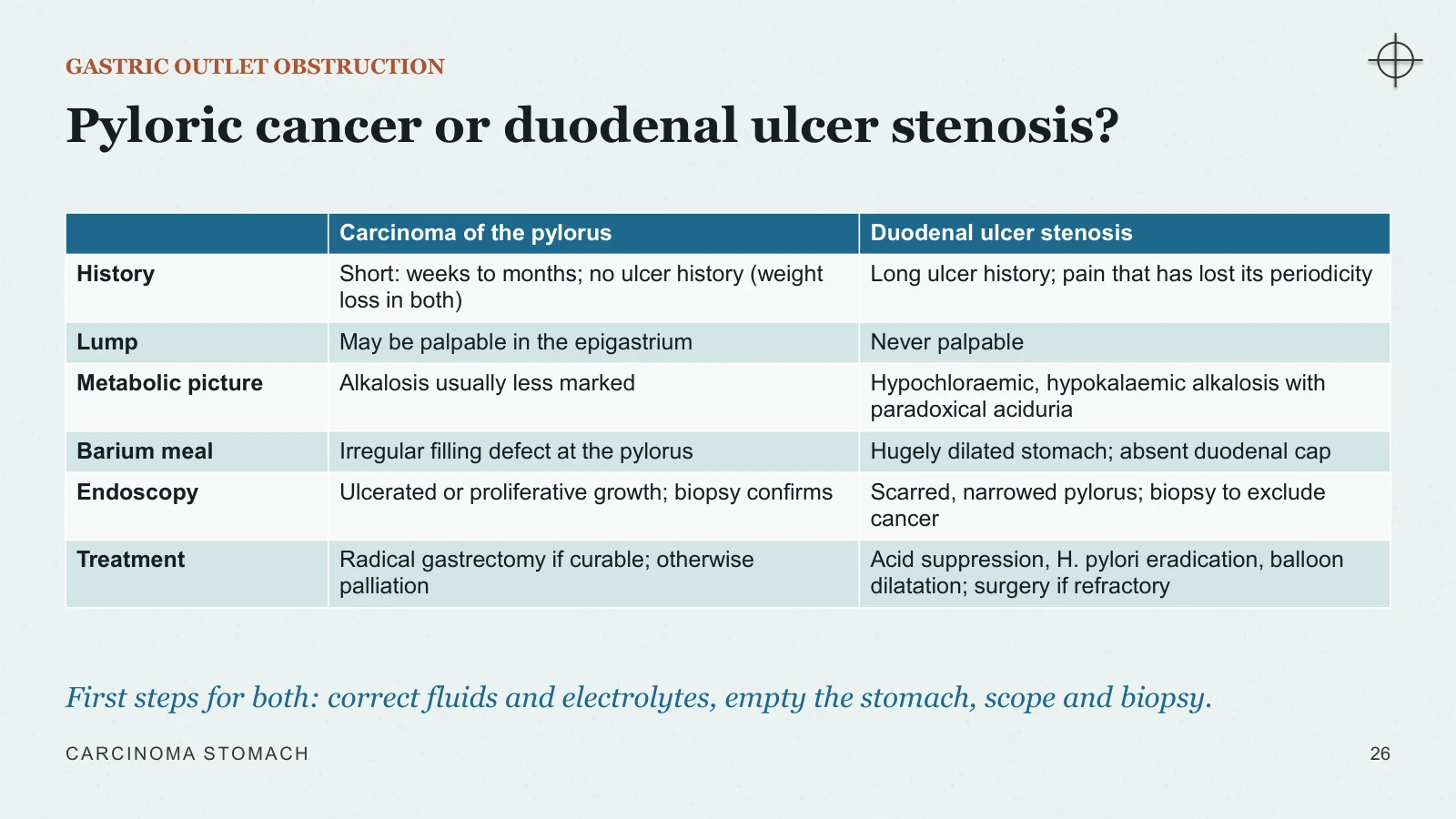
Pyloric cancer or duodenal ulcer stenosis?
GASTRIC OUTLET OBSTRUCTION
First steps for both: correct fluids and electrolytes, empty the stomach, scope and biopsy.
CARCINOMA STOMACH
26
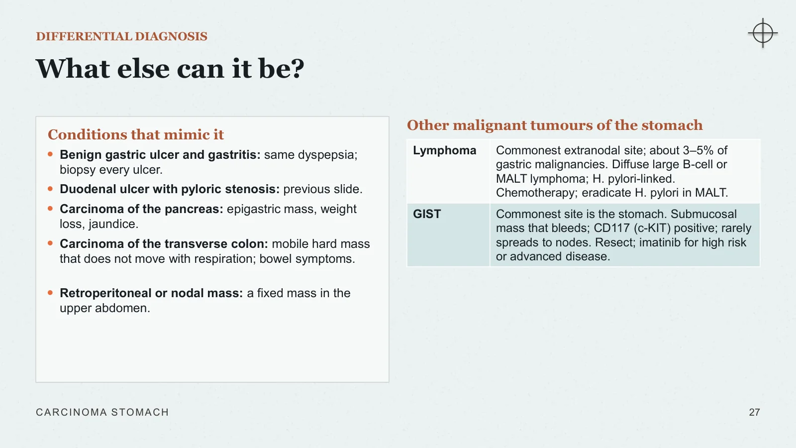
What else can it be?
DIFFERENTIAL DIAGNOSIS
Conditions that mimic it
Benign gastric ulcer and gastritis: same dyspepsia; biopsy every ulcer.
Duodenal ulcer with pyloric stenosis: previous slide.
Carcinoma of the pancreas: epigastric mass, weight loss, jaundice.
Carcinoma of the transverse colon: mobile hard mass that does not move with respiration; bowel symptoms.
Retroperitoneal or nodal mass: a fixed mass in the upper abdomen.
Other malignant tumours of the stomach
CARCINOMA STOMACH
27
Diagnosis and staging
Confirm, stage, decide
IV
The aim of staging is to spare the incurable an operation and to give the curable the right one.
The investigation plan
What endoscopy and barium show
Staging tools: CT, endoscopic ultrasound, PET-CT and laparoscopy
TNM 8th edition, stage groups and the OG junction
CARCINOMA STOMACH
28
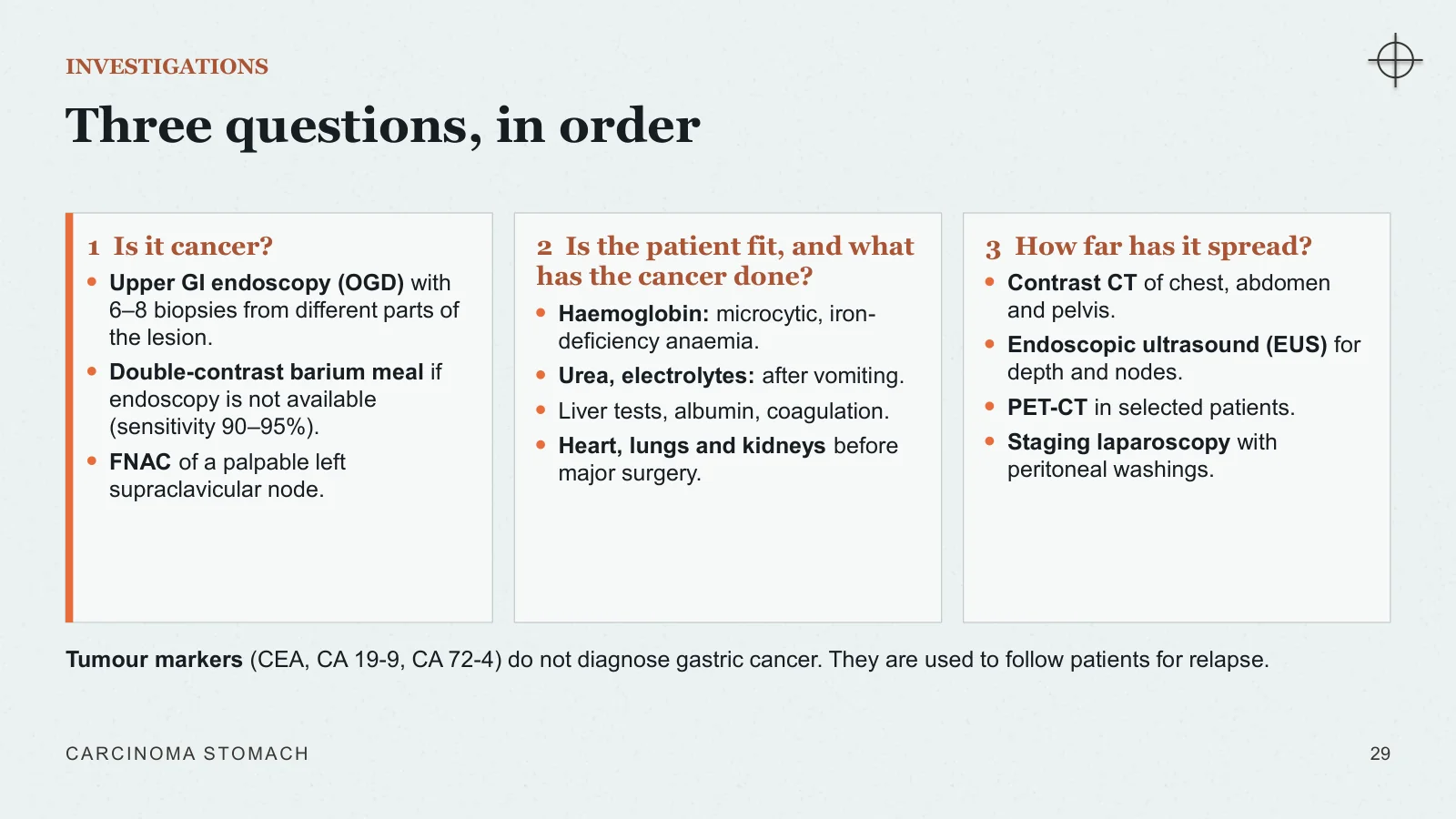
Three questions, in order
INVESTIGATIONS
1 Is it cancer?
Upper GI endoscopy (OGD) with 6–8 biopsies from different parts of the lesion.
Double-contrast barium meal if endoscopy is not available (sensitivity 90–95%).
FNAC of a palpable left supraclavicular node.
2 Is the patient fit, and what has the cancer done?
Haemoglobin: microcytic, iron-deficiency anaemia.
Urea, electrolytes: after vomiting.
Liver tests, albumin, coagulation.
Heart, lungs and kidneys before major surgery.
3 How far has it spread?
Contrast CT of chest, abdomen and pelvis.
Endoscopic ultrasound (EUS) for depth and nodes.
PET-CT in selected patients.
Staging laparoscopy with peritoneal washings.
Tumour markers (CEA, CA 19-9, CA 72-4) do not diagnose gastric cancer. They are used to follow patients for relapse.
CARCINOMA STOMACH
29
Endoscopy and barium meal
SEEING THE TUMOUR
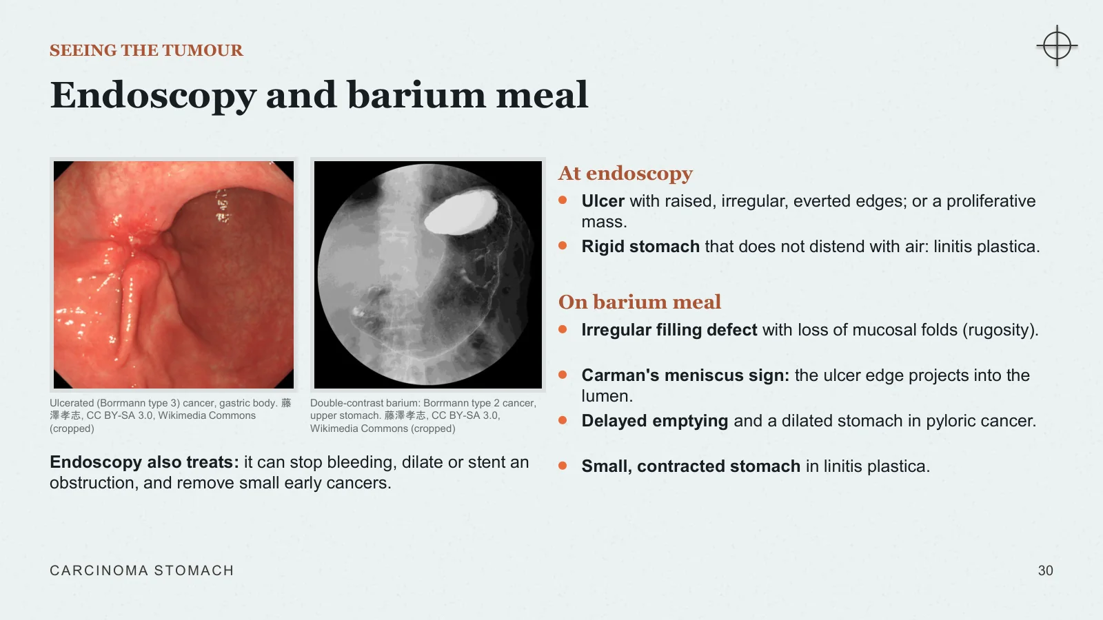
Ulcerated (Borrmann type 3) cancer, gastric body. 藤澤孝志, CC BY-SA 3.0, Wikimedia Commons (cropped)
Double-contrast barium: Borrmann type 2 cancer, upper stomach. 藤澤孝志, CC BY-SA 3.0, Wikimedia Commons (cropped)
Endoscopy also treats: it can stop bleeding, dilate or stent an obstruction, and remove small early cancers.
At endoscopy
Ulcer with raised, irregular, everted edges; or a proliferative mass.
Rigid stomach that does not distend with air: linitis plastica.
On barium meal
Irregular filling defect with loss of mucosal folds (rugosity).
Carman's meniscus sign: the ulcer edge projects into the lumen.
Delayed emptying and a dilated stomach in pyloric cancer.
Small, contracted stomach in linitis plastica.
CARCINOMA STOMACH
30
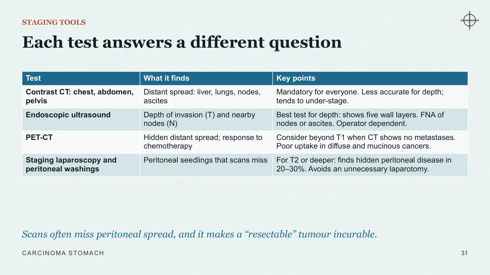
Each test answers a different question
STAGING TOOLS
Scans often miss peritoneal spread, and it makes a “resectable” tumour incurable.
CARCINOMA STOMACH
31
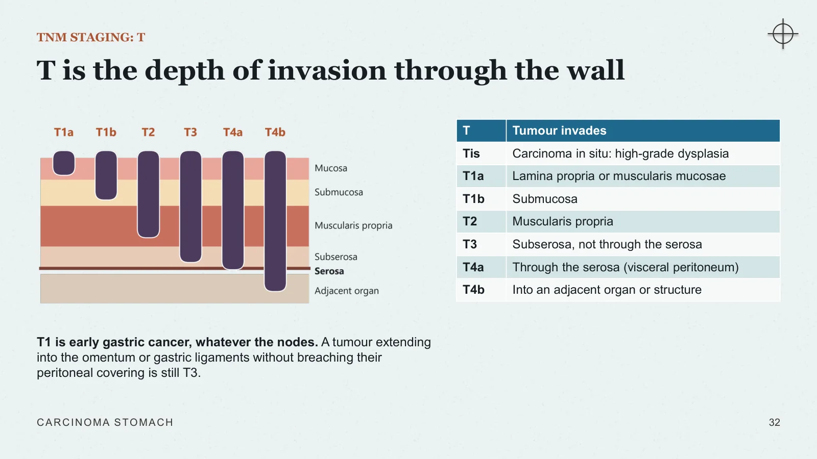
T is the depth of invasion through the wall
TNM STAGING: T
T1 is early gastric cancer, whatever the nodes. A tumour extending into the omentum or gastric ligaments without breaching their peritoneal covering is still T3.
CARCINOMA STOMACH
32
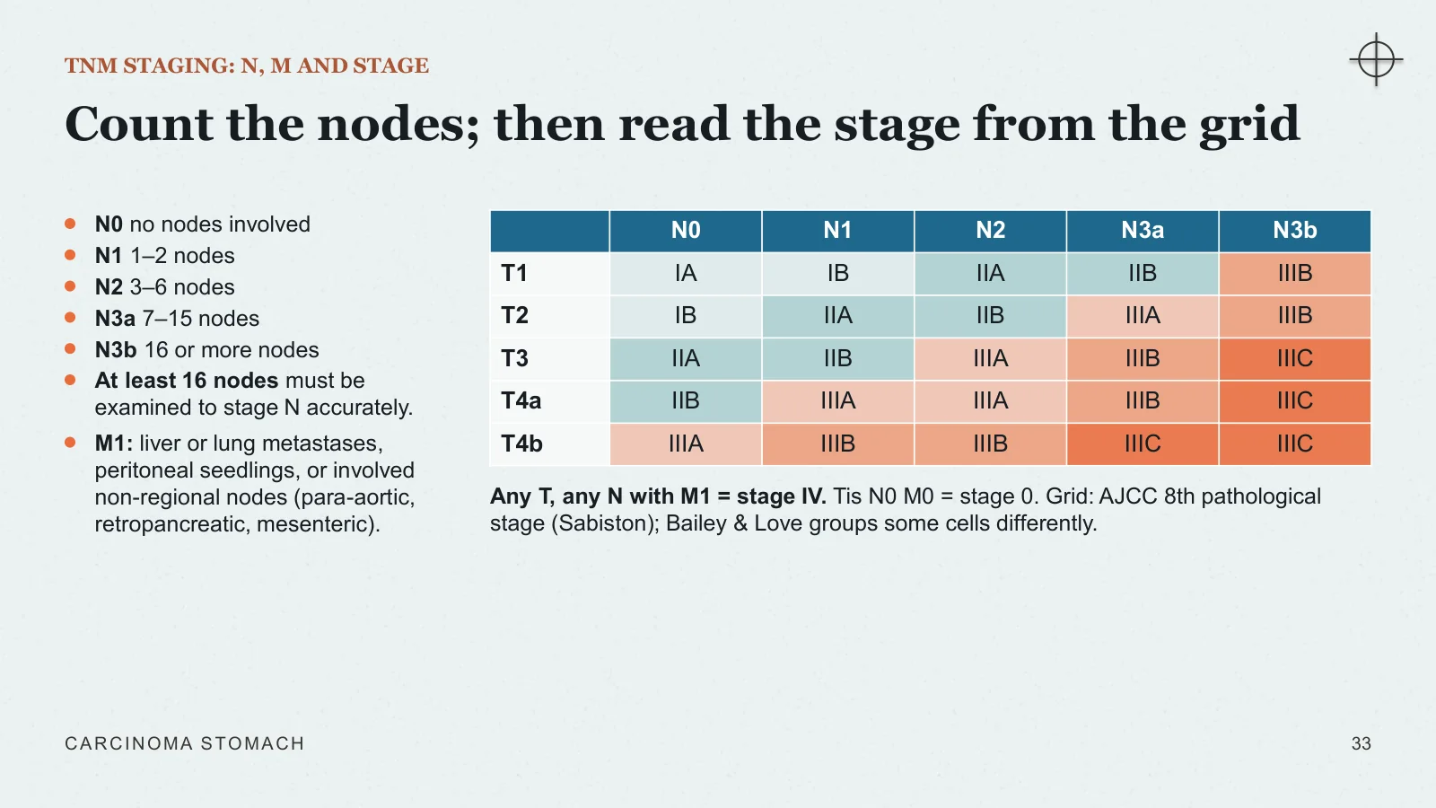
Count the nodes; then read the stage from the grid
TNM STAGING: N, M AND STAGE
N0 no nodes involved
N1 1–2 nodes
N2 3–6 nodes
N3a 7–15 nodes
N3b 16 or more nodes
At least 16 nodes must be examined to stage N accurately.
M1: liver or lung metastases, peritoneal seedlings, or involved non-regional nodes (para-aortic, retropancreatic, mesenteric).
Any T, any N with M1 = stage IV. Tis N0 M0 = stage 0. Grid: AJCC 8th pathological stage (Sabiston); Bailey & Love groups some cells differently.
CARCINOMA STOMACH
33
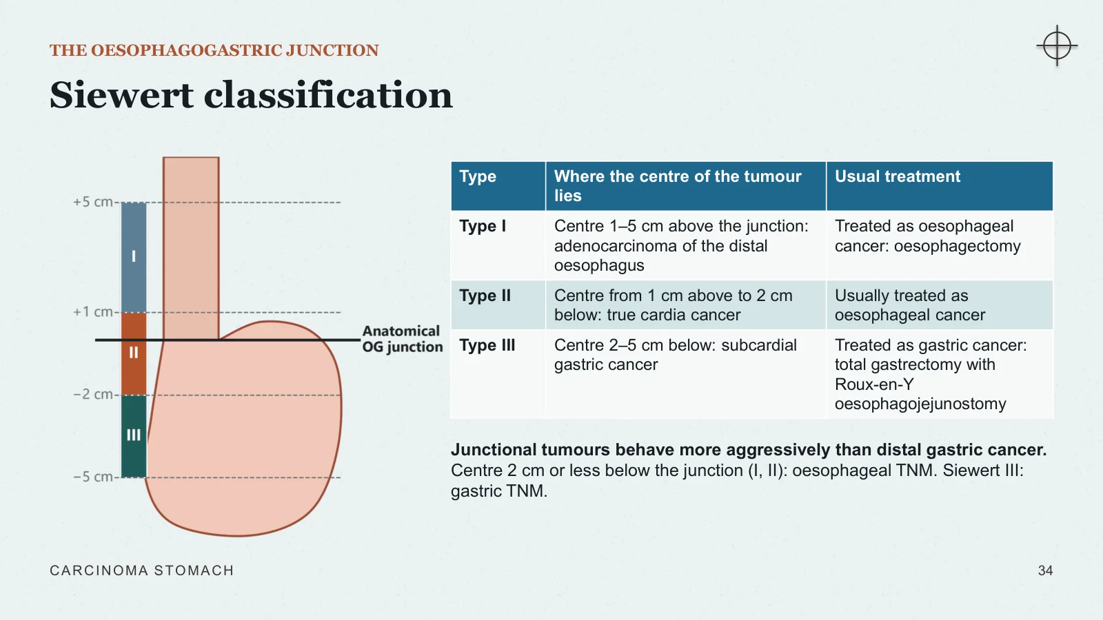
Siewert classification
THE OESOPHAGOGASTRIC JUNCTION
Junctional tumours behave more aggressively than distal gastric cancer. Centre 2 cm or less below the junction (I, II): oesophageal TNM. Siewert III: gastric TNM.
CARCINOMA STOMACH
34
Treatment
Treatment
V
Surgery is the only cure. Chemotherapy makes more of those operations succeed.
The treatment pathway and the multidisciplinary team
Endoscopic resection of early cancer
Radical gastrectomy: margins, D2 lymphadenectomy, reconstruction
Perioperative chemotherapy: the trial evidence
Advanced disease, palliation, complications and follow-up
CARCINOMA STOMACH
35
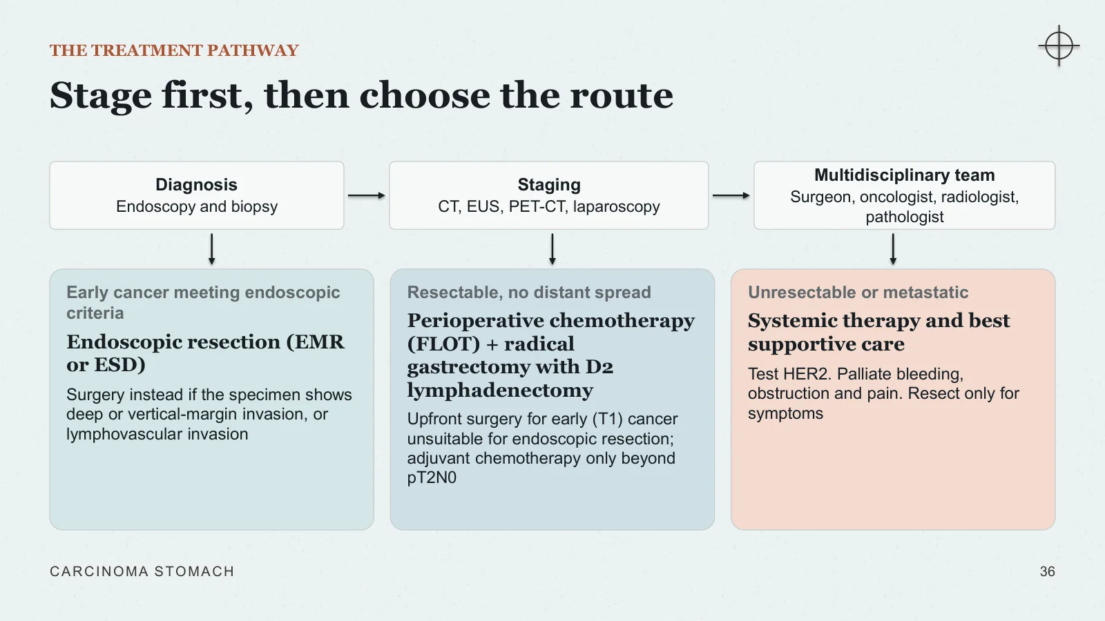
Stage first, then choose the route
THE TREATMENT PATHWAY
Diagnosis Endoscopy and biopsy
Staging CT, EUS, PET-CT, laparoscopy
Multidisciplinary team Surgeon, oncologist, radiologist, pathologist
Early cancer meeting endoscopic criteria
Endoscopic resection (EMR or ESD)
Surgery instead if the specimen shows deep or vertical-margin invasion, or lymphovascular invasion
Resectable, no distant spread
Perioperative chemotherapy (FLOT) + radical gastrectomy with D2 lymphadenectomy
Upfront surgery for early (T1) cancer unsuitable for endoscopic resection; adjuvant chemotherapy only beyond pT2N0
Unresectable or metastatic
Systemic therapy and best supportive care
Test HER2. Palliate bleeding, obstruction and pain. Resect only for symptoms
CARCINOMA STOMACH
36
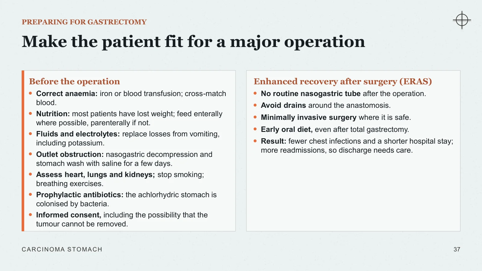
Make the patient fit for a major operation
PREPARING FOR GASTRECTOMY
Before the operation
Correct anaemia: iron or blood transfusion; cross-match blood.
Nutrition: most patients have lost weight; feed enterally where possible, parenterally if not.
Fluids and electrolytes: replace losses from vomiting, including potassium.
Outlet obstruction: nasogastric decompression and stomach wash with saline for a few days.
Assess heart, lungs and kidneys; stop smoking; breathing exercises.
Prophylactic antibiotics: the achlorhydric stomach is colonised by bacteria.
Informed consent, including the possibility that the tumour cannot be removed.
Enhanced recovery after surgery (ERAS)
No routine nasogastric tube after the operation.
Avoid drains around the anastomosis.
Minimally invasive surgery where it is safe.
Early oral diet, even after total gastrectomy.
Result: fewer chest infections and a shorter hospital stay; more readmissions, so discharge needs care.
CARCINOMA STOMACH
37
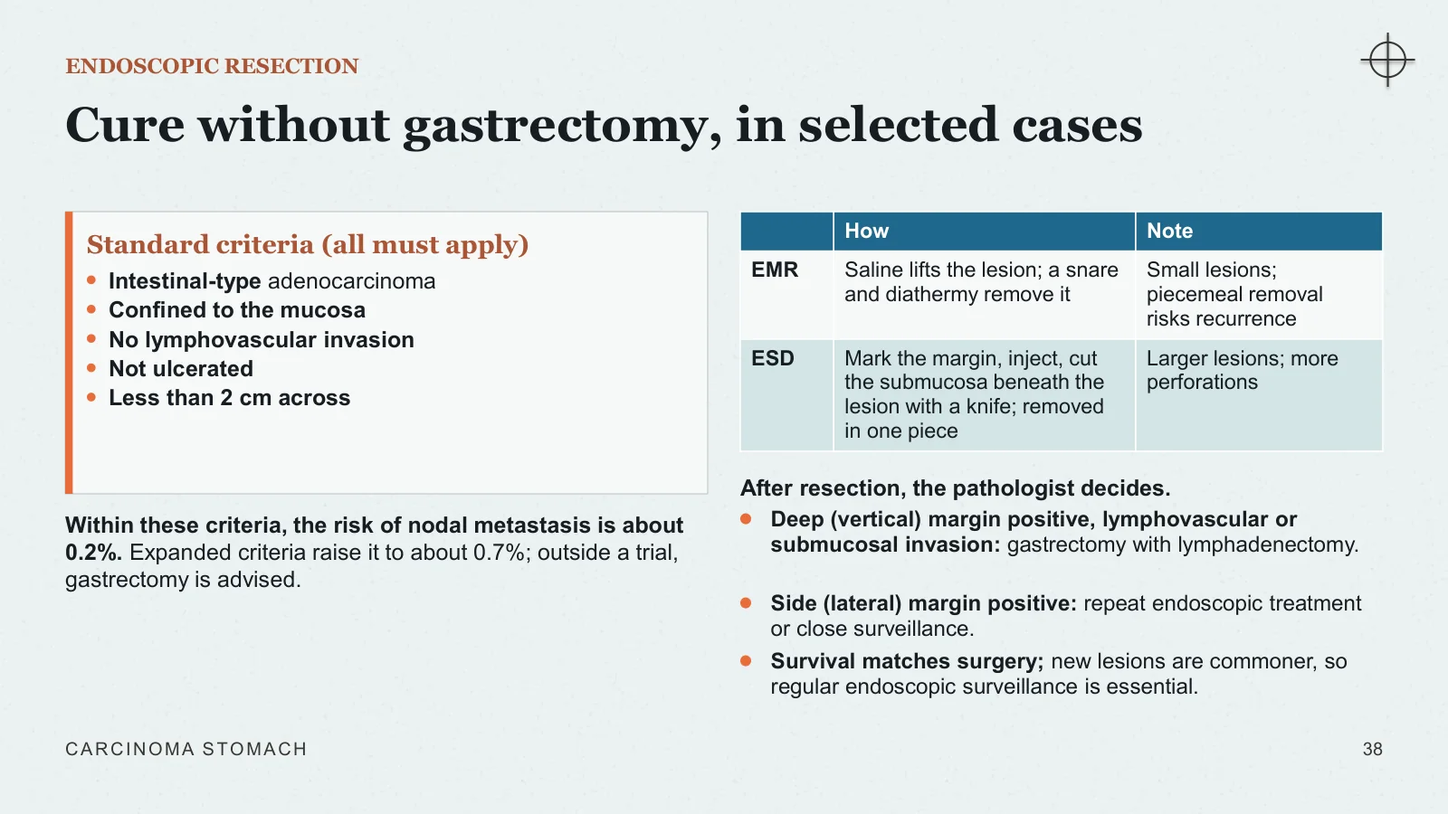
Cure without gastrectomy, in selected cases
ENDOSCOPIC RESECTION
Standard criteria (all must apply)
Intestinal-type adenocarcinoma
Confined to the mucosa
No lymphovascular invasion
Not ulcerated
Less than 2 cm across
Within these criteria, the risk of nodal metastasis is about 0.2%. Expanded criteria raise it to about 0.7%; outside a trial, gastrectomy is advised.
After resection, the pathologist decides.
Deep (vertical) margin positive, lymphovascular or submucosal invasion: gastrectomy with lymphadenectomy.
Side (lateral) margin positive: repeat endoscopic treatment or close surveillance.
Survival matches surgery; new lesions are commoner, so regular endoscopic surveillance is essential.
CARCINOMA STOMACH
38
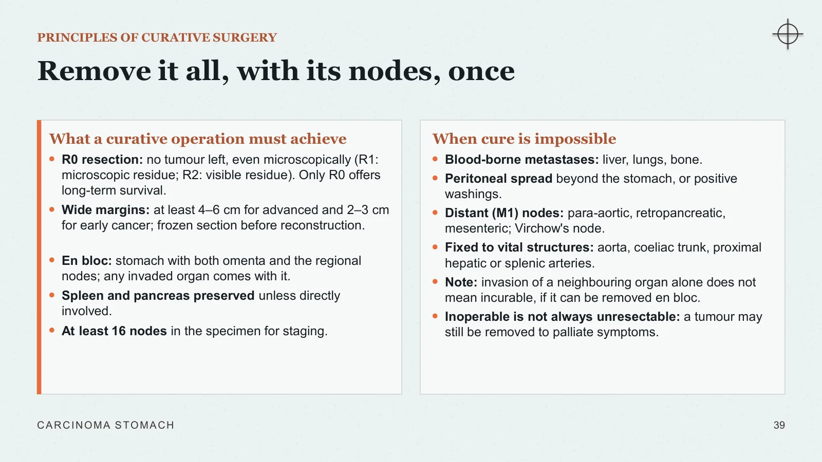
Remove it all, with its nodes, once
PRINCIPLES OF CURATIVE SURGERY
What a curative operation must achieve
R0 resection: no tumour left, even microscopically (R1: microscopic residue; R2: visible residue). Only R0 offers long-term survival.
Wide margins: at least 4–6 cm for advanced and 2–3 cm for early cancer; frozen section before reconstruction.
En bloc: stomach with both omenta and the regional nodes; any invaded organ comes with it.
Spleen and pancreas preserved unless directly involved.
At least 16 nodes in the specimen for staging.
When cure is impossible
Blood-borne metastases: liver, lungs, bone.
Peritoneal spread beyond the stomach, or positive washings.
Distant (M1) nodes: para-aortic, retropancreatic, mesenteric; Virchow's node.
Fixed to vital structures: aorta, coeliac trunk, proximal hepatic or splenic arteries.
Note: invasion of a neighbouring organ alone does not mean incurable, if it can be removed en bloc.
Inoperable is not always unresectable: a tumour may still be removed to palliate symptoms.
CARCINOMA STOMACH
39
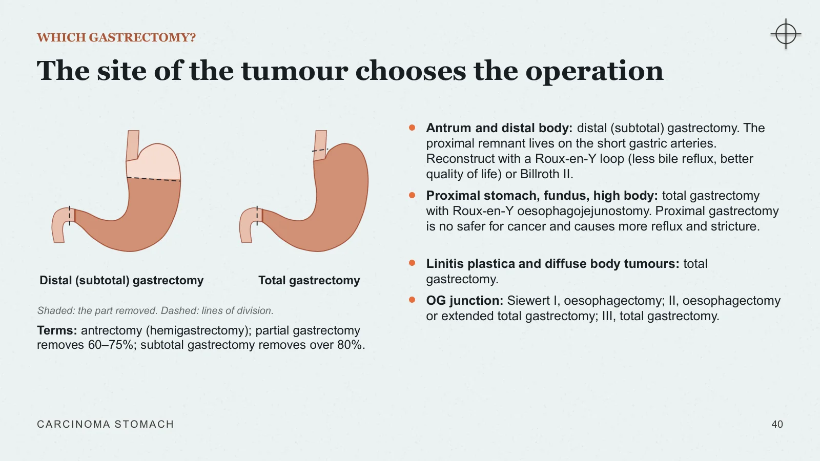
The site of the tumour chooses the operation
WHICH GASTRECTOMY?
Distal (subtotal) gastrectomy
Total gastrectomy
Shaded: the part removed. Dashed: lines of division.
Terms: antrectomy (hemigastrectomy); partial gastrectomy removes 60–75%; subtotal gastrectomy removes over 80%.
Antrum and distal body: distal (subtotal) gastrectomy. The proximal remnant lives on the short gastric arteries. Reconstruct with a Roux-en-Y loop (less bile reflux, better quality of life) or Billroth II.
Proximal stomach, fundus, high body: total gastrectomy with Roux-en-Y oesophagojejunostomy. Proximal gastrectomy is no safer for cancer and causes more reflux and stricture.
Linitis plastica and diffuse body tumours: total gastrectomy.
OG junction: Siewert I, oesophagectomy; II, oesophagectomy or extended total gastrectomy; III, total gastrectomy.
CARCINOMA STOMACH
40
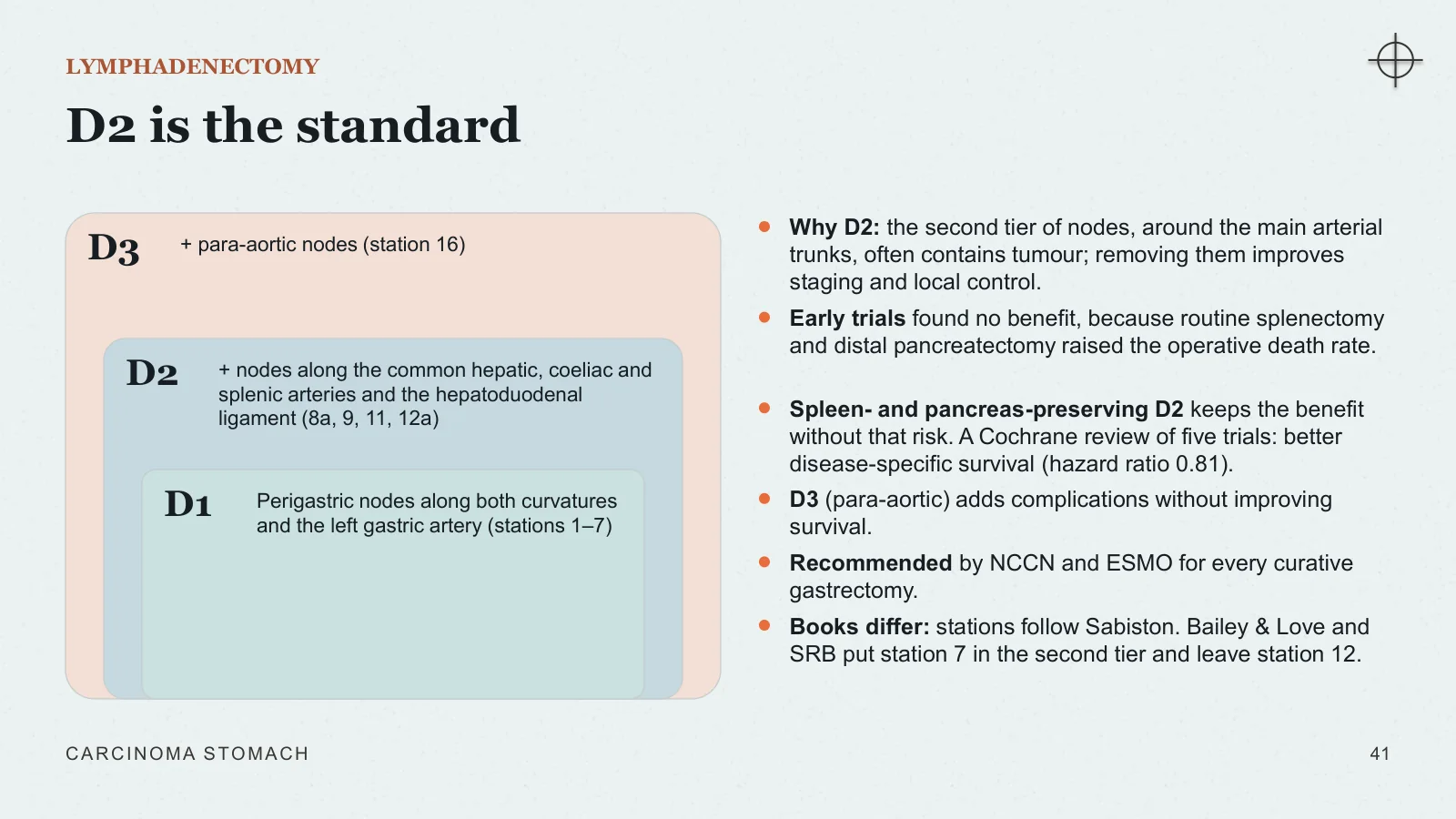
D2 is the standard
LYMPHADENECTOMY
D3
+ para-aortic nodes (station 16)
D2
+ nodes along the common hepatic, coeliac and splenic arteries and the hepatoduodenal ligament (8a, 9, 11, 12a)
D1
Perigastric nodes along both curvatures and the left gastric artery (stations 1–7)
Why D2: the second tier of nodes, around the main arterial trunks, often contains tumour; removing them improves staging and local control.
Early trials found no benefit, because routine splenectomy and distal pancreatectomy raised the operative death rate.
Spleen- and pancreas-preserving D2 keeps the benefit without that risk. A Cochrane review of five trials: better disease-specific survival (hazard ratio 0.81).
D3 (para-aortic) adds complications without improving survival.
Recommended by NCCN and ESMO for every curative gastrectomy.
Books differ: stations follow Sabiston. Bailey & Love and SRB put station 7 in the second tier and leave station 12.
CARCINOMA STOMACH
41
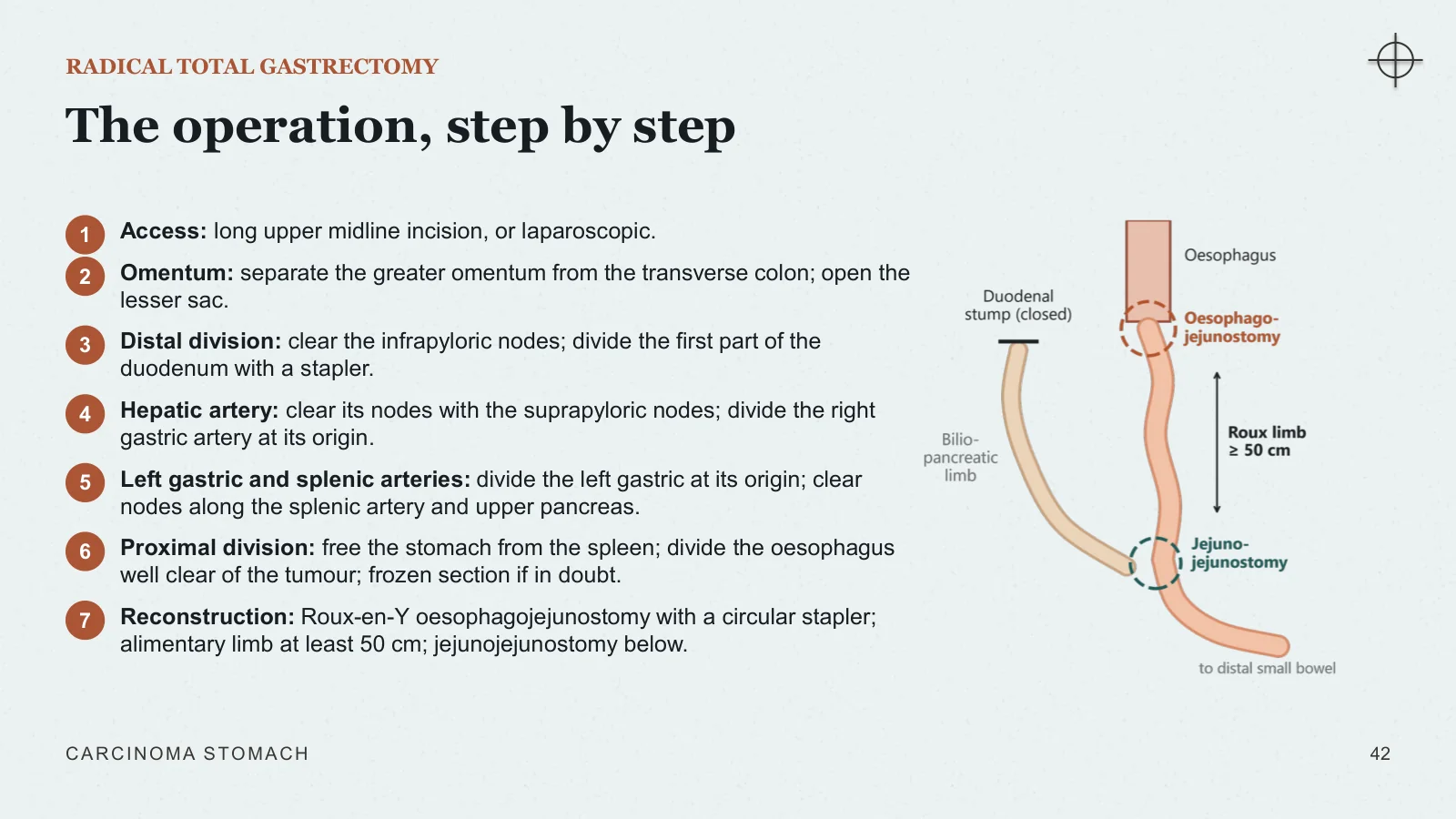
The operation, step by step
RADICAL TOTAL GASTRECTOMY
1
Access: long upper midline incision, or laparoscopic.
2
Omentum: separate the greater omentum from the transverse colon; open the lesser sac.
3
Distal division: clear the infrapyloric nodes; divide the first part of the duodenum with a stapler.
4
Hepatic artery: clear its nodes with the suprapyloric nodes; divide the right gastric artery at its origin.
5
Left gastric and splenic arteries: divide the left gastric at its origin; clear nodes along the splenic artery and upper pancreas.
6
Proximal division: free the stomach from the spleen; divide the oesophagus well clear of the tumour; frozen section if in doubt.
7
Reconstruction: Roux-en-Y oesophagojejunostomy with a circular stapler; alimentary limb at least 50 cm; jejunojejunostomy below.
CARCINOMA STOMACH
42
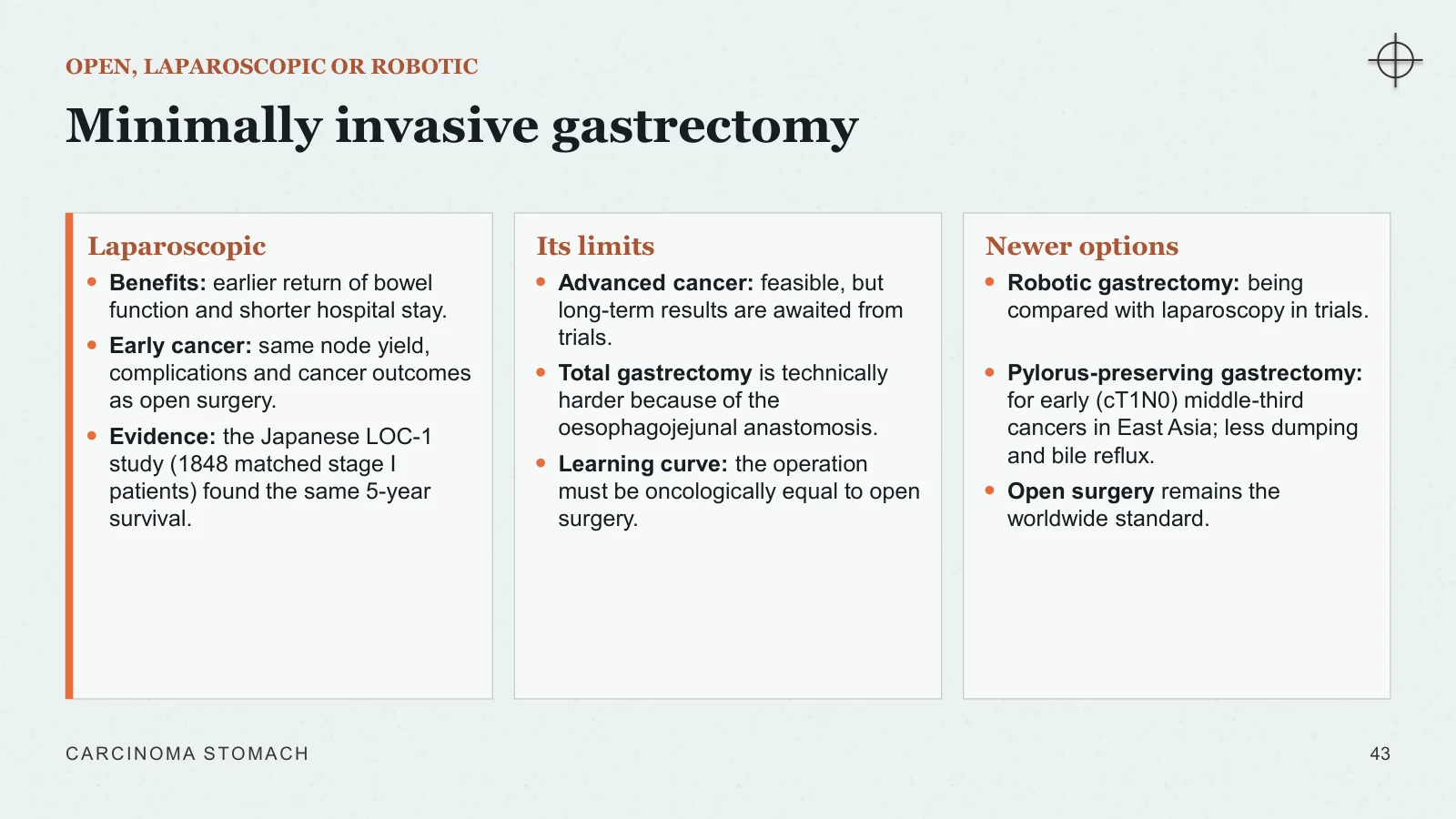
Minimally invasive gastrectomy
OPEN, LAPAROSCOPIC OR ROBOTIC
Laparoscopic
Benefits: earlier return of bowel function and shorter hospital stay.
Early cancer: same node yield, complications and cancer outcomes as open surgery.
Evidence: the Japanese LOC-1 study (1848 matched stage I patients) found the same 5-year survival.
Its limits
Advanced cancer: feasible, but long-term results are awaited from trials.
Total gastrectomy is technically harder because of the oesophagojejunal anastomosis.
Learning curve: the operation must be oncologically equal to open surgery.
Newer options
Robotic gastrectomy: being compared with laparoscopy in trials.
Pylorus-preserving gastrectomy: for early (cT1N0) middle-third cancers in East Asia; less dumping and bile reflux.
Open surgery remains the worldwide standard.
CARCINOMA STOMACH
43
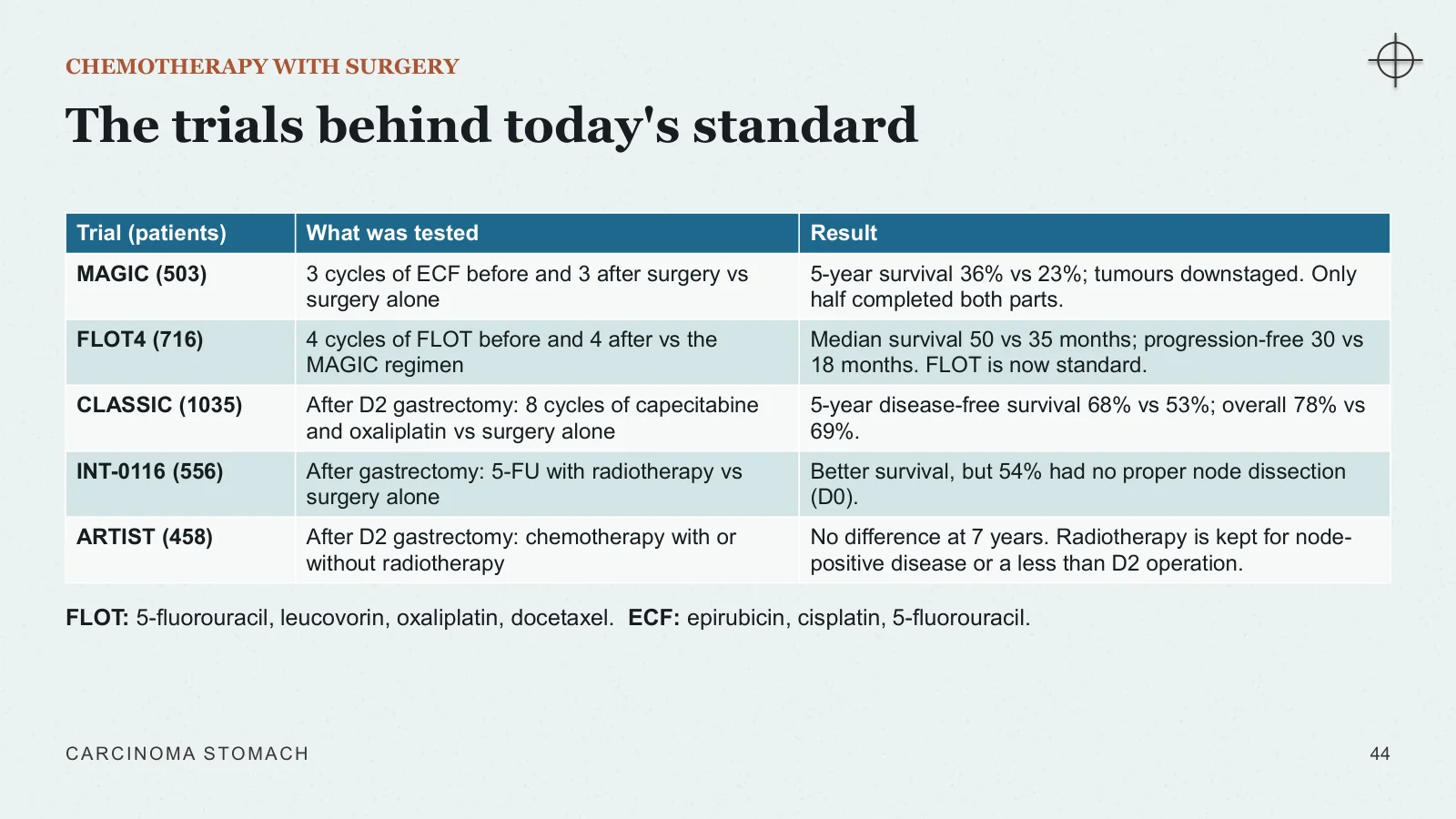
The trials behind today's standard
CHEMOTHERAPY WITH SURGERY
FLOT: 5-fluorouracil, leucovorin, oxaliplatin, docetaxel. ECF: epirubicin, cisplatin, 5-fluorouracil.
CARCINOMA STOMACH
44
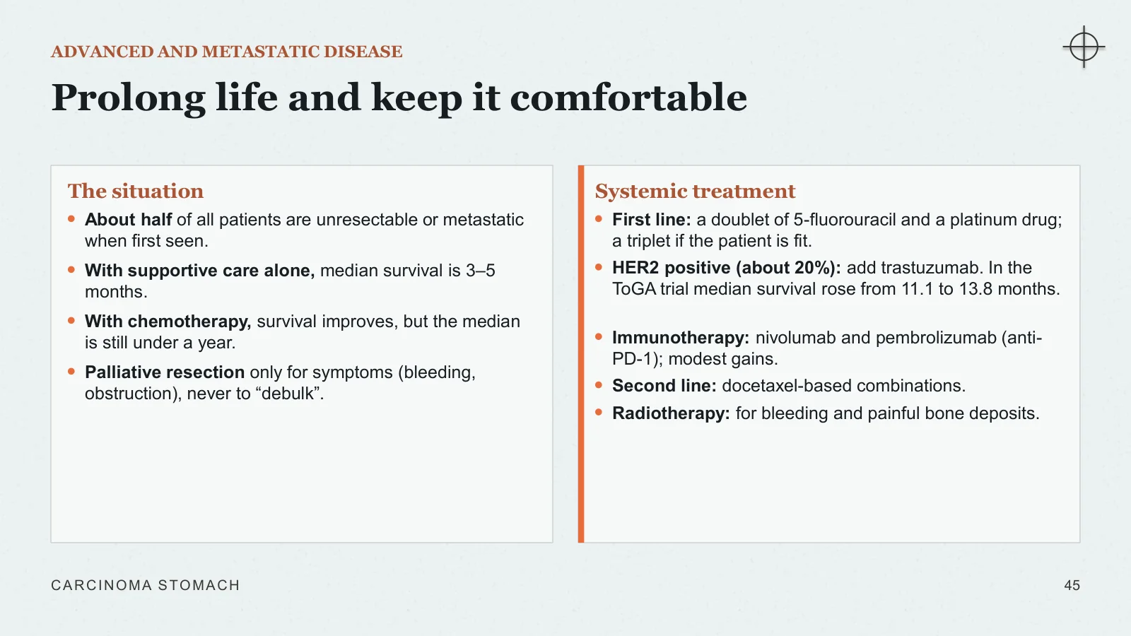
Prolong life and keep it comfortable
ADVANCED AND METASTATIC DISEASE
The situation
About half of all patients are unresectable or metastatic when first seen.
With supportive care alone, median survival is 3–5 months.
With chemotherapy, survival improves, but the median is still under a year.
Palliative resection only for symptoms (bleeding, obstruction), never to “debulk”.
Systemic treatment
First line: a doublet of 5-fluorouracil and a platinum drug; a triplet if the patient is fit.
HER2 positive (about 20%): add trastuzumab. In the ToGA trial median survival rose from 11.1 to 13.8 months.
Immunotherapy: nivolumab and pembrolizumab (anti-PD-1); modest gains.
Second line: docetaxel-based combinations.
Radiotherapy: for bleeding and painful bone deposits.
CARCINOMA STOMACH
45
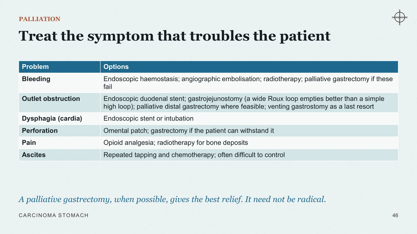
Treat the symptom that troubles the patient
PALLIATION
A palliative gastrectomy, when possible, gives the best relief. It need not be radical.
CARCINOMA STOMACH
46
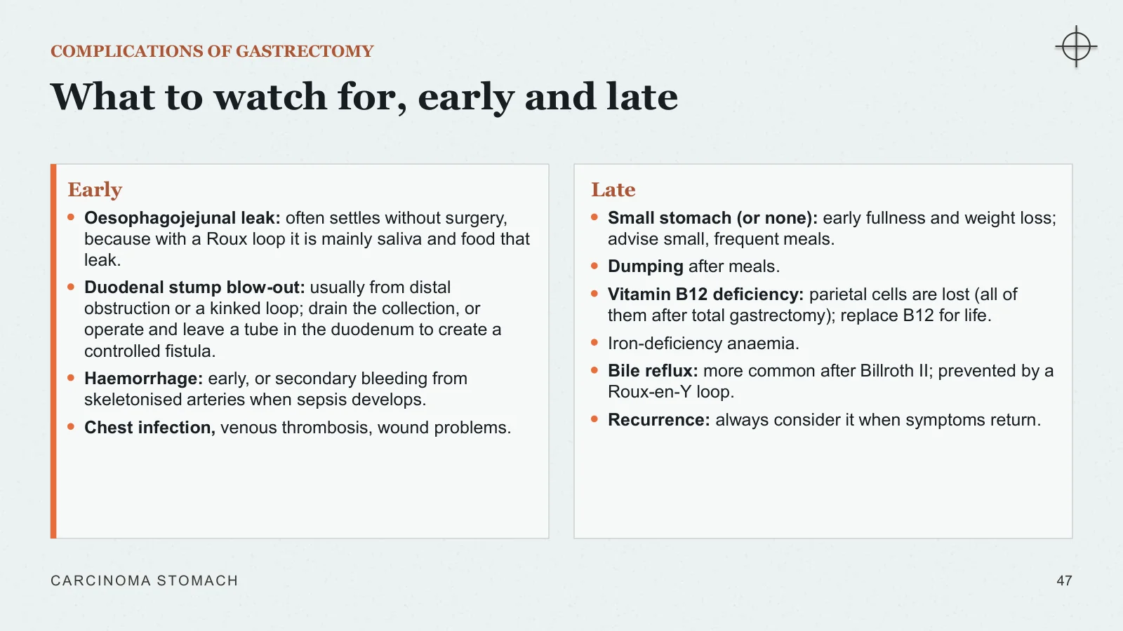
What to watch for, early and late
COMPLICATIONS OF GASTRECTOMY
Early
Oesophagojejunal leak: often settles without surgery, because with a Roux loop it is mainly saliva and food that leak.
Duodenal stump blow-out: usually from distal obstruction or a kinked loop; drain the collection, or operate and leave a tube in the duodenum to create a controlled fistula.
Haemorrhage: early, or secondary bleeding from skeletonised arteries when sepsis develops.
Chest infection, venous thrombosis, wound problems.
Late
Small stomach (or none): early fullness and weight loss; advise small, frequent meals.
Dumping after meals.
Vitamin B12 deficiency: parietal cells are lost (all of them after total gastrectomy); replace B12 for life.
Iron-deficiency anaemia.
Bile reflux: more common after Billroth II; prevented by a Roux-en-Y loop.
Recurrence: always consider it when symptoms return.
CARCINOMA STOMACH
47
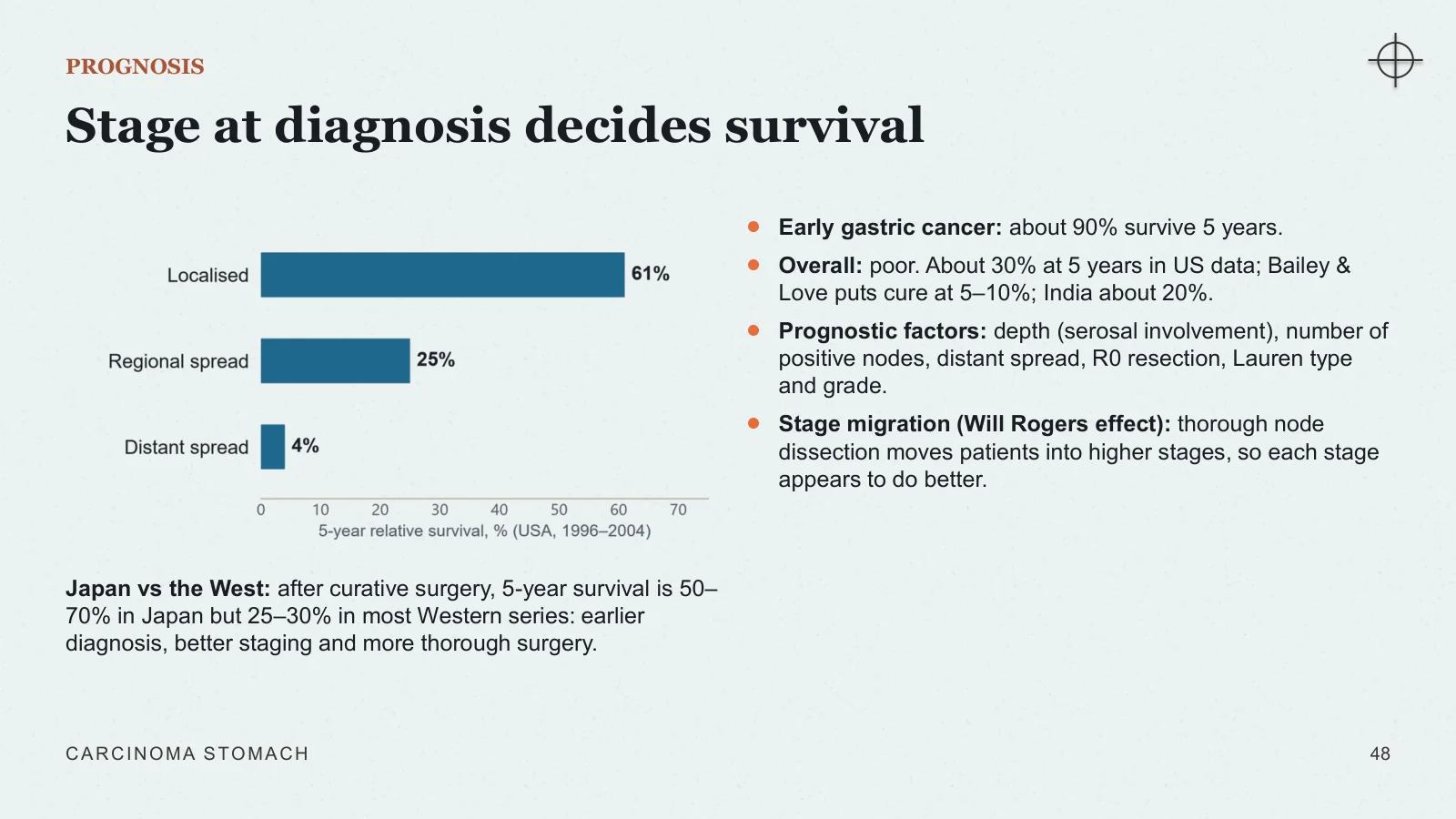
Stage at diagnosis decides survival
PROGNOSIS
Japan vs the West: after curative surgery, 5-year survival is 50–70% in Japan but 25–30% in most Western series: earlier diagnosis, better staging and more thorough surgery.
Early gastric cancer: about 90% survive 5 years.
Overall: poor. About 30% at 5 years in US data; Bailey & Love puts cure at 5–10%; India about 20%.
Prognostic factors: depth (serosal involvement), number of positive nodes, distant spread, R0 resection, Lauren type and grade.
Stage migration (Will Rogers effect): thorough node dissection moves patients into higher stages, so each stage appears to do better.
CARCINOMA STOMACH
48
Most recurrences appear within two years
RECURRENCE AND FOLLOW-UP
Where it comes back
Frequency: 30–90% after gastrectomy, depending on the series.
Timing: mostly within the first two years.
Local (about 40%): gastric bed, the anastomosis or remnant, regional nodes.
Distant: liver and peritoneum; lung and bone usually only after the liver.
Follow-up schedule
History and examination: every 3–6 months for 1–2 years, then every 6–12 months to 5 years, then yearly.
Blood count and liver tests as clinically indicated.
CT or PET-CT when recurrence is suspected.
Yearly endoscopy can be considered after subtotal gastrectomy or endoscopic resection.
Evidence: there is no proof that follow-up improves long-term survival.
CARCINOMA STOMACH
49
Recap and sources
Putting it together
BACK TO OUR PATIENT
1
Diagnosis: carcinoma of the distal stomach (antrum or pylorus) causing gastric outlet obstruction.
2
Resuscitate: IV fluids, potassium and chloride; nasogastric decompression and stomach wash; correct anaemia; start nutrition.
3
Confirm: endoscopy with 6–8 biopsies.
4
Stage: contrast CT of chest, abdomen and pelvis; staging laparoscopy with washings; endoscopic ultrasound if it would change the plan.
5
If curable: multidisciplinary team; FLOT, 4 cycles before and 4 after a radical distal (subtotal) gastrectomy with D2 lymphadenectomy and Roux-en-Y reconstruction.
6
If metastatic: palliative distal gastrectomy if feasible, otherwise stent or gastrojejunostomy; HER2 testing, palliative chemotherapy and supportive care.
7
Afterwards: follow-up for recurrence; vitamin B12, iron and nutrition.
CARCINOMA STOMACH
50
Eight things to remember
TAKE-HOME MESSAGES
1
Early gastric cancer mimics benign dyspepsia: keep a high index of suspicion and scope early.
2
H. pylori, salt and atrophy drive distal, intestinal-type cancer; proximal cancer has different causes.
3
Early cancer is defined by depth (T1), not by size or nodes, and is curable in about 90%.
4
Look for the named signs of spread: Virchow's node, Sister Mary Joseph nodule, Blumer's shelf, Krukenberg tumour.
5
Endoscopy with multiple biopsies confirms it; CT and laparoscopy decide whether it is curable.
6
Curative surgery means R0 resection with wide margins and a spleen-preserving D2 lymphadenectomy.
7
Perioperative FLOT chemotherapy is standard for operable cancer beyond the early stage.
8
Palliate the symptom in incurable disease. After total or subtotal gastrectomy, replace vitamin B12 for life.
CARCINOMA STOMACH
51
Sources and image credits
SOURCES
Textbooks
Bailey & Love's Short Practice of Surgery, 28th edition: chapter 67, The stomach and duodenum.
Sabiston Textbook of Surgery, 21st edition: chapter 49, Stomach.
SRB's Manual of Surgery, 6th edition: Stomach and duodenum.
Roy H. Short Textbook of Surgery: Stomach and duodenum.
Staging: AJCC TNM 8th edition, stage groups from Sabiston Table 49.6.
Diagrams and charts were drawn for this lecture from these sources. The case is illustrative.
Images (Wikimedia Commons and Wellcome Collection)
Cover: Watercolour by William Alfred Delamotte, 1848: hard cancer of the stomach. Wellcome Collection, CC BY 4.0.
Linitis plastica plate: Alfred Kast, plate F19: diffuse colloid (scirrhous) cancer of the stomach. Wellcome Collection, CC BY 4.0.
Early cancer, endoscopy (two images): Med_Chaos, public domain, Wikimedia Commons.
Advanced cancer, endoscopy: GASTROLAB, public domain, Wikimedia Commons.
Ulcerated cancer, endoscopy: 藤澤孝志, CC BY-SA 3.0, Wikimedia Commons (cropped).
Barium meal: 藤澤孝志, CC BY-SA 3.0, Wikimedia Commons (cropped).
Fungating cancer, specimen: Ed Uthman, CC BY 2.0, Wikimedia Commons (cropped).
Linitis plastica, specimen: Narraburra, CC0, Wikimedia Commons (cropped).
Intestinal-type histology: Dept of Pathology, Calicut Medical College, CC BY-SA 4.0, Wikimedia Commons (cropped).
Signet-ring cells: Nominoraspis, CC BY-SA 4.0, Wikimedia Commons (cropped).
Intestinal metaplasia: CoRus13, CC BY-SA 4.0, Wikimedia Commons (cropped).
H. pylori micrograph: Yutaka Tsutsumi, Fujita Health University, free use, Wikimedia Commons.
Krukenberg tumour: Ed Uthman specimen collection, public domain, Wikimedia Commons.
CARCINOMA STOMACH
52
Sources and image credits
Image credits and source details are reproduced from the final slide below.
Sources
- [1] SOURCES
- [2] Sources and image credits
- [3] Textbooks
- [4] Bailey & Love's Short Practice of Surgery, 28th edition: chapter 67, The stomach and duodenum.
- [5] Sabiston Textbook of Surgery, 21st edition: chapter 49, Stomach.
- [6] SRB's Manual of Surgery, 6th edition: Stomach and duodenum.
- [7] Roy H. Short Textbook of Surgery: Stomach and duodenum.
- [8] Staging: AJCC TNM 8th edition, stage groups from Sabiston Table 49.6.
- [9] Diagrams and charts were drawn for this lecture from these sources. The case is illustrative.
- [10] Images (Wikimedia Commons and Wellcome Collection)
- [11] Cover: Watercolour by William Alfred Delamotte, 1848: hard cancer of the stomach. Wellcome Collection, CC BY 4.0.
- [12] Linitis plastica plate: Alfred Kast, plate F19: diffuse colloid (scirrhous) cancer of the stomach. Wellcome Collection, CC BY 4.0.
- [13] Early cancer, endoscopy (two images): Med_Chaos, public domain, Wikimedia Commons.
- [14] Advanced cancer, endoscopy: GASTROLAB, public domain, Wikimedia Commons.
- [15] Ulcerated cancer, endoscopy: 藤澤孝志, CC BY-SA 3.0, Wikimedia Commons (cropped).
- [16] Barium meal: 藤澤孝志, CC BY-SA 3.0, Wikimedia Commons (cropped).
- [17] Fungating cancer, specimen: Ed Uthman, CC BY 2.0, Wikimedia Commons (cropped).
- [18] Linitis plastica, specimen: Narraburra, CC0, Wikimedia Commons (cropped).
- [19] Intestinal-type histology: Dept of Pathology, Calicut Medical College, CC BY-SA 4.0, Wikimedia Commons (cropped).
- [20] Signet-ring cells: Nominoraspis, CC BY-SA 4.0, Wikimedia Commons (cropped).
- [21] Intestinal metaplasia: CoRus13, CC BY-SA 4.0, Wikimedia Commons (cropped).
- [22] H. pylori micrograph: Yutaka Tsutsumi, Fujita Health University, free use, Wikimedia Commons.
- [23] Krukenberg tumour: Ed Uthman specimen collection, public domain, Wikimedia Commons.