Colorectal · Stomas · 47 slides
Ileostomy and colostomy.
Follow the bowel to understand what a stoma changes: from the reason for diversion to postoperative care, complications and life with a stoma.
About the downloads
PowerPoint without speaker notes. SHA-256 checksums verify the downloaded files.
Contents · 6 parts
Understanding stomas
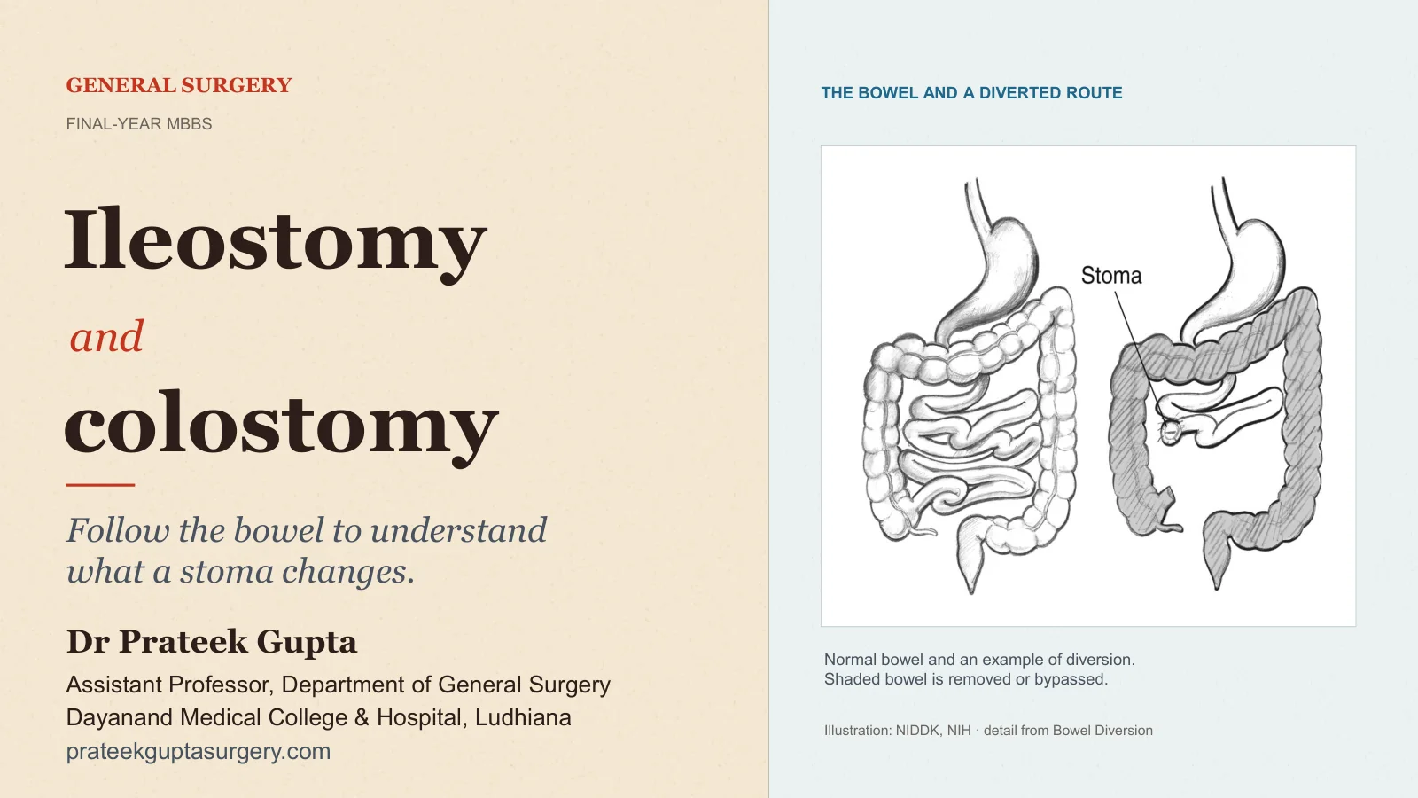
Ileostomy and Colostomy
GENERAL SURGERY
FINAL-YEAR MBBS
Ileostomy
and
colostomy
Follow the bowel to understand what a stoma changes.
THE BOWEL AND A DIVERTED ROUTE
Normal bowel and an example of diversion. Shaded bowel is removed or bypassed.
Illustration: NIDDK, NIH · detail from Bowel Diversion
Dr Prateek Gupta
Assistant Professor, Department of General Surgery
Dayanand Medical College & Hospital, Ludhiana
prateekguptasurgery.com
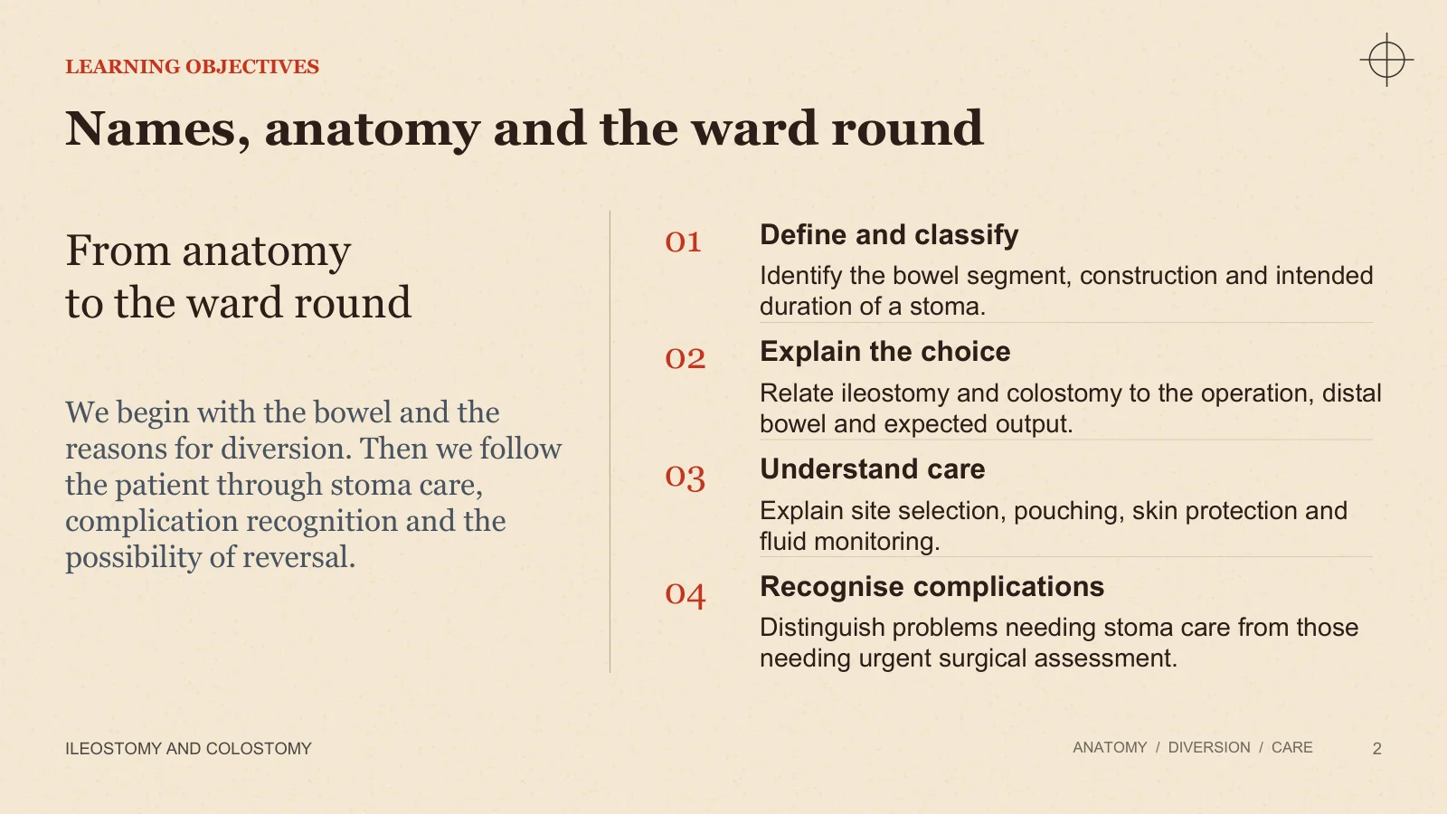
Names, anatomy and the ward round
LEARNING OBJECTIVES
ILEOSTOMY AND COLOSTOMY
ANATOMY / DIVERSION / CARE
2
From anatomy to the ward round
We begin with the bowel and the reasons for diversion. Then we follow the patient through stoma care, complication recognition and the possibility of reversal.
01
Define and classify
Identify the bowel segment, construction and intended duration of a stoma.
02
Explain the choice
Relate ileostomy and colostomy to the operation, distal bowel and expected output.
03
Understand care
Explain site selection, pouching, skin protection and fluid monitoring.
04
Recognise complications
Distinguish problems needing stoma care from those needing urgent surgical assessment.
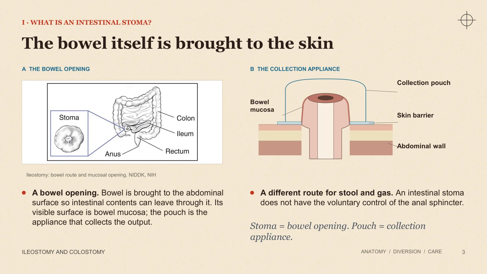
The bowel itself is brought to the skin
I · WHAT IS AN INTESTINAL STOMA?
ILEOSTOMY AND COLOSTOMY
ANATOMY / DIVERSION / CARE
3
A THE BOWEL OPENING
Ileostomy: bowel route and mucosal opening. NIDDK, NIH
B THE COLLECTION APPLIANCE
Collection pouch
Bowel mucosa
Skin barrier
Abdominal wall
A bowel opening. Bowel is brought to the abdominal surface so intestinal contents can leave through it. Its visible surface is bowel mucosa; the pouch is the appliance that collects the output.
A different route for stool and gas. An intestinal stoma does not have the voluntary control of the anal sphincter.
Stoma = bowel opening. Pouch = collection appliance.
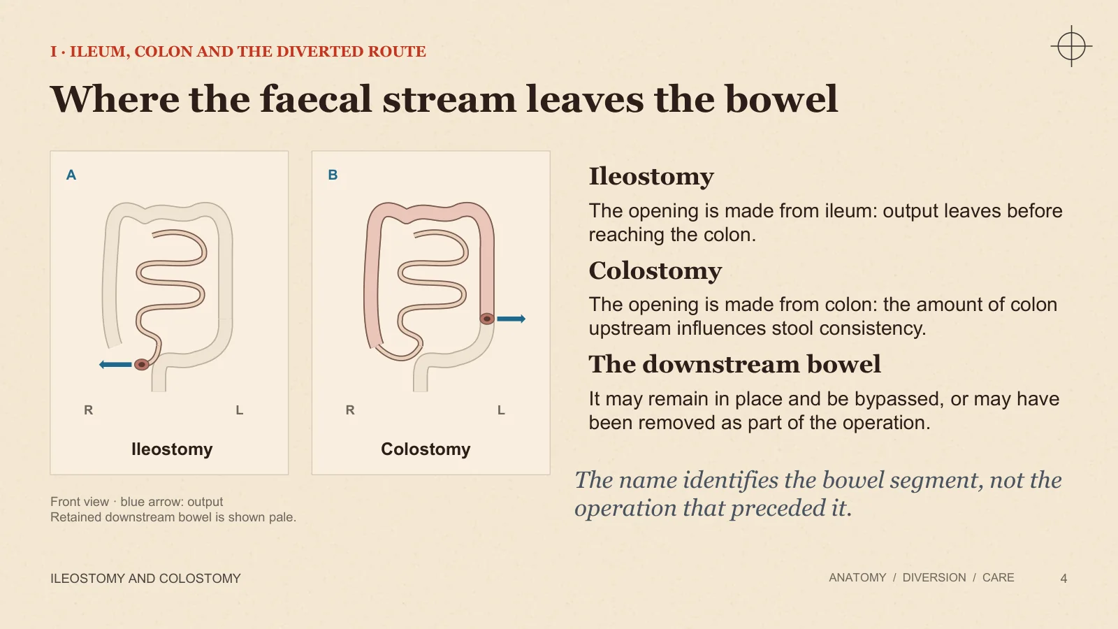
Where the faecal stream leaves the bowel
I · ILEUM, COLON AND THE DIVERTED ROUTE
ILEOSTOMY AND COLOSTOMY
ANATOMY / DIVERSION / CARE
4
A
B
R
L
R
L
Ileostomy
Colostomy
Front view · blue arrow: output Retained downstream bowel is shown pale.
Ileostomy
The opening is made from ileum: output leaves before reaching the colon.
Colostomy
The opening is made from colon: the amount of colon upstream influences stool consistency.
The downstream bowel
It may remain in place and be bypassed, or may have been removed as part of the operation.
The name identifies the bowel segment, not the operation that preceded it.
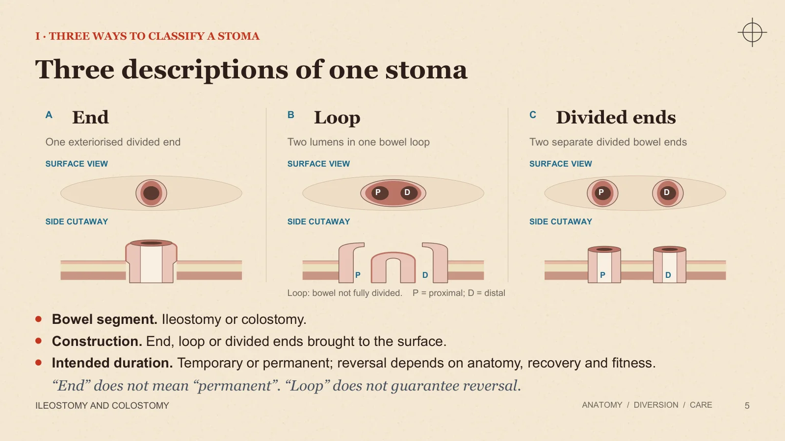
Three descriptions of one stoma
I · THREE WAYS TO CLASSIFY A STOMA
ILEOSTOMY AND COLOSTOMY
ANATOMY / DIVERSION / CARE
5
A
End
One exteriorised divided end
SURFACE VIEW
SIDE CUTAWAY
B
Loop
Two lumens in one bowel loop
SURFACE VIEW
P
D
SIDE CUTAWAY
P
D
C
Divided ends
Two separate divided bowel ends
SURFACE VIEW
P
D
SIDE CUTAWAY
P
D
Loop: bowel not fully divided. P = proximal; D = distal
Bowel segment. Ileostomy or colostomy.
Construction. End, loop or divided ends brought to the surface.
Intended duration. Temporary or permanent; reversal depends on anatomy, recovery and fitness.
“End” does not mean “permanent”. “Loop” does not guarantee reversal.
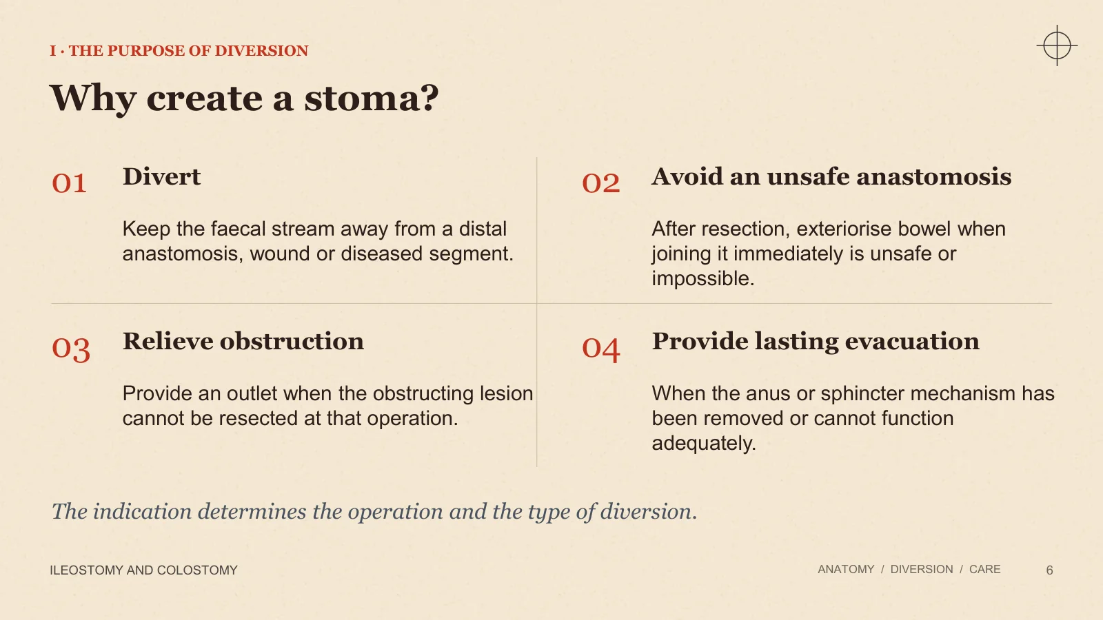
Why create a stoma?
I · THE PURPOSE OF DIVERSION
ILEOSTOMY AND COLOSTOMY
ANATOMY / DIVERSION / CARE
6
01
Divert
Keep the faecal stream away from a distal anastomosis, wound or diseased segment.
02
Avoid an unsafe anastomosis
After resection, exteriorise bowel when joining it immediately is unsafe or impossible.
03
Relieve obstruction
Provide an outlet when the obstructing lesion cannot be resected at that operation.
04
Provide lasting evacuation
When the anus or sphincter mechanism has been removed or cannot function adequately.
The indication determines the operation and the type of diversion.
Temporary and permanent stomas
I · DURATION IS AN INTENTION
ILEOSTOMY AND COLOSTOMY
ANATOMY / DIVERSION / CARE
7
LATER CONTINUITY MAY BE POSSIBLE
Temporary intention
A later operation may restore bowel continuity after the original problem has resolved.
LONG-TERM STOMA IS PLANNED
Permanent intention
Long-term evacuation through the stoma is planned when a usable distal outlet is absent or unsuitable.
Intention is not a promise. Complications, disease progression or reduced fitness can prevent a planned reversal.
Counselling must explain both the intended plan and the possibility that it changes.
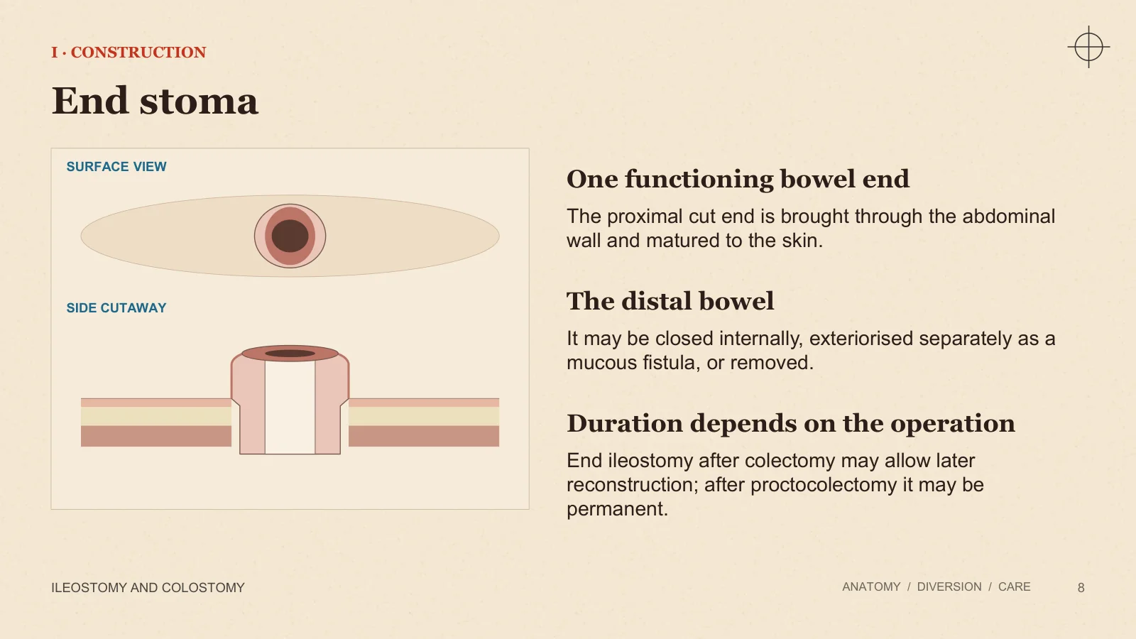
End stoma
I · CONSTRUCTION
ILEOSTOMY AND COLOSTOMY
ANATOMY / DIVERSION / CARE
8
SURFACE VIEW
SIDE CUTAWAY
One functioning bowel end
The proximal cut end is brought through the abdominal wall and matured to the skin.
The distal bowel
It may be closed internally, exteriorised separately as a mucous fistula, or removed.
Duration depends on the operation
End ileostomy after colectomy may allow later reconstruction; after proctocolectomy it may be permanent.
Loop stoma
I · CONSTRUCTION
ILEOSTOMY AND COLOSTOMY
ANATOMY / DIVERSION / CARE
9
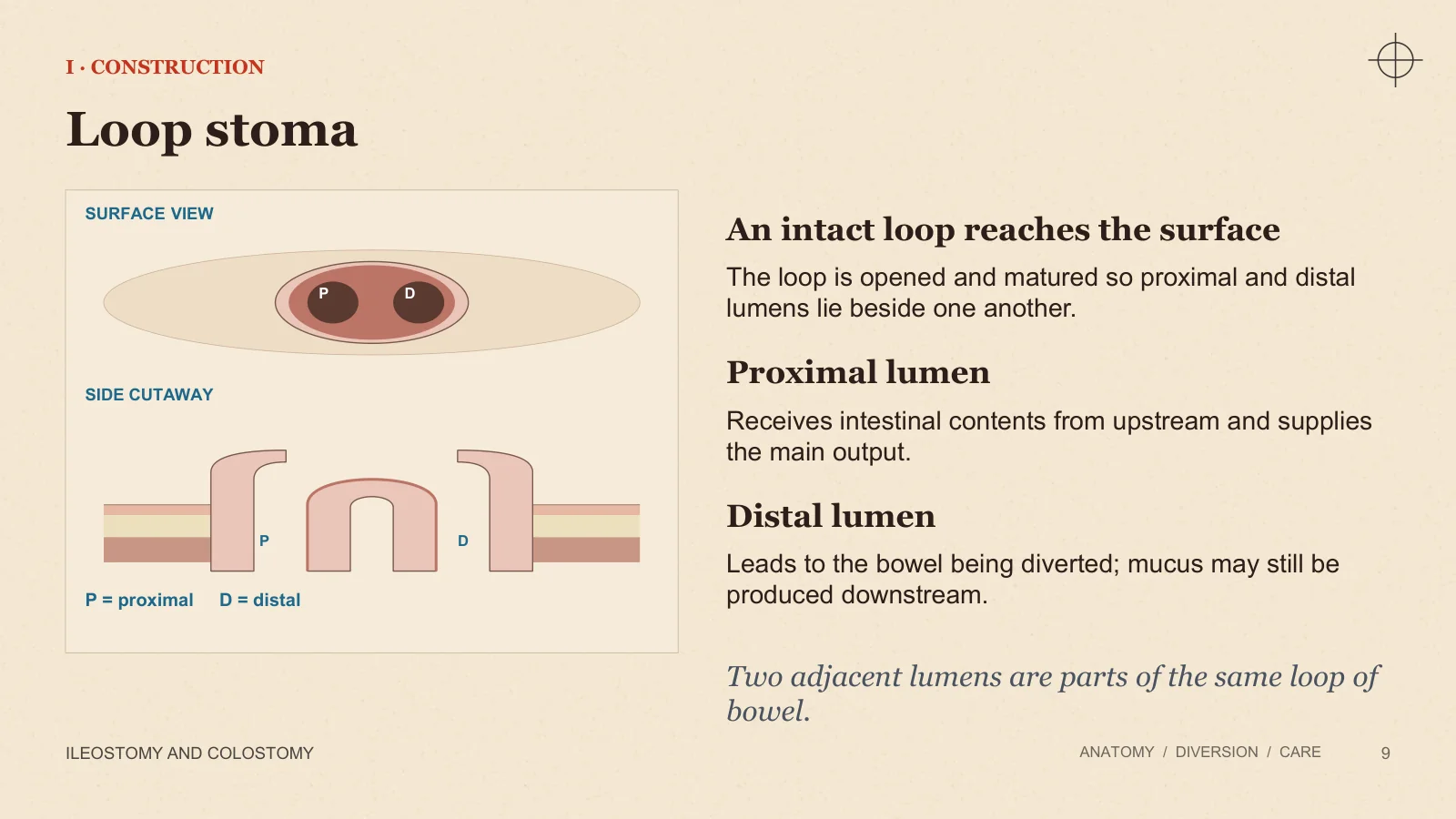
SURFACE VIEW
P
D
SIDE CUTAWAY
P
D
An intact loop reaches the surface
The loop is opened and matured so proximal and distal lumens lie beside one another.
Proximal lumen
Receives intestinal contents from upstream and supplies the main output.
Distal lumen
Leads to the bowel being diverted; mucus may still be produced downstream.
P = proximal D = distal
Two adjacent lumens are parts of the same loop of bowel.
Double-barrelled stoma and mucous fistula
I · CONSTRUCTION
ILEOSTOMY AND COLOSTOMY
ANATOMY / DIVERSION / CARE
10
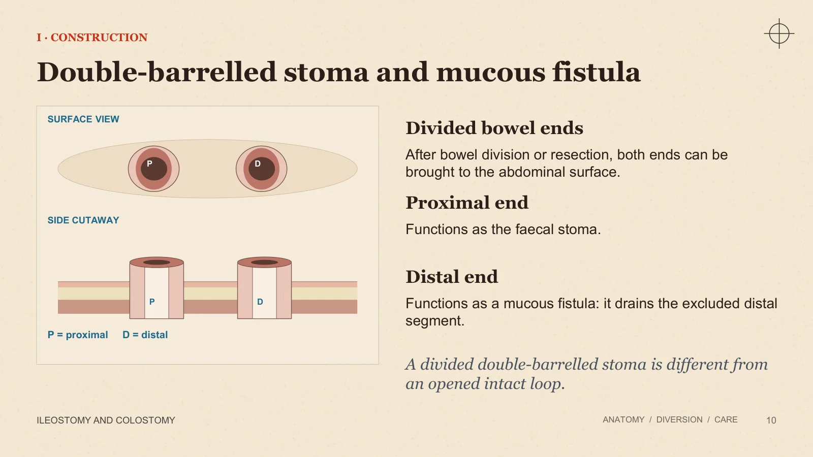
SURFACE VIEW
P
D
SIDE CUTAWAY
P
D
Divided bowel ends
After bowel division or resection, both ends can be brought to the abdominal surface.
Proximal end
Functions as the faecal stoma.
Distal end
Functions as a mucous fistula: it drains the excluded distal segment.
P = proximal D = distal
A divided double-barrelled stoma is different from an opened intact loop.
Ileostomy and colostomy
Ileostomy: indications
II · ILEOSTOMY
ILEOSTOMY AND COLOSTOMY
ANATOMY / DIVERSION / CARE
11
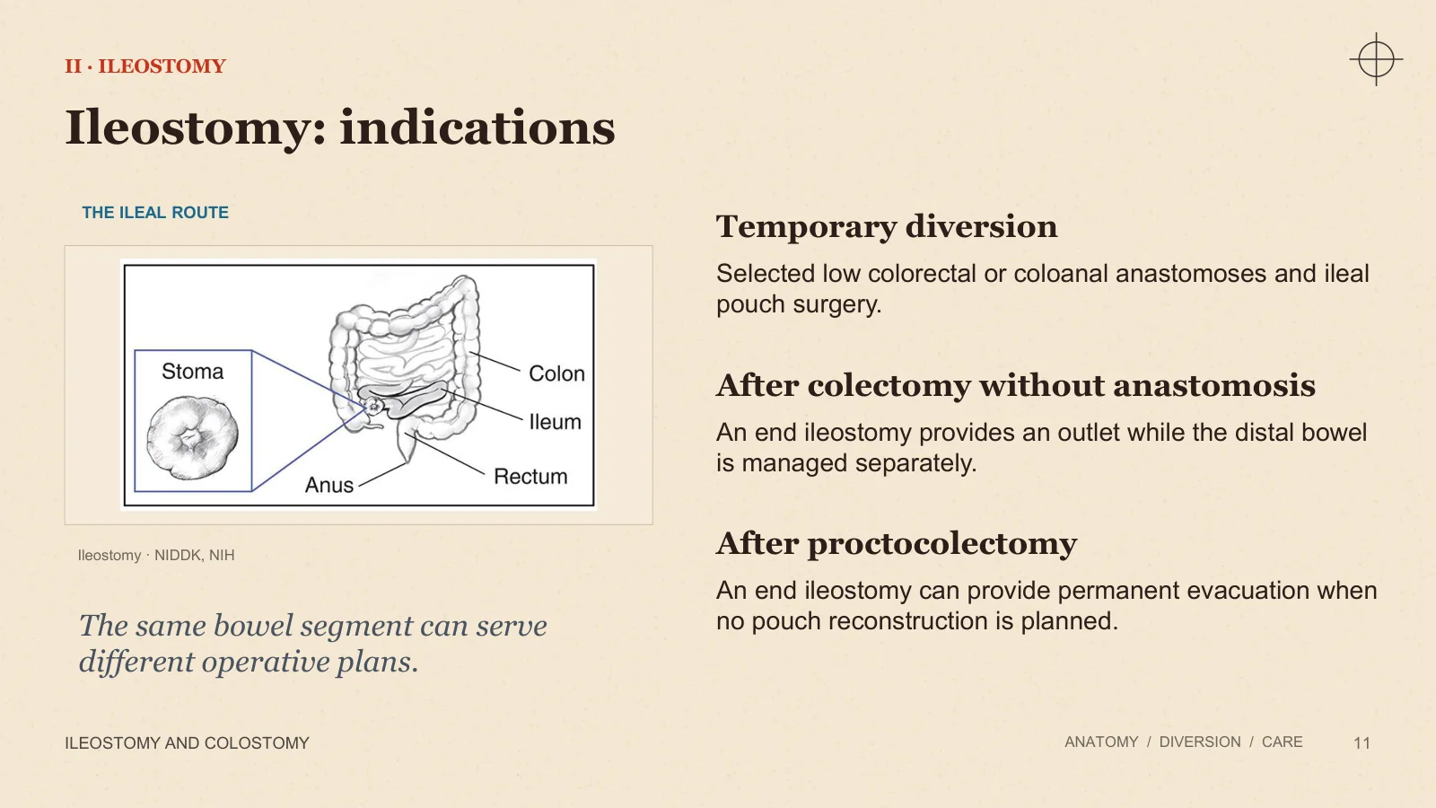
THE ILEAL ROUTE
Ileostomy · NIDDK, NIH
The same bowel segment can serve different operative plans.
Temporary diversion
Selected low colorectal or coloanal anastomoses and ileal pouch surgery.
After colectomy without anastomosis
An end ileostomy provides an outlet while the distal bowel is managed separately.
After proctocolectomy
An end ileostomy can provide permanent evacuation when no pouch reconstruction is planned.
Loop ileostomy and a distal anastomosis
II · DIVERSION AND HEALING
ILEOSTOMY AND COLOSTOMY
ANATOMY / DIVERSION / CARE
12
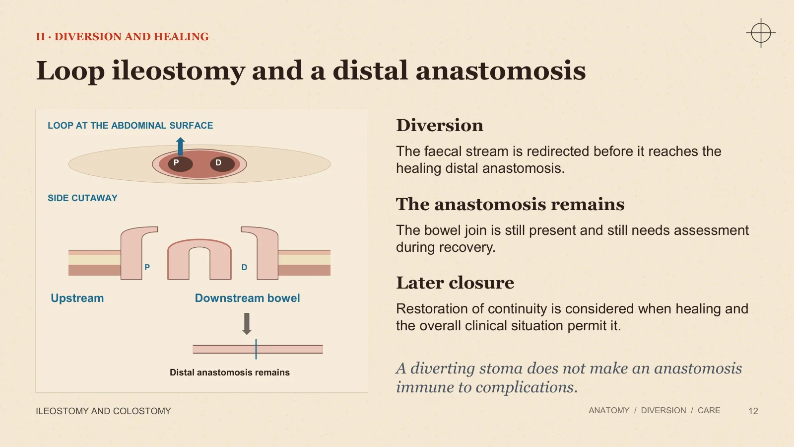
LOOP AT THE ABDOMINAL SURFACE
P
D
SIDE CUTAWAY
P
D
Upstream
Downstream bowel
Distal anastomosis remains
Diversion
The faecal stream is redirected before it reaches the healing distal anastomosis.
The anastomosis remains
The bowel join is still present and still needs assessment during recovery.
Later closure
Restoration of continuity is considered when healing and the overall clinical situation permit it.
A diverting stoma does not make an anastomosis immune to complications.
End ileostomy
II · ILEOSTOMY CONSTRUCTION
ILEOSTOMY AND COLOSTOMY
ANATOMY / DIVERSION / CARE
13
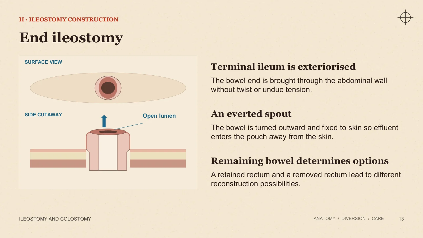
SURFACE VIEW
SIDE CUTAWAY
Open lumen
Terminal ileum is exteriorised
The bowel end is brought through the abdominal wall without twist or undue tension.
An everted spout
The bowel is turned outward and fixed to skin so effluent enters the pouch away from the skin.
Remaining bowel determines options
A retained rectum and a removed rectum lead to different reconstruction possibilities.
Ileostomy output and adaptation
II · THE PHYSIOLOGY OF ILEOSTOMY
ILEOSTOMY AND COLOSTOMY
ANATOMY / DIVERSION / CARE
14
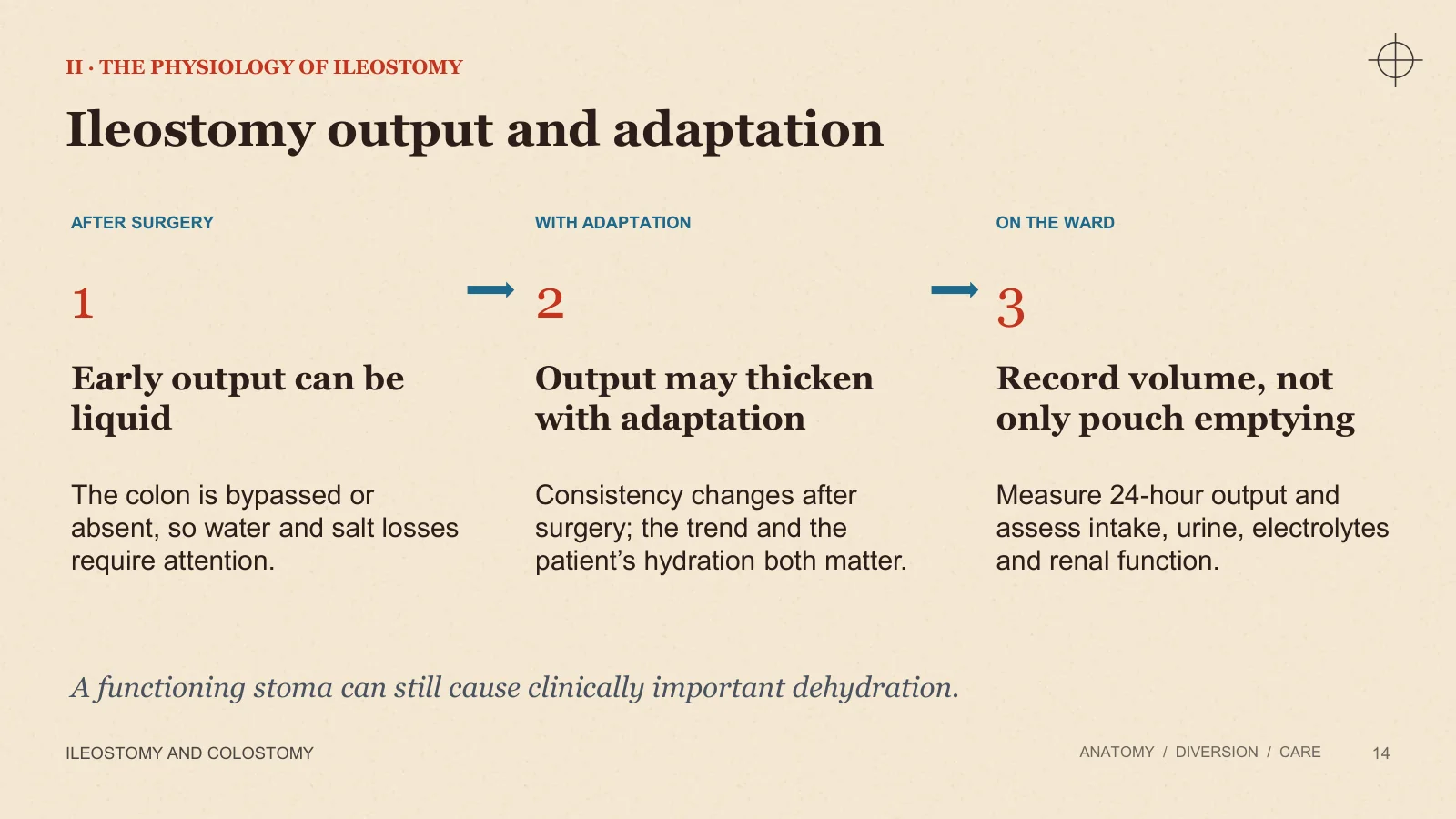
AFTER SURGERY
WITH ADAPTATION
ON THE WARD
1
Early output can be liquid
The colon is bypassed or absent, so water and salt losses require attention.
2
Output may thicken with adaptation
Consistency changes after surgery; the trend and the patient’s hydration both matter.
3
Record volume, not only pouch emptying
Measure 24-hour output and assess intake, urine, electrolytes and renal function.
A functioning stoma can still cause clinically important dehydration.
Colostomy: indications
II · COLOSTOMY
ILEOSTOMY AND COLOSTOMY
ANATOMY / DIVERSION / CARE
15
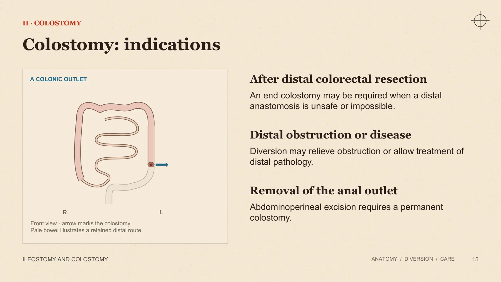
A COLONIC OUTLET
R
L
Front view · arrow marks the colostomy Pale bowel illustrates a retained distal route.
After distal colorectal resection
An end colostomy may be required when a distal anastomosis is unsafe or impossible.
Distal obstruction or disease
Diversion may relieve obstruction or allow treatment of distal pathology.
Removal of the anal outlet
Abdominoperineal excision requires a permanent colostomy.
The colonic level changes the output
II · COLOSTOMY OUTPUT
ILEOSTOMY AND COLOSTOMY
ANATOMY / DIVERSION / CARE
16
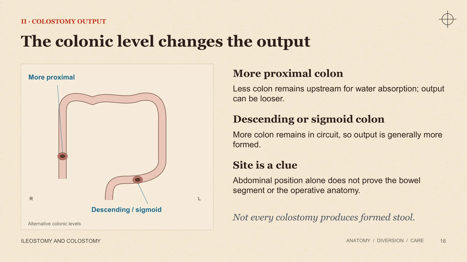
More proximal
Descending / sigmoid
R
L
Alternative colonic levels
More proximal colon
Less colon remains upstream for water absorption; output can be looser.
Descending or sigmoid colon
More colon remains in circuit, so output is generally more formed.
Site is a clue
Abdominal position alone does not prove the bowel segment or the operative anatomy.
Not every colostomy produces formed stool.
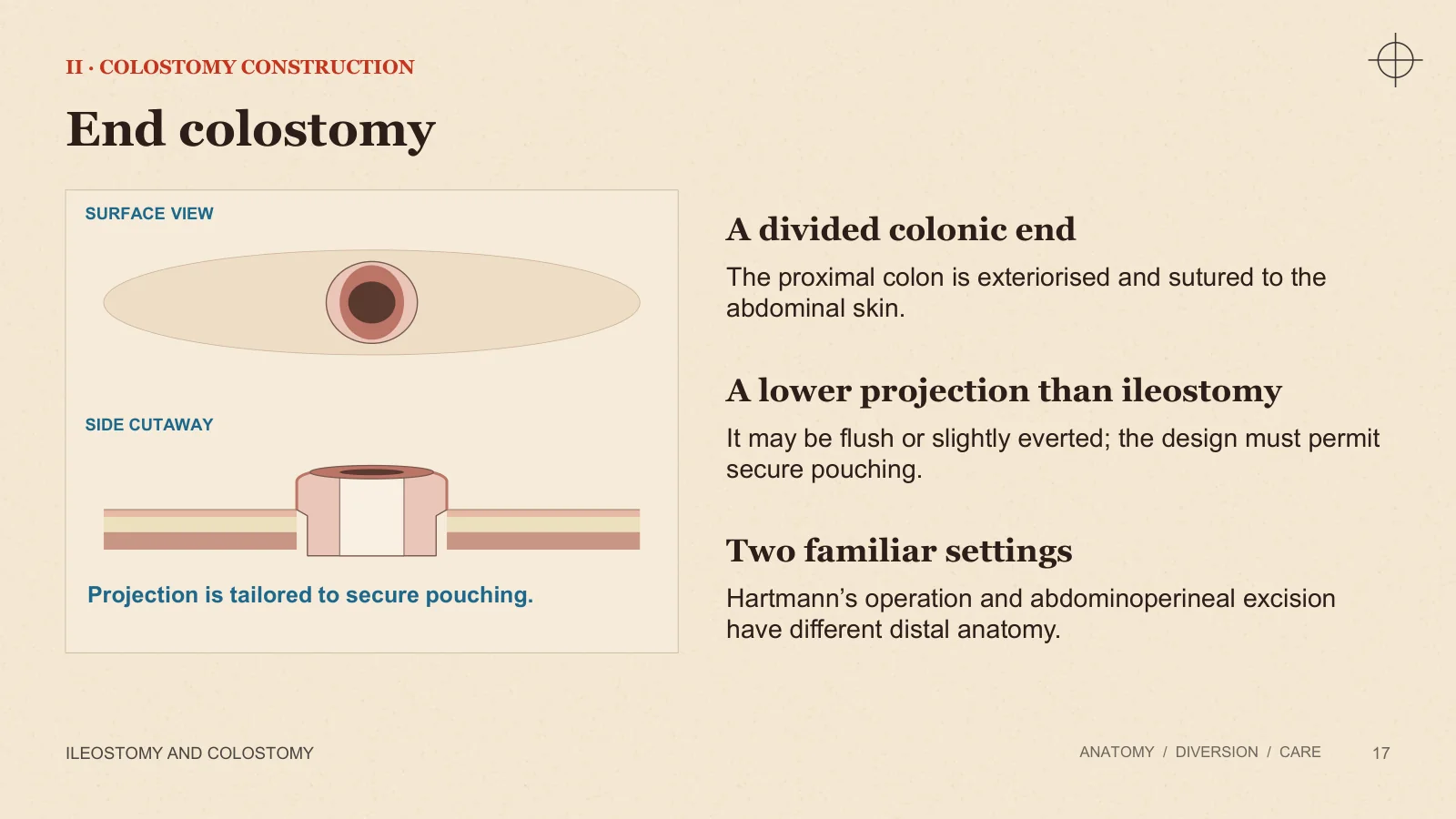
End colostomy
II · COLOSTOMY CONSTRUCTION
ILEOSTOMY AND COLOSTOMY
ANATOMY / DIVERSION / CARE
17
SURFACE VIEW
SIDE CUTAWAY
Projection is tailored to secure pouching.
A divided colonic end
The proximal colon is exteriorised and sutured to the abdominal skin.
A lower projection than ileostomy
It may be flush or slightly everted; the design must permit secure pouching.
Two familiar settings
Hartmann’s operation and abdominoperineal excision have different distal anatomy.
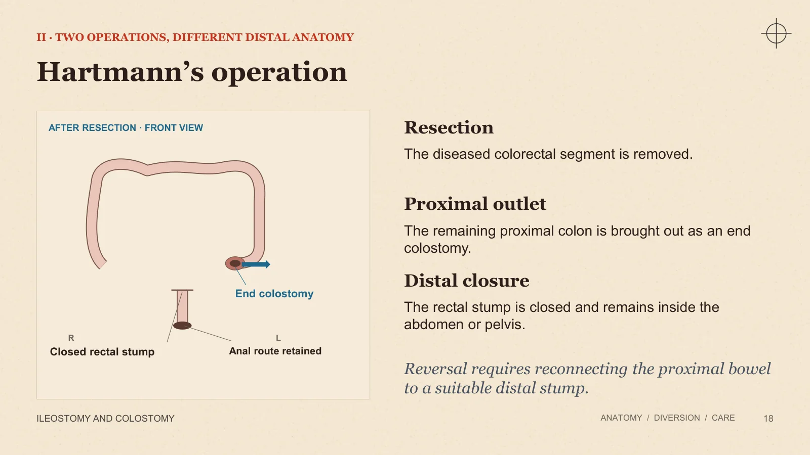
Hartmann’s operation
II · TWO OPERATIONS, DIFFERENT DISTAL ANATOMY
ILEOSTOMY AND COLOSTOMY
ANATOMY / DIVERSION / CARE
18
AFTER RESECTION · FRONT VIEW
R
L
End colostomy
Closed rectal stump
Anal route retained
Resection
The diseased colorectal segment is removed.
Proximal outlet
The remaining proximal colon is brought out as an end colostomy.
Distal closure
The rectal stump is closed and remains inside the abdomen or pelvis.
Reversal requires reconnecting the proximal bowel to a suitable distal stump.
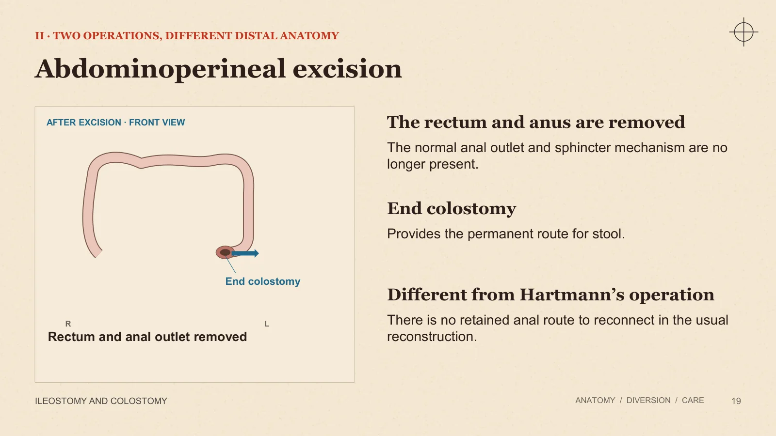
Abdominoperineal excision
II · TWO OPERATIONS, DIFFERENT DISTAL ANATOMY
ILEOSTOMY AND COLOSTOMY
ANATOMY / DIVERSION / CARE
19
AFTER EXCISION · FRONT VIEW
R
L
End colostomy
Rectum and anal outlet removed
The rectum and anus are removed
The normal anal outlet and sphincter mechanism are no longer present.
End colostomy
Provides the permanent route for stool.
Different from Hartmann’s operation
There is no retained anal route to reconnect in the usual reconstruction.
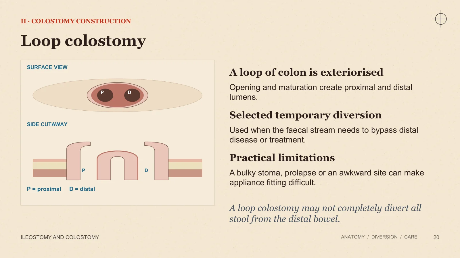
Loop colostomy
II · COLOSTOMY CONSTRUCTION
ILEOSTOMY AND COLOSTOMY
ANATOMY / DIVERSION / CARE
20
SURFACE VIEW
P
D
SIDE CUTAWAY
P
D
P = proximal D = distal
A loop of colon is exteriorised
Opening and maturation create proximal and distal lumens.
Selected temporary diversion
Used when the faecal stream needs to bypass distal disease or treatment.
Practical limitations
A bulky stoma, prolapse or an awkward site can make appliance fitting difficult.
A loop colostomy may not completely divert all stool from the distal bowel.
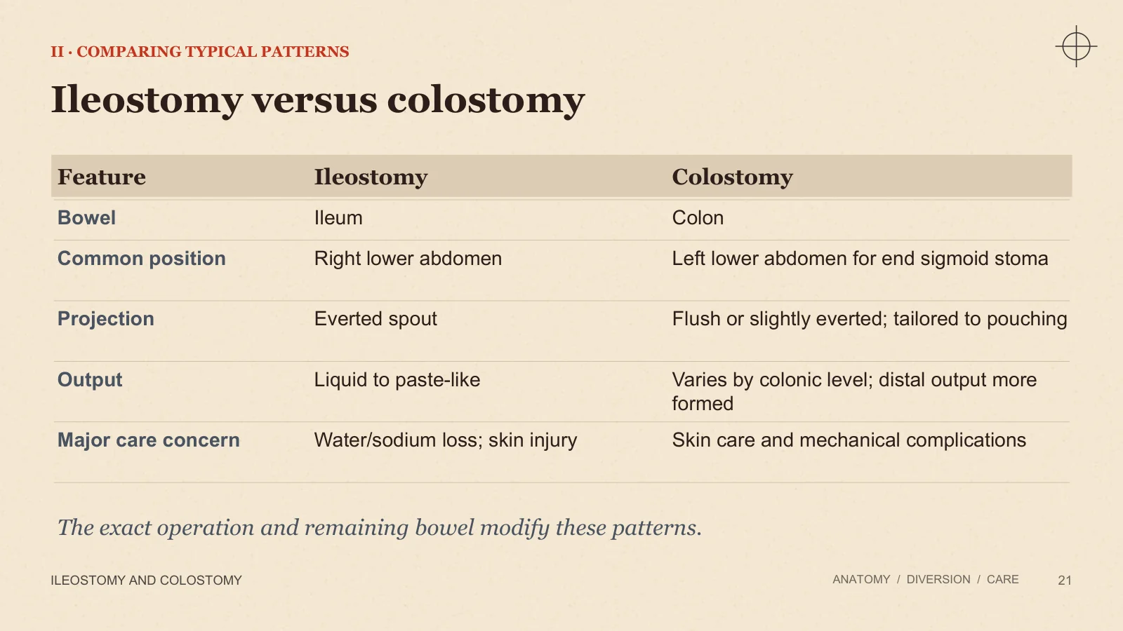
Ileostomy versus colostomy
II · COMPARING TYPICAL PATTERNS
ILEOSTOMY AND COLOSTOMY
ANATOMY / DIVERSION / CARE
21
Feature
Ileostomy
Colostomy
Bowel
Ileum
Colon
Common position
Right lower abdomen
Left lower abdomen for end sigmoid stoma
Projection
Everted spout
Flush or slightly everted; tailored to pouching
Output
Liquid to paste-like
Varies by colonic level; distal output more formed
Major care concern
Water/sodium loss; skin injury
Skin care and mechanical complications
The exact operation and remaining bowel modify these patterns.
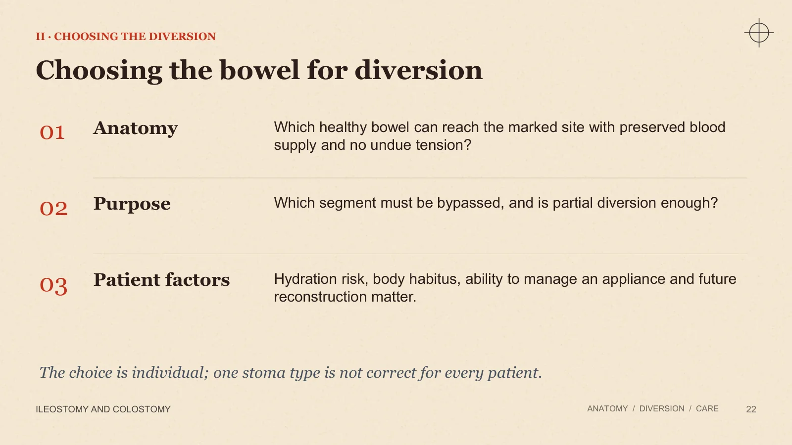
Choosing the bowel for diversion
II · CHOOSING THE DIVERSION
ILEOSTOMY AND COLOSTOMY
ANATOMY / DIVERSION / CARE
22
01
Anatomy
Which healthy bowel can reach the marked site with preserved blood supply and no undue tension?
02
Purpose
Which segment must be bypassed, and is partial diversion enough?
03
Patient factors
Hydration risk, body habitus, ability to manage an appliance and future reconstruction matter.
The choice is individual; one stoma type is not correct for every patient.
Planning and care
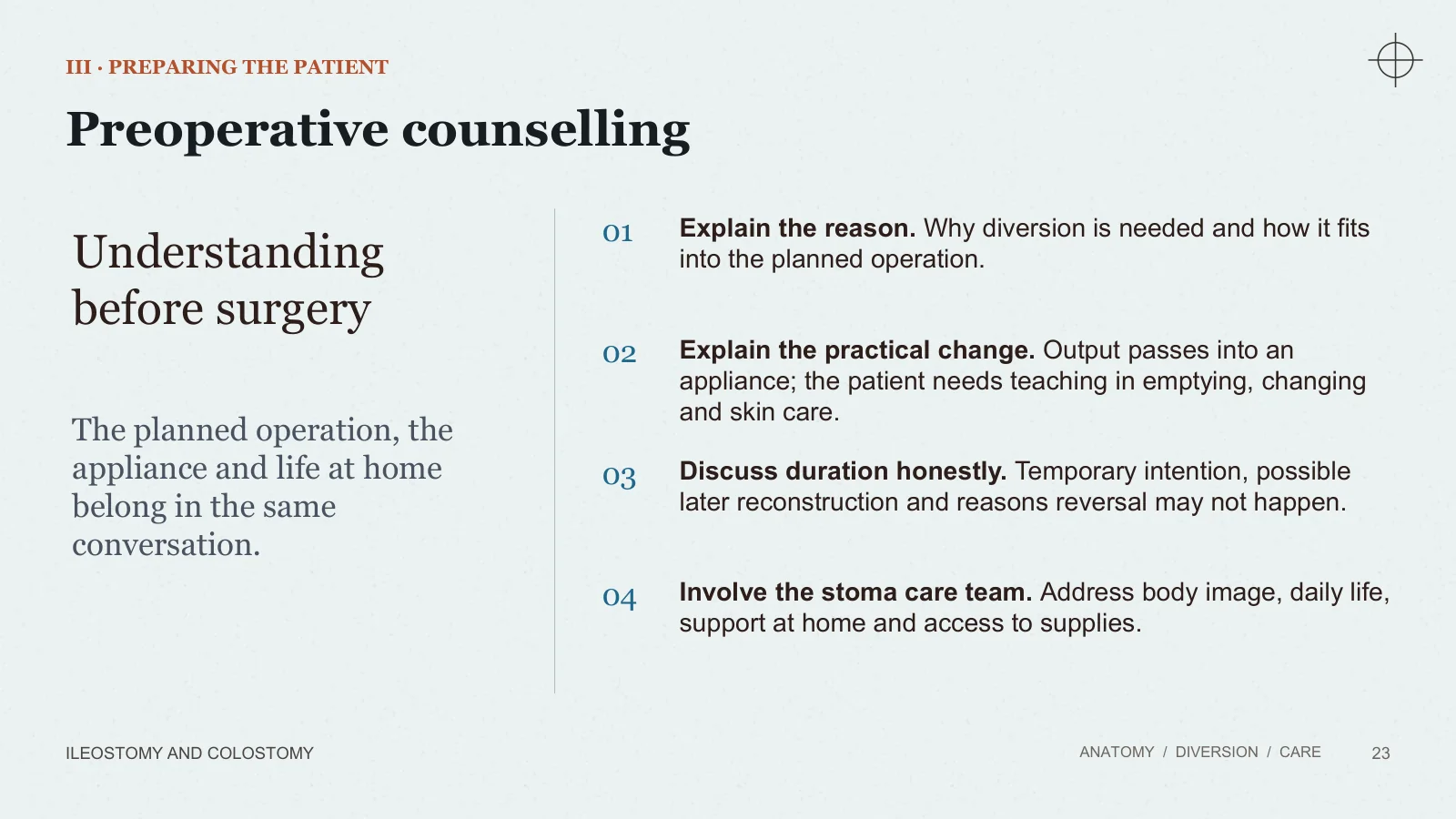
Preoperative counselling
III · PREPARING THE PATIENT
ILEOSTOMY AND COLOSTOMY
ANATOMY / DIVERSION / CARE
23
Understanding before surgery
The planned operation, the appliance and life at home belong in the same conversation.
01
Explain the reason. Why diversion is needed and how it fits into the planned operation.
02
Explain the practical change. Output passes into an appliance; the patient needs teaching in emptying, changing and skin care.
03
Discuss duration honestly. Temporary intention, possible later reconstruction and reasons reversal may not happen.
04
Involve the stoma care team. Address body image, daily life, support at home and access to supplies.
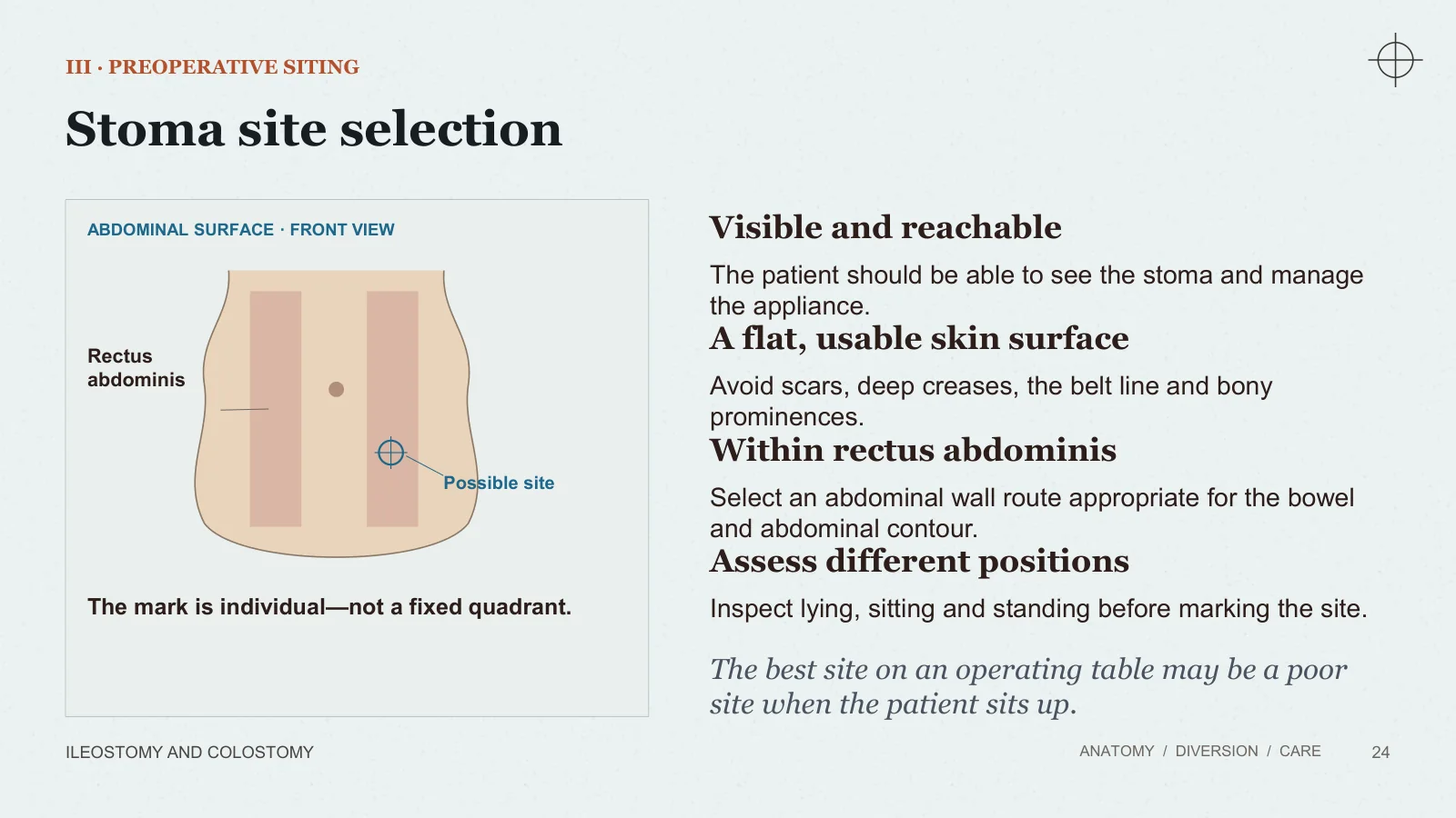
Stoma site selection
III · PREOPERATIVE SITING
ILEOSTOMY AND COLOSTOMY
ANATOMY / DIVERSION / CARE
24
ABDOMINAL SURFACE · FRONT VIEW
Rectus abdominis
Possible site
The mark is individual—not a fixed quadrant.
Visible and reachable
The patient should be able to see the stoma and manage the appliance.
A flat, usable skin surface
Avoid scars, deep creases, the belt line and bony prominences.
Within rectus abdominis
Select an abdominal wall route appropriate for the bowel and abdominal contour.
Assess different positions
Inspect lying, sitting and standing before marking the site.
The best site on an operating table may be a poor site when the patient sits up.
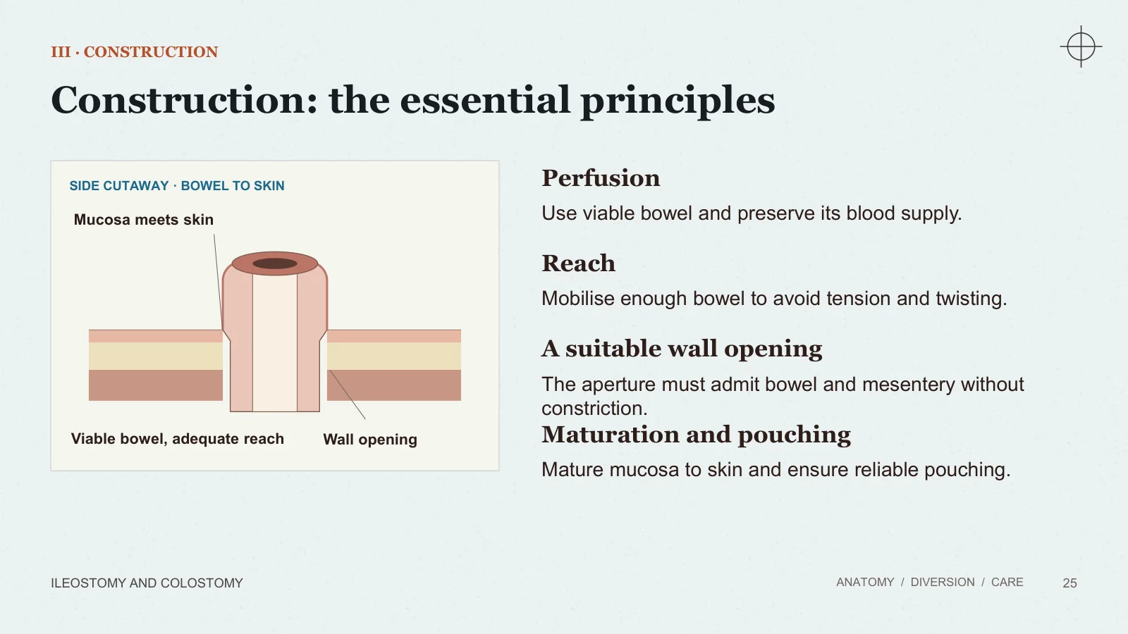
Construction: the essential principles
III · CONSTRUCTION
ILEOSTOMY AND COLOSTOMY
ANATOMY / DIVERSION / CARE
25
SIDE CUTAWAY · BOWEL TO SKIN
Mucosa meets skin
Wall opening
Viable bowel, adequate reach
Perfusion
Use viable bowel and preserve its blood supply.
Reach
Mobilise enough bowel to avoid tension and twisting.
A suitable wall opening
The aperture must admit bowel and mesentery without constriction.
Maturation and pouching
Mature mucosa to skin and ensure reliable pouching.
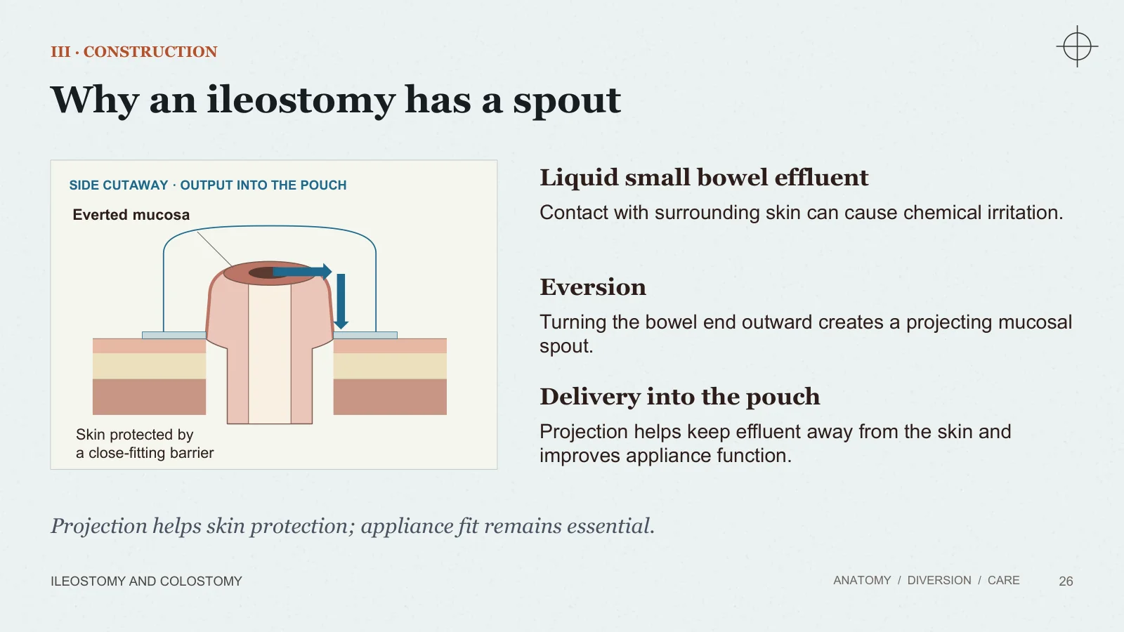
Why an ileostomy has a spout
III · CONSTRUCTION
ILEOSTOMY AND COLOSTOMY
ANATOMY / DIVERSION / CARE
26
SIDE CUTAWAY · OUTPUT INTO THE POUCH
Everted mucosa
Skin protected by a close-fitting barrier
Liquid small bowel effluent
Contact with surrounding skin can cause chemical irritation.
Eversion
Turning the bowel end outward creates a projecting mucosal spout.
Delivery into the pouch
Projection helps keep effluent away from the skin and improves appliance function.
Projection helps skin protection; appliance fit remains essential.
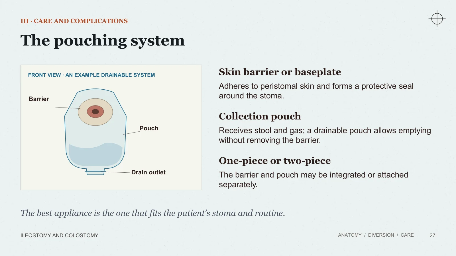
The pouching system
III · CARE AND COMPLICATIONS
ILEOSTOMY AND COLOSTOMY
ANATOMY / DIVERSION / CARE
27
FRONT VIEW · AN EXAMPLE DRAINABLE SYSTEM
Barrier
Pouch
Drain outlet
Skin barrier or baseplate
Adheres to peristomal skin and forms a protective seal around the stoma.
Collection pouch
Receives stool and gas; a drainable pouch allows emptying without removing the barrier.
One-piece or two-piece
The barrier and pouch may be integrated or attached separately.
The best appliance is the one that fits the patient’s stoma and routine.
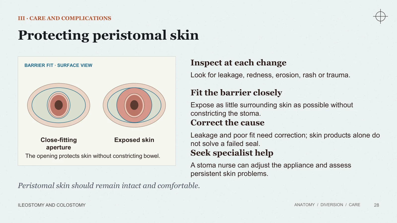
Protecting peristomal skin
III · CARE AND COMPLICATIONS
ILEOSTOMY AND COLOSTOMY
ANATOMY / DIVERSION / CARE
28
BARRIER FIT · SURFACE VIEW
Close-fitting aperture
Exposed skin
The opening protects skin without constricting bowel.
Inspect at each change
Look for leakage, redness, erosion, rash or trauma.
Fit the barrier closely
Expose as little surrounding skin as possible without constricting the stoma.
Correct the cause
Leakage and poor fit need correction; skin products alone do not solve a failed seal.
Seek specialist help
A stoma nurse can adjust the appliance and assess persistent skin problems.
Peristomal skin should remain intact and comfortable.
Early postoperative assessment
III · CARE AND COMPLICATIONS
ILEOSTOMY AND COLOSTOMY
ANATOMY / DIVERSION / CARE
29
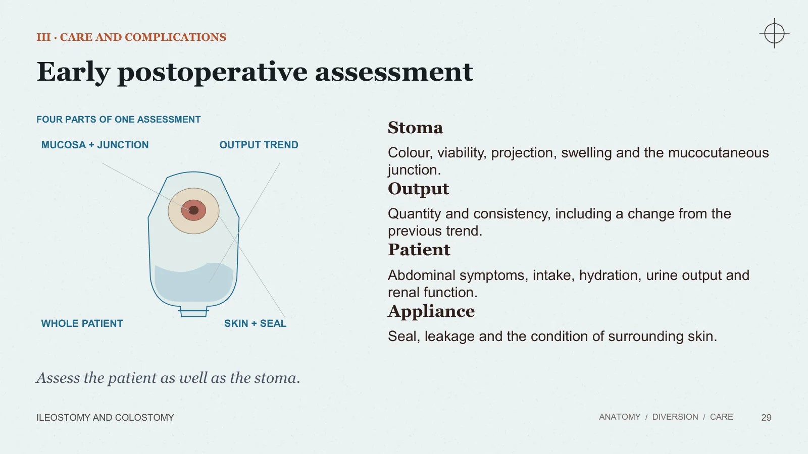
FOUR PARTS OF ONE ASSESSMENT
MUCOSA + JUNCTION
OUTPUT TREND
WHOLE PATIENT
SKIN + SEAL
Stoma
Colour, viability, projection, swelling and the mucocutaneous junction.
Output
Quantity and consistency, including a change from the previous trend.
Patient
Abdominal symptoms, intake, hydration, urine output and renal function.
Appliance
Seal, leakage and the condition of surrounding skin.
Assess the patient as well as the stoma.
Complications
Complications: an overview
III · CARE AND COMPLICATIONS
ILEOSTOMY AND COLOSTOMY
ANATOMY / DIVERSION / CARE
30
Early or immediate problems
01
Ischaemia or necrosis
02
Retraction or mucocutaneous separation
03
Bleeding and appliance leakage
04
High output and dehydration
Later or persistent problems
01
Prolapse
02
Stenosis
03
Parastomal hernia
04
Persistent skin problems or fistulation
Timing overlaps: any complication is interpreted in its clinical setting.
Stoma ischaemia and necrosis
III · CARE AND COMPLICATIONS
ILEOSTOMY AND COLOSTOMY
ANATOMY / DIVERSION / CARE
31
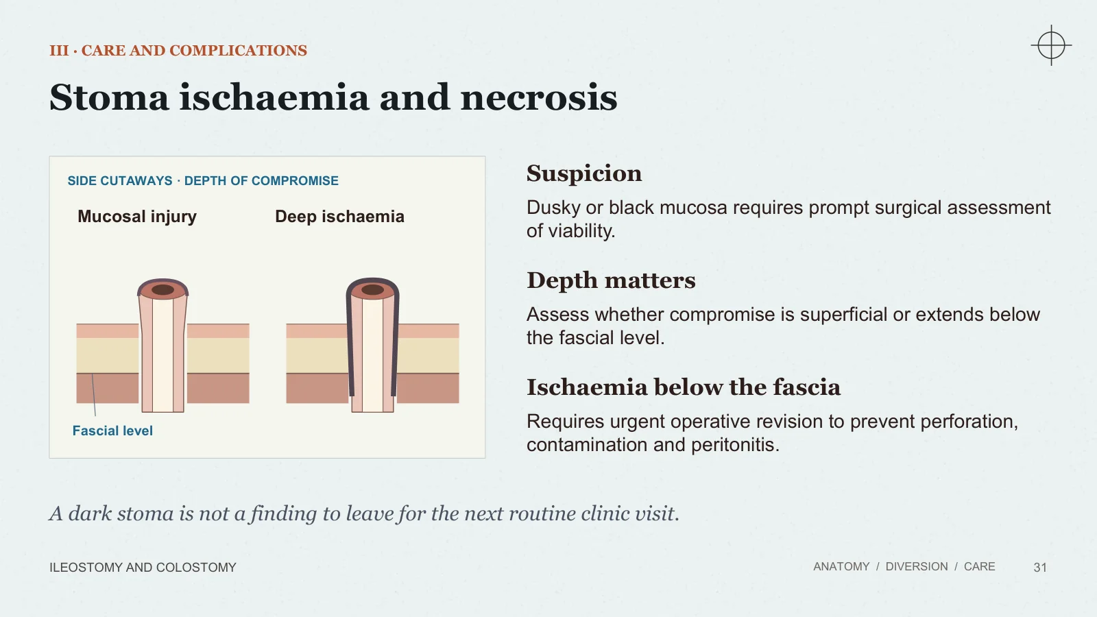
SIDE CUTAWAYS · DEPTH OF COMPROMISE
Mucosal injury
Deep ischaemia
Fascial level
Suspicion
Dusky or black mucosa requires prompt surgical assessment of viability.
Depth matters
Assess whether compromise is superficial or extends below the fascial level.
Ischaemia below the fascia
Requires urgent operative revision to prevent perforation, contamination and peritonitis.
A dark stoma is not a finding to leave for the next routine clinic visit.
Retraction and mucocutaneous separation
III · CARE AND COMPLICATIONS
ILEOSTOMY AND COLOSTOMY
ANATOMY / DIVERSION / CARE
32
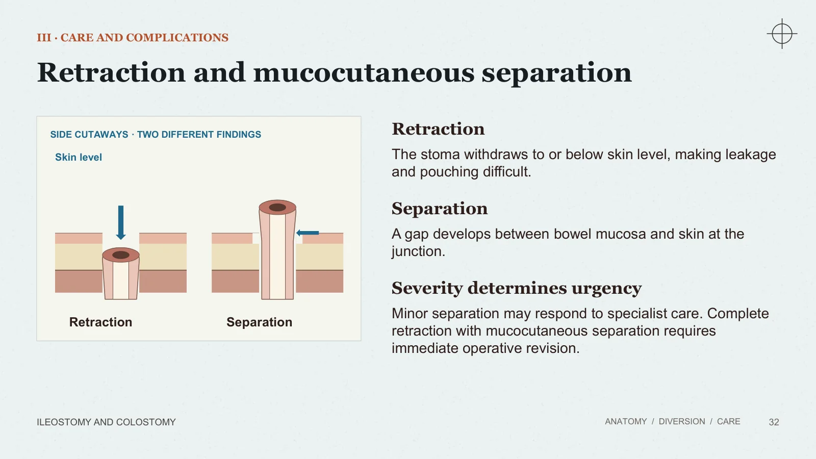
SIDE CUTAWAYS · TWO DIFFERENT FINDINGS
Retraction
Separation
Skin level
Retraction
The stoma withdraws to or below skin level, making leakage and pouching difficult.
Separation
A gap develops between bowel mucosa and skin at the junction.
Severity determines urgency
Minor separation may respond to specialist care. Complete retraction with mucocutaneous separation requires immediate operative revision.
High-output ileostomy
III · CARE AND COMPLICATIONS
ILEOSTOMY AND COLOSTOMY
ANATOMY / DIVERSION / CARE
33
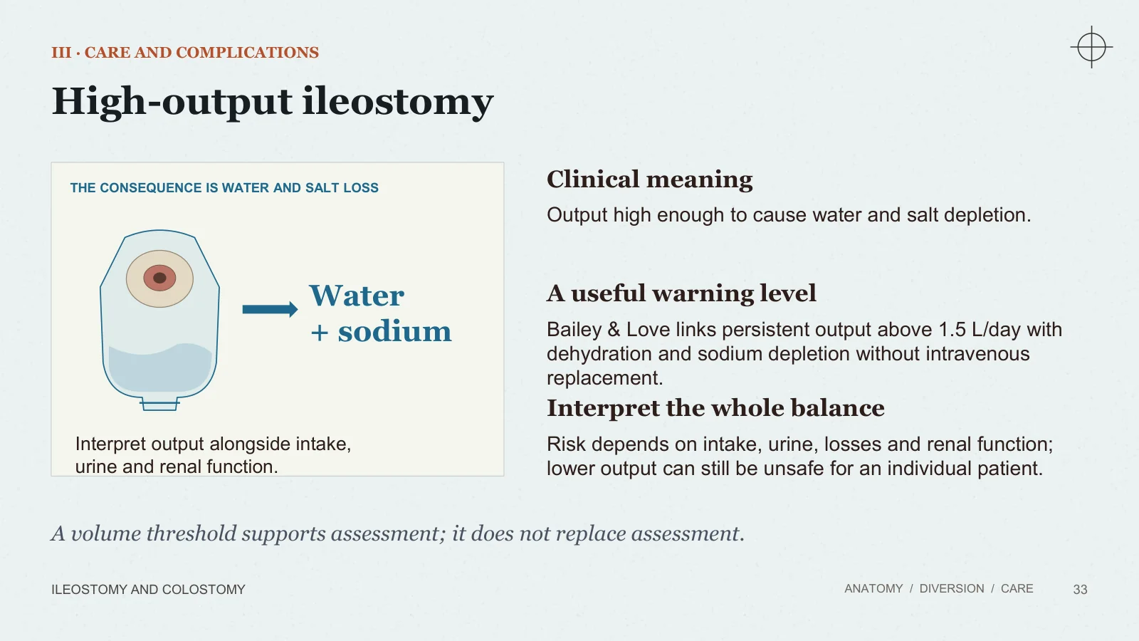
THE CONSEQUENCE IS WATER AND SALT LOSS
Water + sodium
Interpret output alongside intake, urine and renal function.
Clinical meaning
Output high enough to cause water and salt depletion.
A useful warning level
Bailey & Love links persistent output above 1.5 L/day with dehydration and sodium depletion without intravenous replacement.
Interpret the whole balance
Risk depends on intake, urine, losses and renal function; lower output can still be unsafe for an individual patient.
A volume threshold supports assessment; it does not replace assessment.
Recognising dehydration
III · CARE AND COMPLICATIONS
ILEOSTOMY AND COLOSTOMY
ANATOMY / DIVERSION / CARE
34
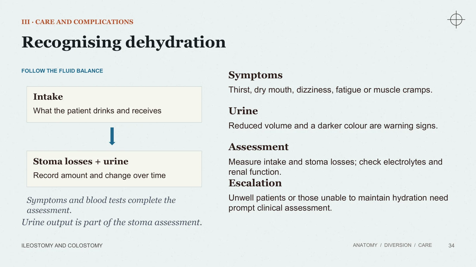
FOLLOW THE FLUID BALANCE
Intake
What the patient drinks and receives
Stoma losses + urine
Record amount and change over time
Symptoms and blood tests complete the assessment.
Symptoms
Thirst, dry mouth, dizziness, fatigue or muscle cramps.
Urine
Reduced volume and a darker colour are warning signs.
Assessment
Measure intake and stoma losses; check electrolytes and renal function.
Escalation
Unwell patients or those unable to maintain hydration need prompt clinical assessment.
Urine output is part of the stoma assessment.
High output: management principles
III · CARE AND COMPLICATIONS
ILEOSTOMY AND COLOSTOMY
ANATOMY / DIVERSION / CARE
35
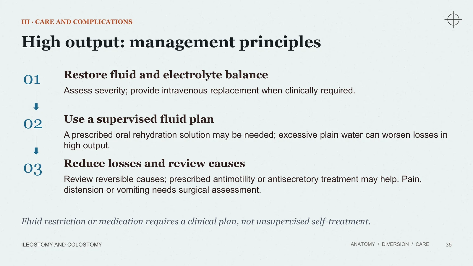
01
Restore fluid and electrolyte balance
Assess severity; provide intravenous replacement when clinically required.
02
Use a supervised fluid plan
A prescribed oral rehydration solution may be needed; excessive plain water can worsen losses in high output.
03
Reduce losses and review causes
Review reversible causes; prescribed antimotility or antisecretory treatment may help. Pain, distension or vomiting needs surgical assessment.
Fluid restriction or medication requires a clinical plan, not unsupervised self-treatment.
Obstruction and stenosis
III · CARE AND COMPLICATIONS
ILEOSTOMY AND COLOSTOMY
ANATOMY / DIVERSION / CARE
36
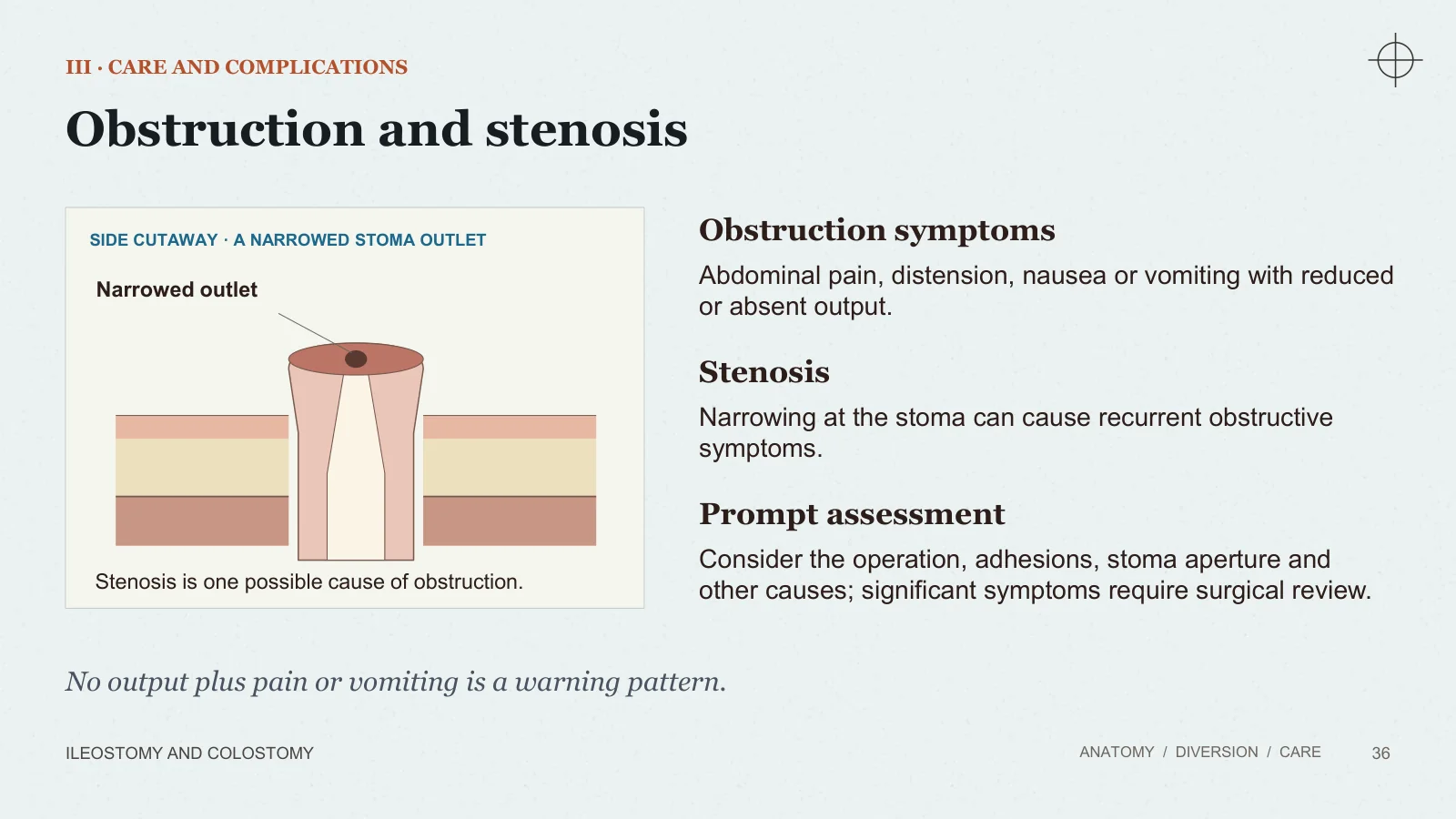
SIDE CUTAWAY · A NARROWED STOMA OUTLET
Narrowed outlet
Stenosis is one possible cause of obstruction.
Obstruction symptoms
Abdominal pain, distension, nausea or vomiting with reduced or absent output.
Stenosis
Narrowing at the stoma can cause recurrent obstructive symptoms.
Prompt assessment
Consider the operation, adhesions, stoma aperture and other causes; significant symptoms require surgical review.
No output plus pain or vomiting is a warning pattern.
Stoma prolapse
III · CARE AND COMPLICATIONS
ILEOSTOMY AND COLOSTOMY
ANATOMY / DIVERSION / CARE
37
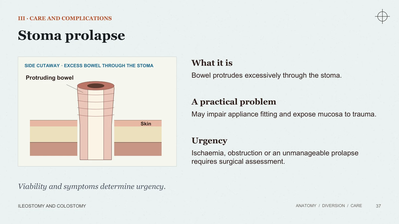
SIDE CUTAWAY · EXCESS BOWEL THROUGH THE STOMA
Protruding bowel
Skin
What it is
Bowel protrudes excessively through the stoma.
A practical problem
May impair appliance fitting and expose mucosa to trauma.
Urgency
Ischaemia, obstruction or an unmanageable prolapse requires surgical assessment.
Viability and symptoms determine urgency.
Parastomal hernia
III · CARE AND COMPLICATIONS
ILEOSTOMY AND COLOSTOMY
ANATOMY / DIVERSION / CARE
38
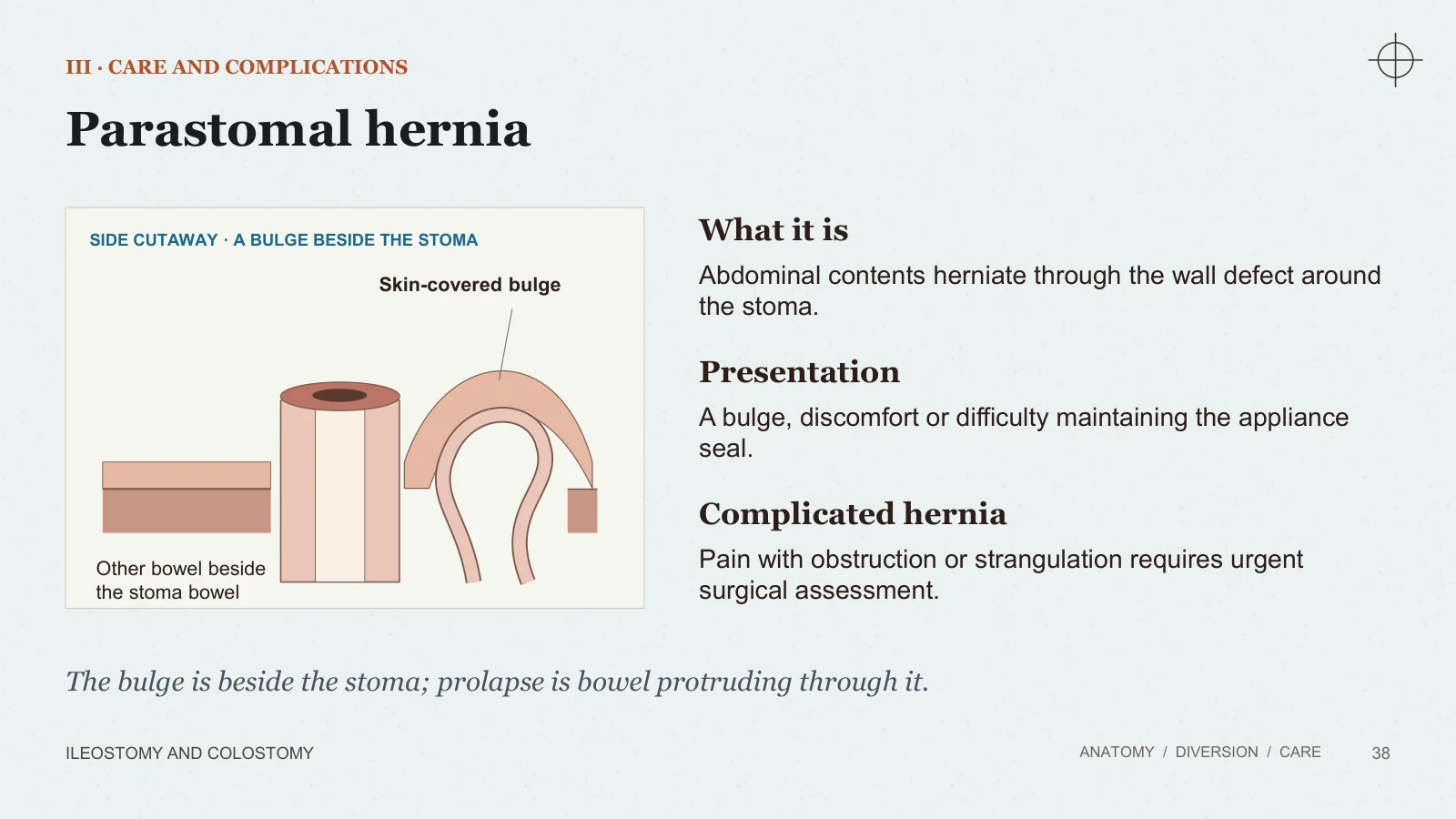
SIDE CUTAWAY · A BULGE BESIDE THE STOMA
Skin-covered bulge
Other bowel beside the stoma bowel
What it is
Abdominal contents herniate through the wall defect around the stoma.
Presentation
A bulge, discomfort or difficulty maintaining the appliance seal.
Complicated hernia
Pain with obstruction or strangulation requires urgent surgical assessment.
The bulge is beside the stoma; prolapse is bowel protruding through it.
Bleeding and skin problems
III · CARE AND COMPLICATIONS
ILEOSTOMY AND COLOSTOMY
ANATOMY / DIVERSION / CARE
39
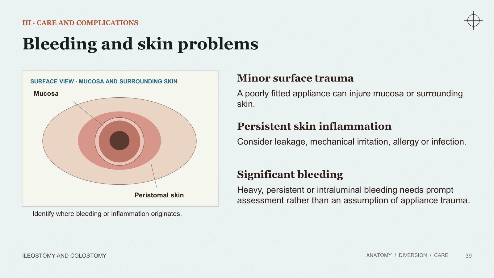
SURFACE VIEW · MUCOSA AND SURROUNDING SKIN
Mucosa
Peristomal skin
Identify where bleeding or inflammation originates.
Minor surface trauma
A poorly fitted appliance can injure mucosa or surrounding skin.
Persistent skin inflammation
Consider leakage, mechanical irritation, allergy or infection.
Significant bleeding
Heavy, persistent or intraluminal bleeding needs prompt assessment rather than an assumption of appliance trauma.
Recovery and reversal
Independence is part of recovery.
III · CARE AND COMPLICATIONS
Living with a stoma
ILEOSTOMY AND COLOSTOMY
ANATOMY / DIVERSION / CARE
40
A secure appliance, usable supplies and respectful support.
Practical independence
Teach emptying, changing, skin inspection and carrying spare supplies.
Food and fluids
Use individual advice; adjust intake and hydration to output and clinical needs.
Daily life
Return to activities is supported by recovery, a secure appliance and confidence in self-care.
Psychological support
Body image, relationships and fear of leakage deserve direct, respectful discussion.
Stoma care includes the person’s life, not only the opening on the abdomen.
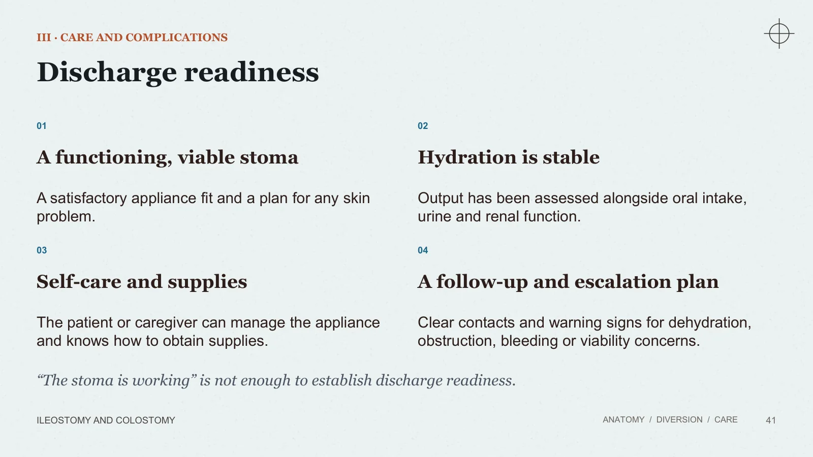
Discharge readiness
III · CARE AND COMPLICATIONS
ILEOSTOMY AND COLOSTOMY
ANATOMY / DIVERSION / CARE
41
01
A functioning, viable stoma
A satisfactory appliance fit and a plan for any skin problem.
02
Hydration is stable
Output has been assessed alongside oral intake, urine and renal function.
03
Self-care and supplies
The patient or caregiver can manage the appliance and knows how to obtain supplies.
04
A follow-up and escalation plan
Clear contacts and warning signs for dehydration, obstruction, bleeding or viability concerns.
“The stoma is working” is not enough to establish discharge readiness.
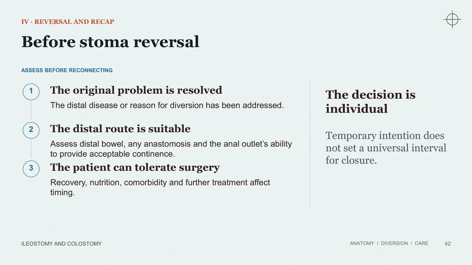
Before stoma reversal
IV · REVERSAL AND RECAP
ILEOSTOMY AND COLOSTOMY
ANATOMY / DIVERSION / CARE
42
ASSESS BEFORE RECONNECTING
1
The original problem is resolved
The distal disease or reason for diversion has been addressed.
2
The distal route is suitable
Assess distal bowel, any anastomosis and the anal outlet’s ability to provide acceptable continence.
3
The patient can tolerate surgery
Recovery, nutrition, comorbidity and further treatment affect timing.
The decision is individual
Temporary intention does not set a universal interval for closure.
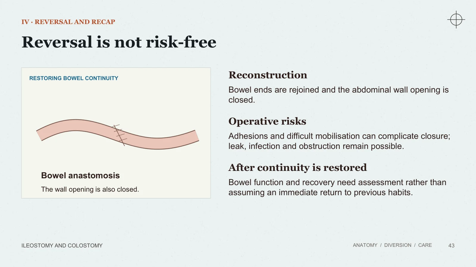
Reversal is not risk-free
IV · REVERSAL AND RECAP
ILEOSTOMY AND COLOSTOMY
ANATOMY / DIVERSION / CARE
43
RESTORING BOWEL CONTINUITY
Bowel anastomosis
The wall opening is also closed.
Reconstruction
Bowel ends are rejoined and the abdominal wall opening is closed.
Operative risks
Adhesions and difficult mobilisation can complicate closure; leak, infection and obstruction remain possible.
After continuity is restored
Bowel function and recovery need assessment rather than assuming an immediate return to previous habits.
Recap and sources
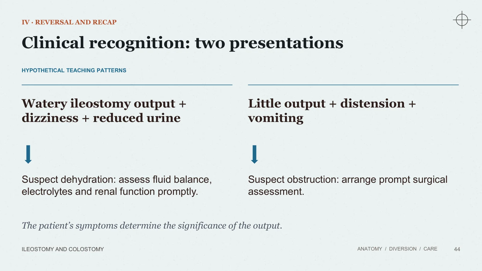
Clinical recognition: two presentations
IV · REVERSAL AND RECAP
ILEOSTOMY AND COLOSTOMY
ANATOMY / DIVERSION / CARE
44
HYPOTHETICAL TEACHING PATTERNS
Watery ileostomy output + dizziness + reduced urine
Suspect dehydration: assess fluid balance, electrolytes and renal function promptly.
Little output + distension + vomiting
Suspect obstruction: arrange prompt surgical assessment.
The patient’s symptoms determine the significance of the output.
Describing a stoma in an examination
IV · REVERSAL AND RECAP
ILEOSTOMY AND COLOSTOMY
ANATOMY / DIVERSION / CARE
45
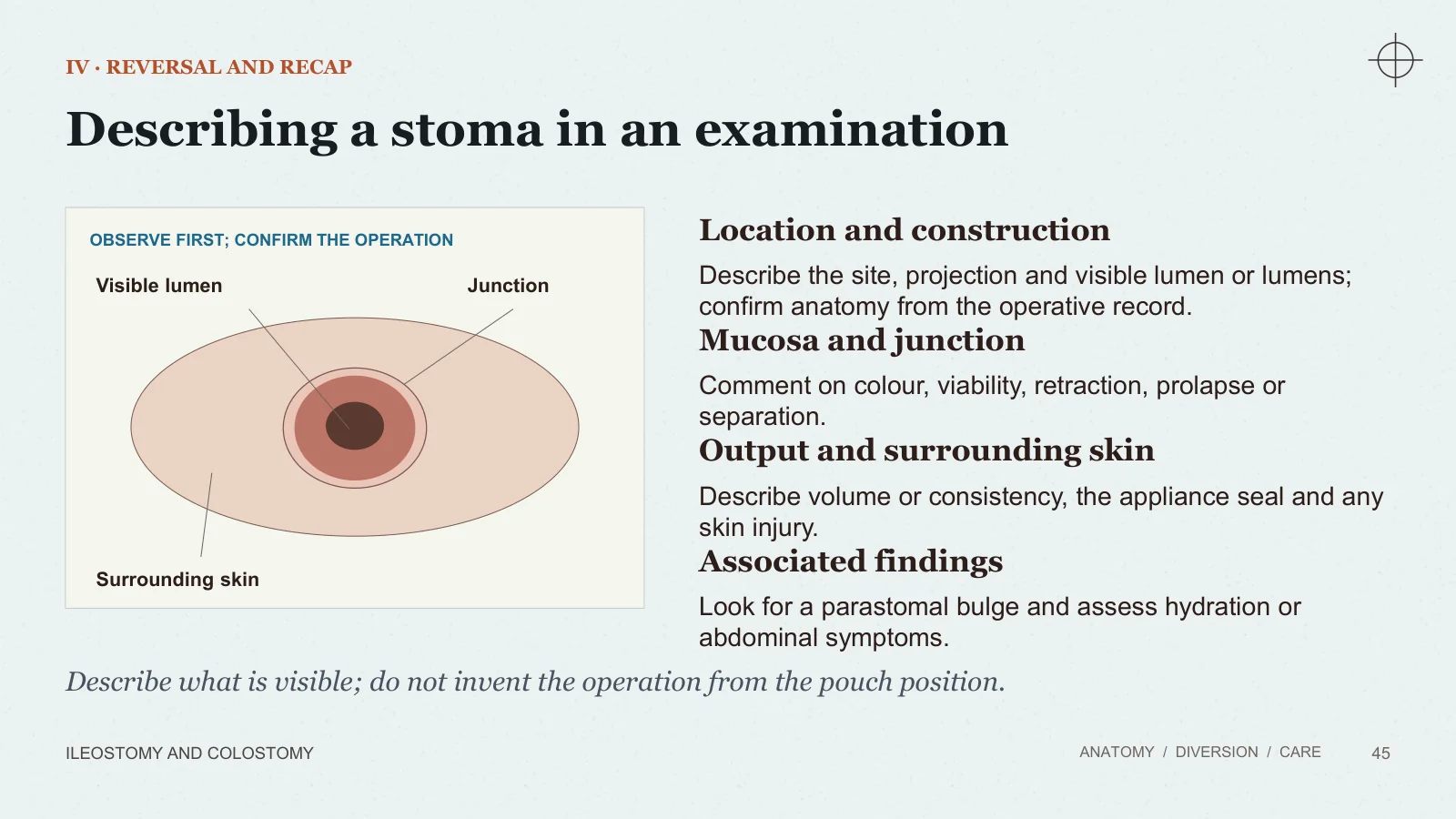
OBSERVE FIRST; CONFIRM THE OPERATION
Visible lumen
Junction
Surrounding skin
Location and construction
Describe the site, projection and visible lumen or lumens; confirm anatomy from the operative record.
Mucosa and junction
Comment on colour, viability, retraction, prolapse or separation.
Output and surrounding skin
Describe volume or consistency, the appliance seal and any skin injury.
Associated findings
Look for a parastomal bulge and assess hydration or abdominal symptoms.
Describe what is visible; do not invent the operation from the pouch position.
Take-home points
THE FOUR ESSENTIALS
ILEOSTOMY AND COLOSTOMY
ANATOMY / DIVERSION / CARE
46
01
Name the bowel and the construction
Ileostomy/colostomy and end/loop describe different aspects.
02
Understand the reason
Diversion, unsafe continuity, obstruction or loss of the anal outlet.
03
Protect function and independence
Good siting, a secure appliance, intact skin and individual teaching.
04
Recognise danger
Ischaemia, dehydration, obstruction and significant bleeding need prompt assessment.
Sources and further reading
READING AND ILLUSTRATION CREDITS
ILEOSTOMY AND COLOSTOMY
ANATOMY / DIVERSION / CARE
47
Bailey & Love’s Short Practice of Surgery · 28th edition
Ch. 74, The small intestine: Stomas, pp. 1313–1314. Ch. 75, Inflammatory bowel disease, pp. 1323–1324. Ch. 77, The large intestine: Colostomies, pp. 1372–1373. Ch. 79, The rectum, pp. 1413–1414.
Sabiston Textbook of Surgery · 21st edition
Ch. 52, Colon and Rectum: Planning intestinal stomas, pp. 1334–1336. Ch. 12, Surgical Complications, pp. 267–268.
Public patient and clinical information
Royal Free London: High output stoma. ASCRS: Ostomy Expanded Information. NIDDK: Ostomy Surgery of the Bowel. NHS: Complications of an ileostomy. Cancer Research UK: Having a colostomy or ileostomy. Torbay and South Devon NHS: Stoma closure.
Illustrations: credited NIDDK, NIH illustrations and teaching diagrams.
Sources and image credits
Image credits and source details are reproduced from the final slide below.
Sources
- [1] READING AND ILLUSTRATION CREDITS
- [2] Sources and further reading
- [3] ANATOMY / DIVERSION / CARE
- [4] Bailey & Love’s Short Practice of Surgery · 28th edition
- [5] Ch. 74, The small intestine: Stomas, pp. 1313–1314. Ch. 75, Inflammatory bowel disease, pp. 1323–1324. Ch. 77, The large intestine: Colostomies, pp. 1372–1373. Ch. 79, The rectum, pp. 1413–1414.
- [6] Sabiston Textbook of Surgery · 21st edition
- [7] Ch. 52, Colon and Rectum: Planning intestinal stomas, pp. 1334–1336. Ch. 12, Surgical Complications, pp. 267–268.
- [8] Public patient and clinical information
- [9] Royal Free London: High output stoma. ASCRS: Ostomy Expanded Information. NIDDK: Ostomy Surgery of the Bowel. NHS: Complications of an ileostomy. Cancer Research UK: Having a colostomy or ileostomy. Torbay and South Devon NHS: Stoma closure.
- [10] Illustrations: credited NIDDK, NIH illustrations and teaching diagrams.