Lecture · 55 slides · 14 animated scenes
The story of surgery.
How we learned to operate, and how we learn to care: one patient before anaesthesia, one admitted last night, and the 180 years between them.
PowerPoint · 47 MB · with the animated scenes · no speaker notes · PDF, 15 MB · SHA-256 checksums
Opening
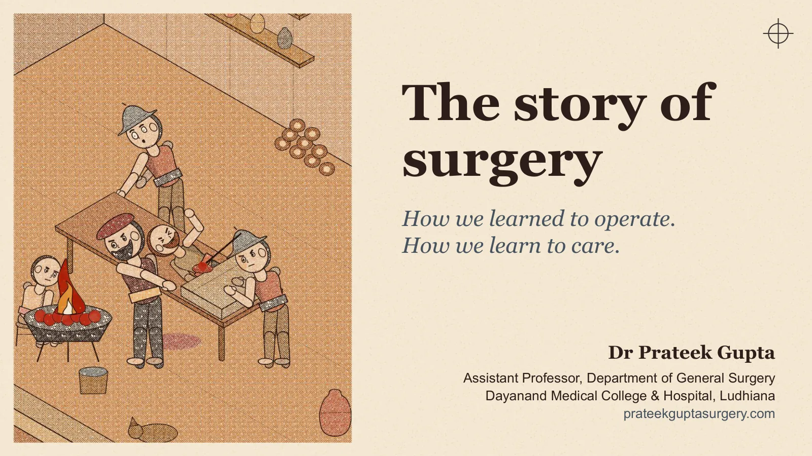
The story of surgery
How we learned to operate. How we learn to care.
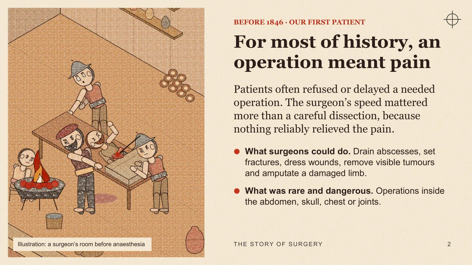
BEFORE 1846 · OUR FIRST PATIENT
For most of history, an operation meant pain
Patients often refused or delayed a needed operation. The surgeon’s speed mattered more than a careful dissection, because nothing reliably relieved the pain.
- What surgeons could do Drain abscesses, set fractures, dress wounds, remove visible tumours and amputate a damaged limb.
- What was rare and dangerous Operations inside the abdomen, skull, chest or joints.
Illustration: a surgeon’s room before anaesthesia
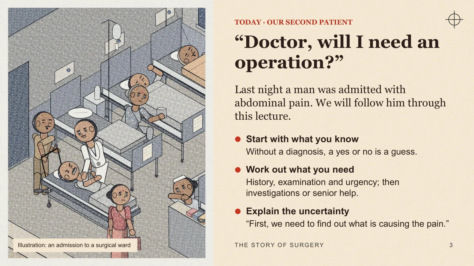
TODAY · OUR SECOND PATIENT
“Doctor, will I need an operation?”
Last night a man was admitted with abdominal pain. We will follow him through this lecture.
- Start with what you know Without a diagnosis, a yes or no is a guess.
- Work out what you need History, examination and urgency; then investigations or senior help.
- Explain the uncertainty “First, we need to find out what is causing the pain.”
Illustration: an admission to a surgical ward
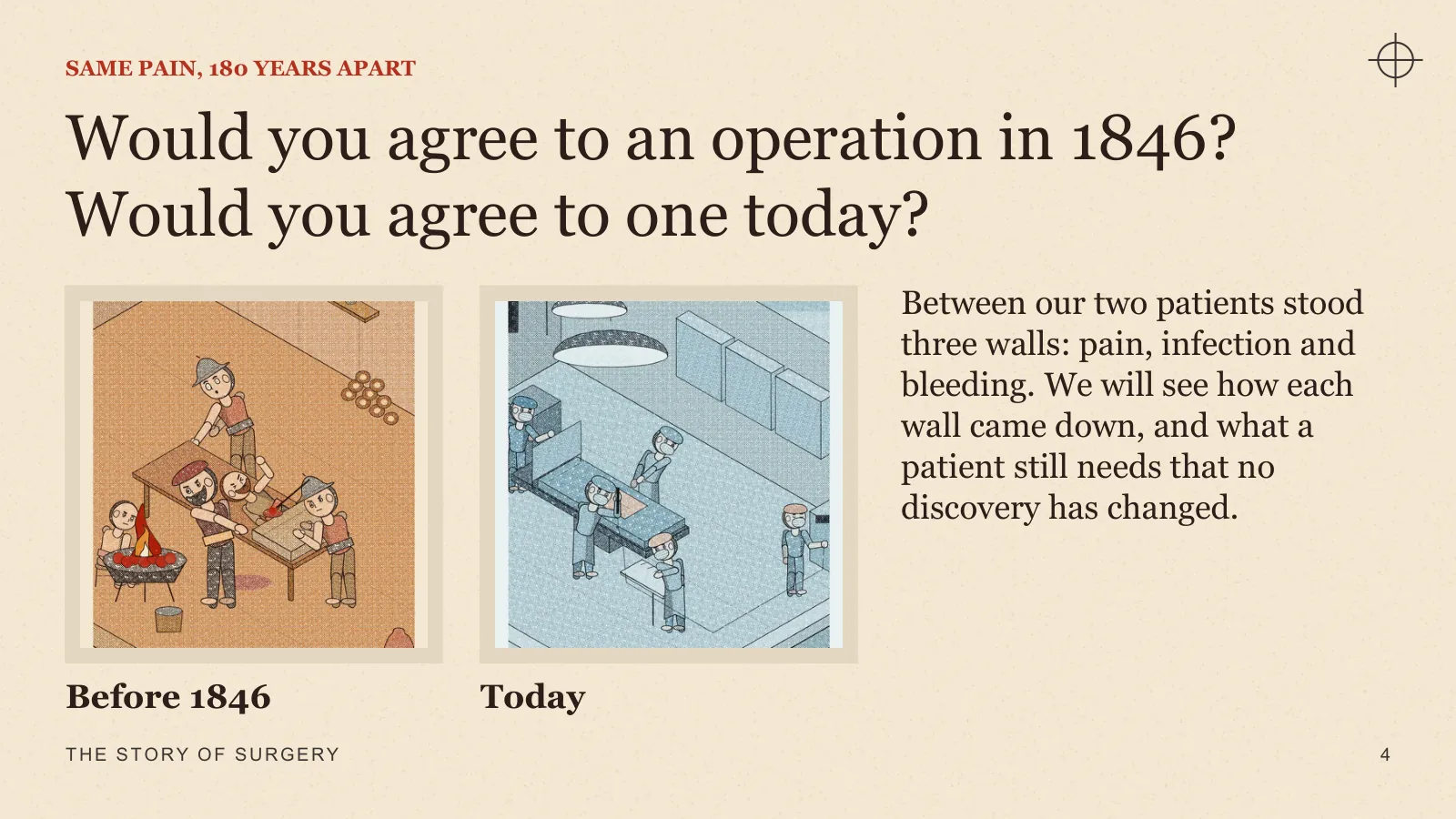
SAME PAIN, 180 YEARS APART
Would you agree to an operation in 1846? Would you agree to one today?
Between our two patients stood three walls: pain, infection and bleeding. We will see how each wall came down, and what a patient still needs that no discovery has changed.
A WORKING DEFINITION
What surgery actually includes
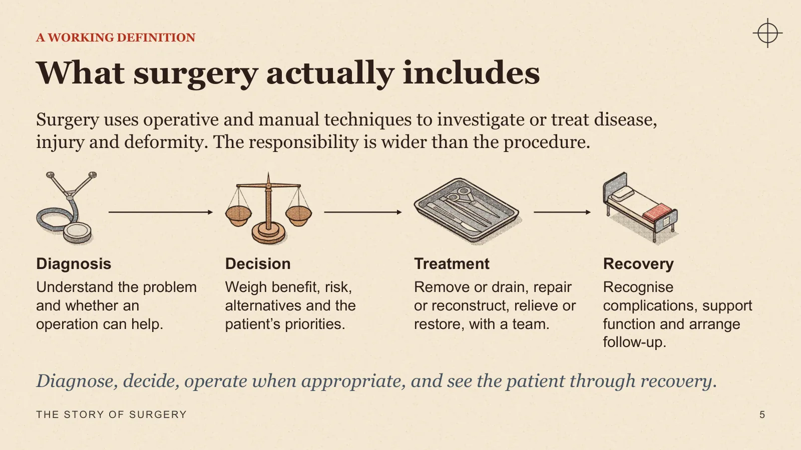
Surgery uses operative and manual techniques to investigate or treat disease, injury and deformity. The responsibility is wider than the procedure.
- Diagnosis Understand the problem and whether an operation can help.
- Decision Weigh benefit, risk, alternatives and the patient’s priorities.
- Treatment Remove or drain, repair or reconstruct, relieve or restore, with a team.
- Recovery Recognise complications, support function and arrange follow-up.
Diagnose, decide, operate when appropriate, and see the patient through recovery.
Act I · Learning to look
ACT I
Learning to look
For thousands of years, surgeons treated what they could see. Progress came from people willing to look again, even when the textbook disagreed.
I · c. 1600 BCE
Surgery began with visible problems
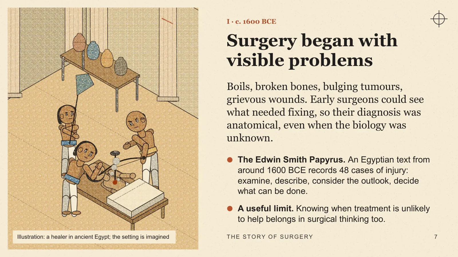
Boils, broken bones, bulging tumours, grievous wounds. Early surgeons could see what needed fixing, so their diagnosis was anatomical, even when the biology was unknown.
- The Edwin Smith Papyrus An Egyptian text from around 1600 BCE records 48 cases of injury: examine, describe, consider the outlook, decide what can be done.
- A useful limit Knowing when treatment is unlikely to help belongs in surgical thinking too.
Illustration: a healer in ancient Egypt; the setting is imagined
I · GREECE AND ROME · GALEN, 130–200 CE
Authority, observation and error
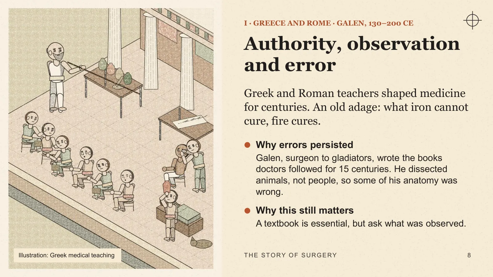
Greek and Roman teachers shaped medicine for centuries. An old adage: what iron cannot cure, fire cures.
- Why errors persisted Galen, surgeon to gladiators, wrote the books doctors followed for 15 centuries. He dissected animals, not people, so some of his anatomy was wrong.
- Why this still matters A textbook is essential, but ask what was observed.
Illustration: Greek medical teaching
I · ANCIENT INDIA
Sushruta and the Samhita
A surgical text revised again and again over centuries. Its 1907 translator dated the final version to the 2nd century BCE.
- Instruments 101 yantras, “of which the hand is the most important”, and 20 sharp instruments.
- Sutures Cotton, hemp, horse-hair or sinew; round, three-edged and curved needles.
- Clean first Do not suture a wound until it is thoroughly cleansed.
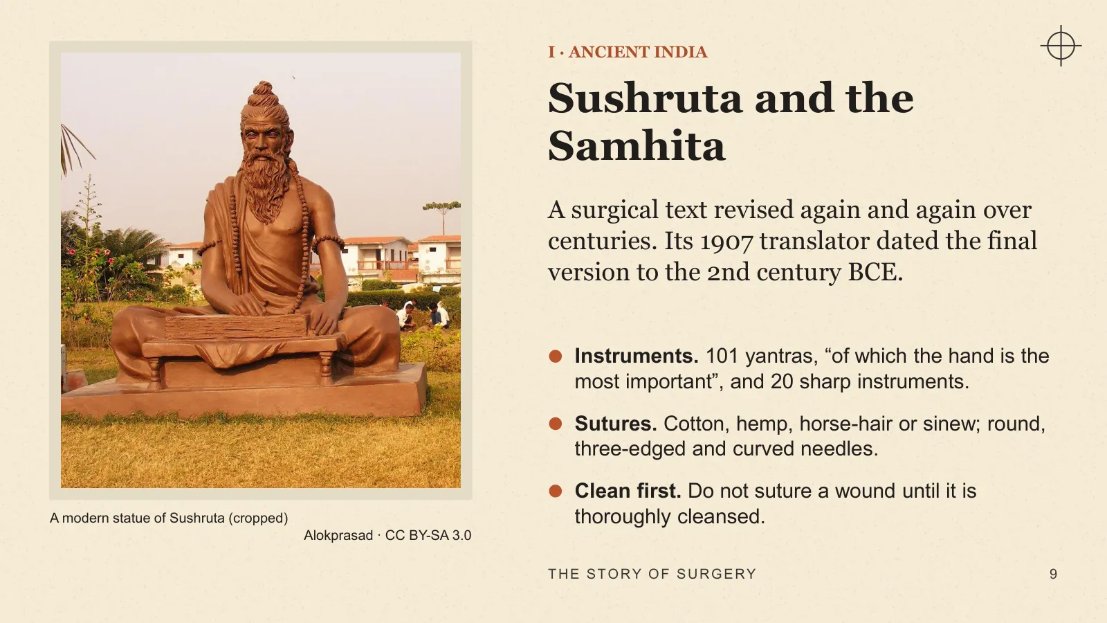
A modern statue of Sushruta (cropped) · Alokprasad · CC BY-SA 3.0
I · SUSHRUTA SAMHITA, SUTRASTHANA IX
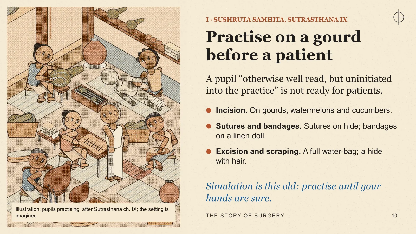
Practise on a gourd before a patient
A pupil “otherwise well read, but uninitiated into the practice” is not ready for patients.
- Incision On gourds, watermelons and cucumbers.
- Sutures and bandages Sutures on hide; bandages on a linen doll.
- Excision and scraping A full water-bag; a hide with hair.
Simulation is this old: practise until your hands are sure.
Illustration: pupils practising, after Sutrasthana ch. IX; the setting is imagined
I · CRAFT AND MEDICINE
The barber-surgeon
For centuries, practical surgery belonged to craftsmen, while university-educated physicians avoided working with their hands.
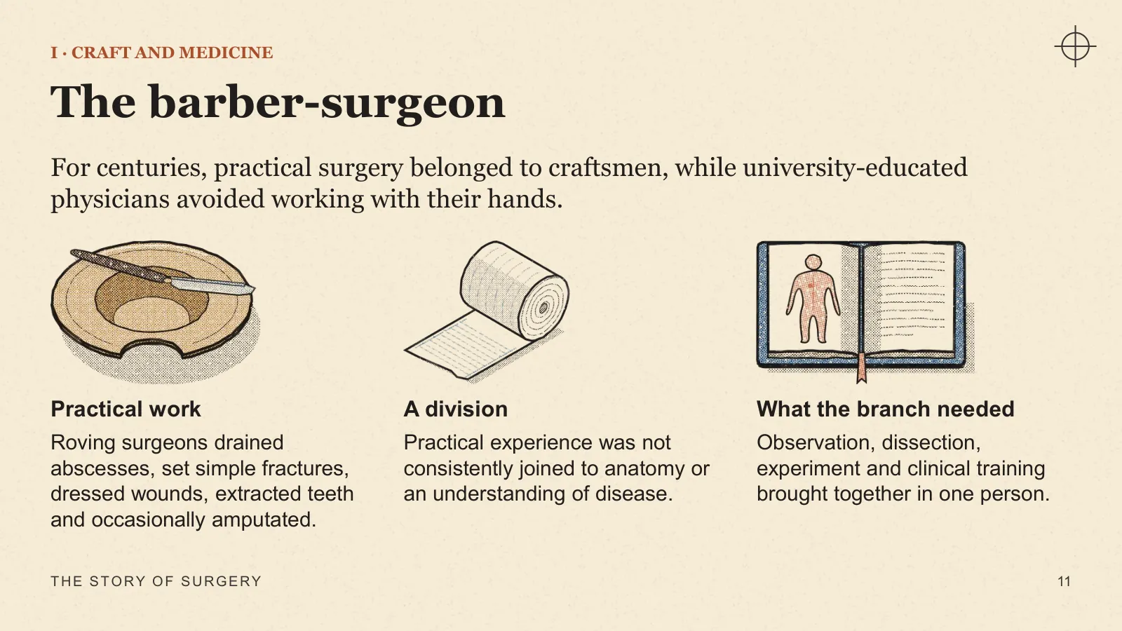
- Practical work Roving surgeons drained abscesses, set simple fractures, dressed wounds, extracted teeth and occasionally amputated.
- A division Practical experience was not consistently joined to anatomy or an understanding of disease.
- What the branch needed Observation, dissection, experiment and clinical training brought together in one person.
I · 1537 · NEAR TURIN · EVENING
The oil runs out
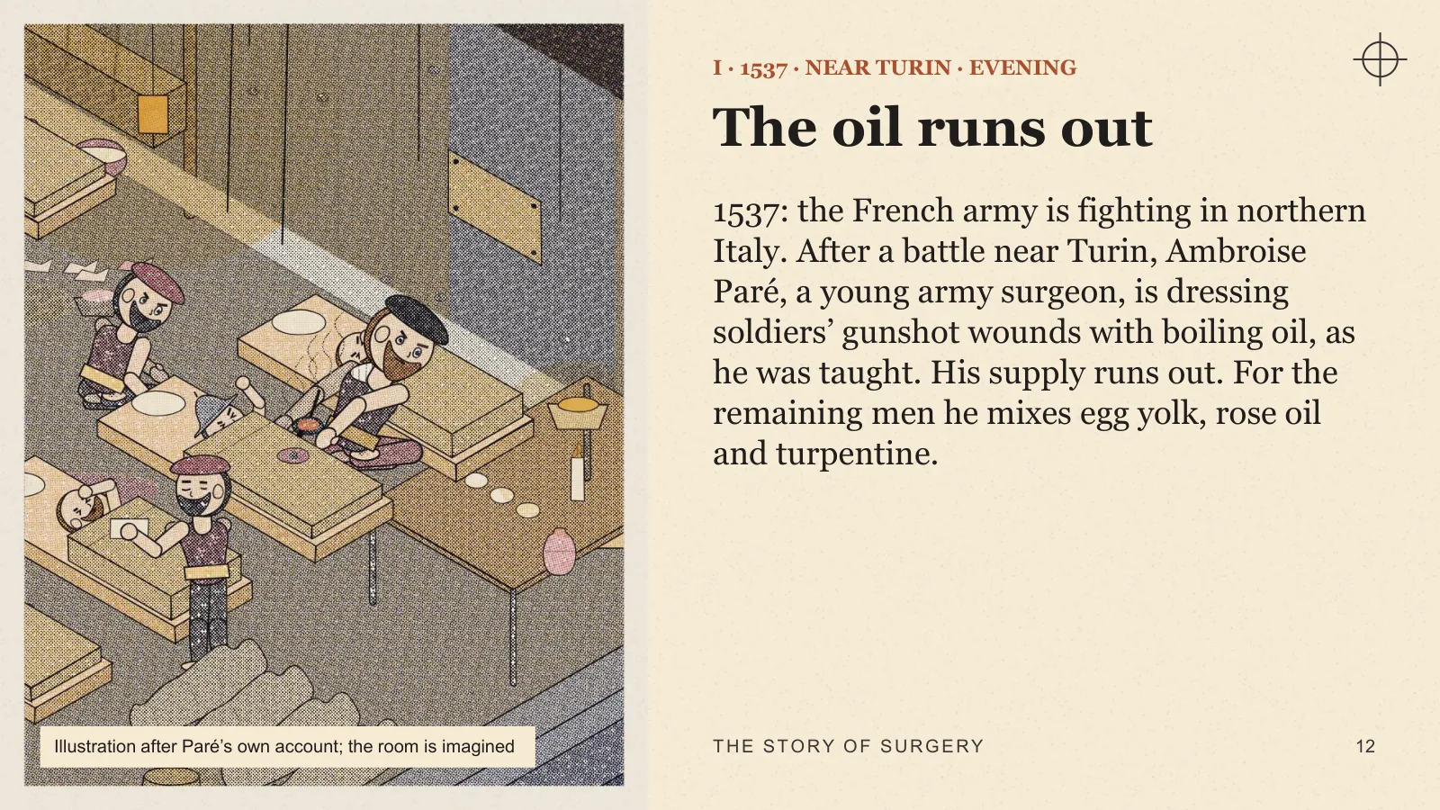
1537: the French army is fighting in northern Italy. After a battle near Turin, Ambroise Paré, a young army surgeon, is dressing soldiers’ gunshot wounds with boiling oil, as he was taught. His supply runs out. For the remaining men he mixes egg yolk, rose oil and turpentine.
Illustration after Paré’s own account; the room is imagined
I · 1537 · THE NEXT MORNING
Paré gives up boiling oil
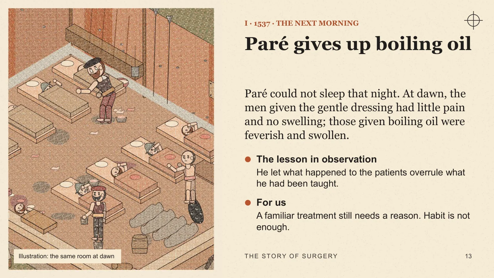
Paré could not sleep that night. At dawn, the men given the gentle dressing had little pain and no swelling; those given boiling oil were feverish and swollen.
- The lesson in observation He let what happened to the patients overrule what he had been taught.
- For us A familiar treatment still needs a reason. Habit is not enough.
Illustration: the same room at dawn
I · 1543
Vesalius looks for himself
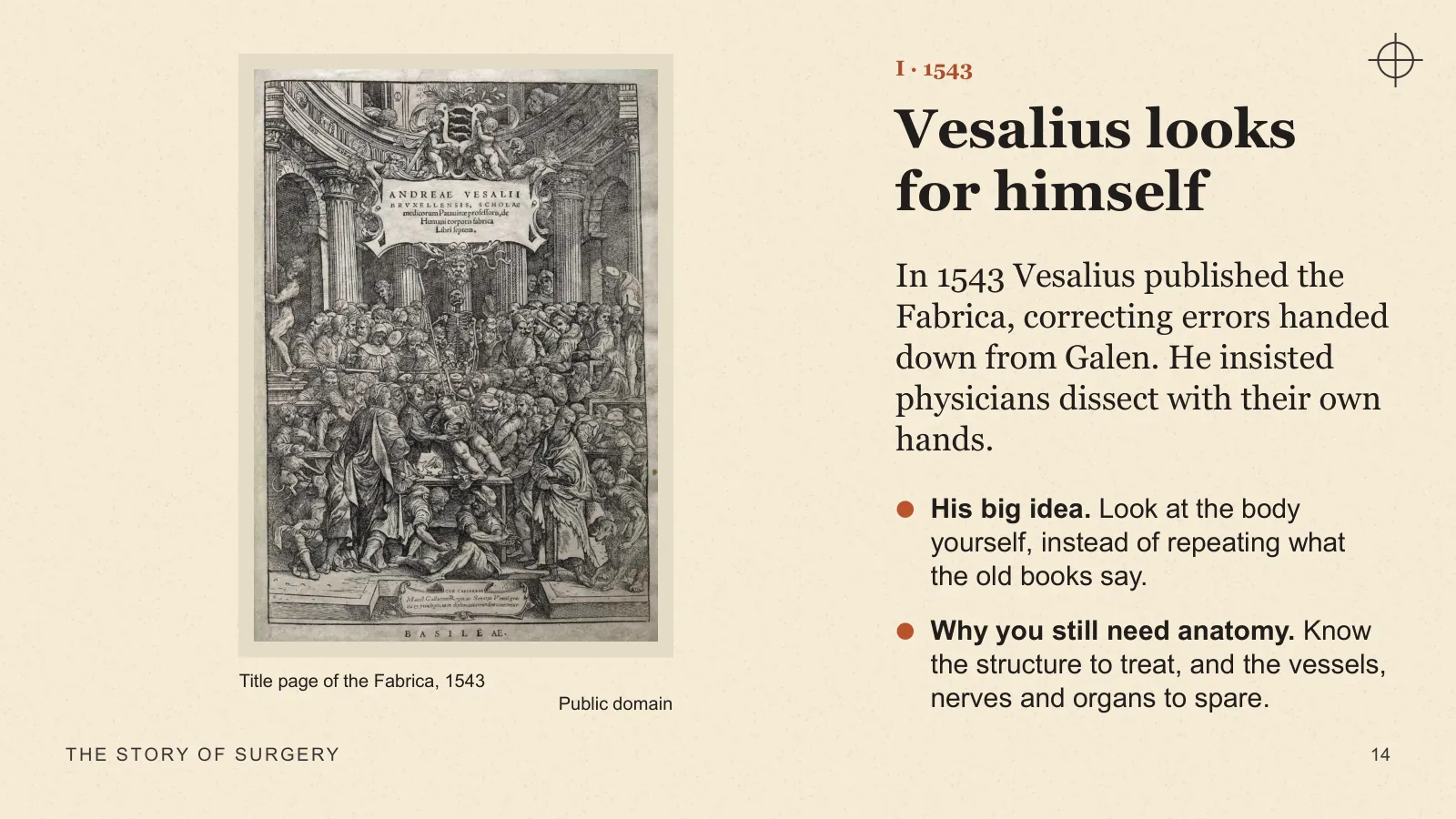
In 1543 Vesalius published the Fabrica, correcting errors handed down from Galen. He insisted physicians dissect with their own hands.
- His big idea Look at the body yourself, instead of repeating what the old books say.
- Why you still need anatomy Know the structure to treat, and the vessels, nerves and organs to spare.
Title page of the Fabrica, 1543 · Public domain
I · THE EIGHTEENTH CENTURY
Hunter asks how living tissue works
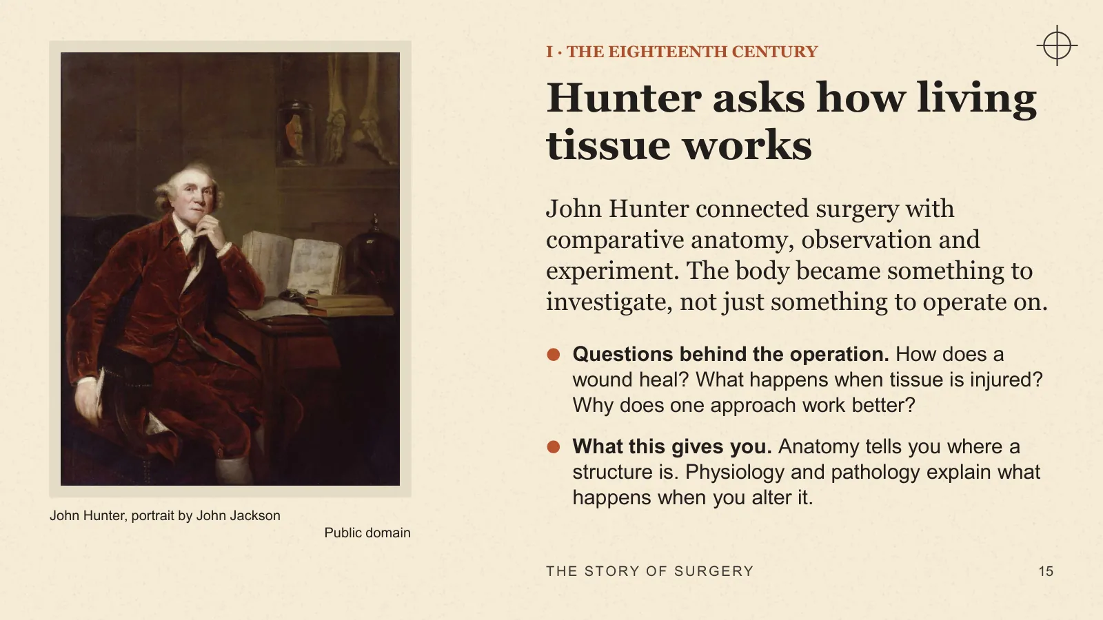
John Hunter connected surgery with comparative anatomy, observation and experiment. The body became something to investigate, not just something to operate on.
- Questions behind the operation How does a wound heal? What happens when tissue is injured? Why does one approach work better?
- What this gives you Anatomy tells you where a structure is. Physiology and pathology explain what happens when you alter it.
John Hunter, portrait by John Jackson · Public domain
I · NEAR POONA, 1790s
A nose cut to pattern
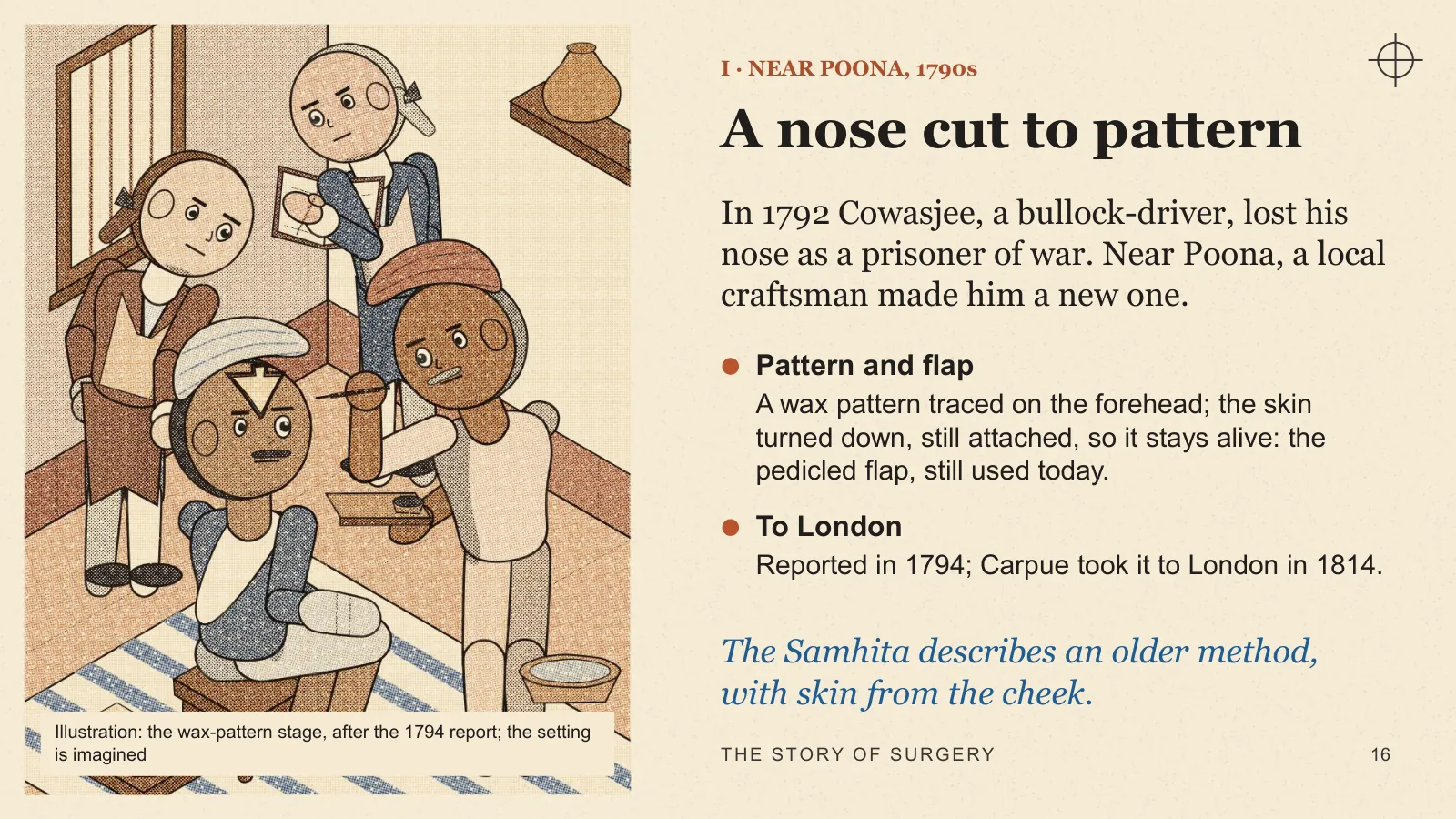
In 1792 Cowasjee, a bullock-driver, lost his nose as a prisoner of war. Near Poona, a local craftsman made him a new one.
- Pattern and flap A wax pattern traced on the forehead; the skin turned down, still attached, so it stays alive: the pedicled flap, still used today.
- To London Reported in 1794; Carpue took it to London in 1814.
The Samhita describes an older method, with skin from the cheek.
Illustration: the wax-pattern stage, after the 1794 report; the setting is imagined
Act II · Three walls: pain, infection, bleeding
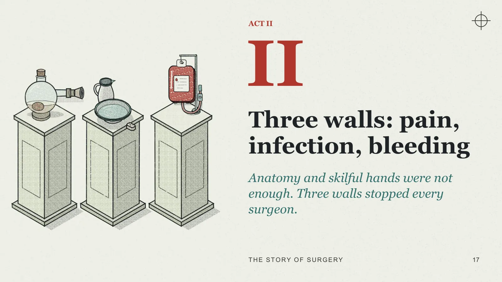
ACT II
Three walls: pain, infection, bleeding
Anatomy and skilful hands were not enough. Three walls stopped every surgeon.
II · THE THREE WALLS
Three walls every surgeon faced
- Pain Without reliable anaesthesia, suffering limited the time and complexity of an operation.
- Infection An apparently successful operation could be followed by a fatal wound infection.
- Bleeding The surgeon needed both control of haemorrhage and ways to replace blood when necessary.
These risks are better controlled today. They have not disappeared.
II · WALL ONE: PAIN · 16 OCTOBER 1846
Ether Day, 180 years ago
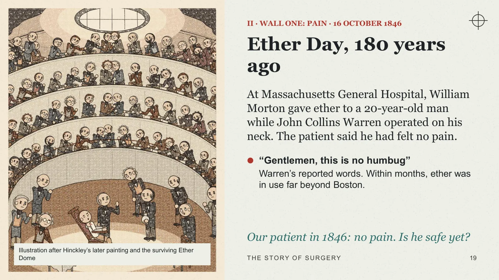
At Massachusetts General Hospital, William Morton gave ether to a 20-year-old man while John Collins Warren operated on his neck. The patient said he had felt no pain.
- “Gentlemen, this is no humbug” Warren’s reported words. Within months, ether was in use far beyond Boston.
Our patient in 1846: no pain. Is he safe yet?
Illustration after Hinckley’s later painting and the surviving Ether Dome
II · WALL TWO: INFECTION · 1847
Semmelweis and the washed hands
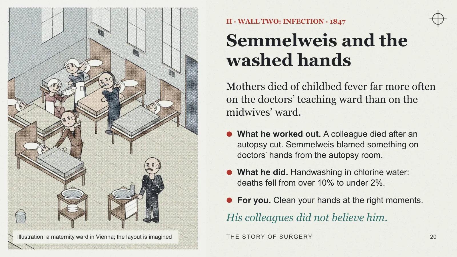
Mothers died of childbed fever far more often on the doctors’ teaching ward than on the midwives’ ward.
- What he worked out A colleague died after an autopsy cut. Semmelweis blamed something on doctors’ hands from the autopsy room.
- What he did Handwashing in chlorine water: deaths fell from over 10% to under 2%.
- For you Clean your hands at the right moments.
His colleagues did not believe him.
Illustration: a maternity ward in Vienna; the layout is imagined
II · LISTER · GLASGOW · 1865–67
Connecting germs and wound infection
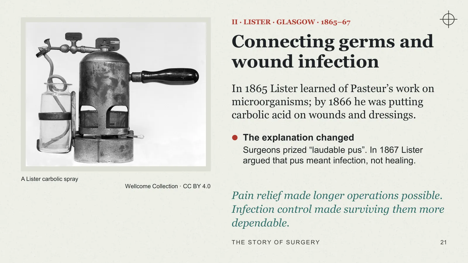
In 1865 Lister learned of Pasteur’s work on microorganisms; by 1866 he was putting carbolic acid on wounds and dressings.
- The explanation changed Surgeons prized “laudable pus”. In 1867 Lister argued that pus meant infection, not healing.
Pain relief made longer operations possible. Infection control made surviving them more dependable.
A Lister carbolic spray · Wellcome Collection · CC BY 4.0
II · LOOK CLOSELY
Can you see the change?
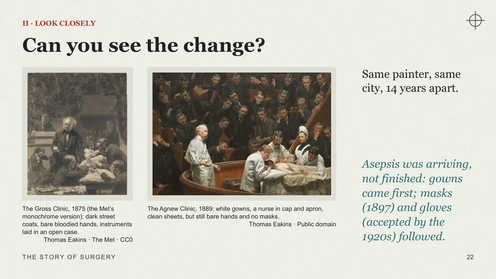
Same painter, same city, 14 years apart.
Asepsis was arriving, not finished: gowns came first; masks (1897) and gloves (accepted by the 1920s) followed.
The Gross Clinic, 1875 (the Met’s monochrome version): dark street coats, bare bloodied hands, instruments laid in an open case. · Thomas Eakins · The Met · CC0
The Agnew Clinic, 1889: white gowns, a nurse in cap and apron, clean sheets, but still bare hands and no masks. · Thomas Eakins · Public domain
II · KEEPING GERMS OUT
Antisepsis, then asepsis
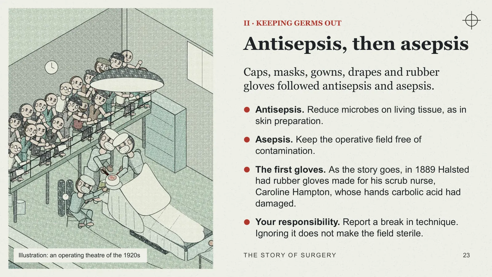
Caps, masks, gowns, drapes and rubber gloves followed antisepsis and asepsis.
- Antisepsis Reduce microbes on living tissue, as in skin preparation.
- Asepsis Keep the operative field free of contamination.
- The first gloves As the story goes, in 1889 Halsted had rubber gloves made for his scrub nurse, Caroline Hampton, whose hands carbolic acid had damaged.
- Your responsibility Report a break in technique. Ignoring it does not make the field sterile.
Illustration: an operating theatre of the 1920s
II · WALL TWO: INFECTION · 1928
Antibiotics
Penicillin is credited to Alexander Fleming in 1928, but it was not isolated for clinical use until 1941, by Florey and Chain.
- Still true today Antibiotics treat infection. A collection of pus still needs drainage.
Antibiotics don’t drain pus. Surgeons do.
II · WALL THREE: BLEEDING
Wall three: stopping the bleeding
Before blood could be replaced, the surgeon had to stop losing it.
- The ligature Paré promoted tying off vessels during amputation.
- The tourniquet Petit’s screw tourniquet, early 1700s, controlled bleeding in a limb.
- Electrosurgery, 1926 Cushing and Bovie used high-frequency current to cut and seal tissue. It remains a fundamental tool.
“What iron cannot cure, fire cures”: fire returns, this time as a controlled current.
II · WALL THREE: BLEEDING · 1901
Replacing the blood
- Before 1901 Scattered, risky transfusions. Halsted once transfused his own blood into his sister.
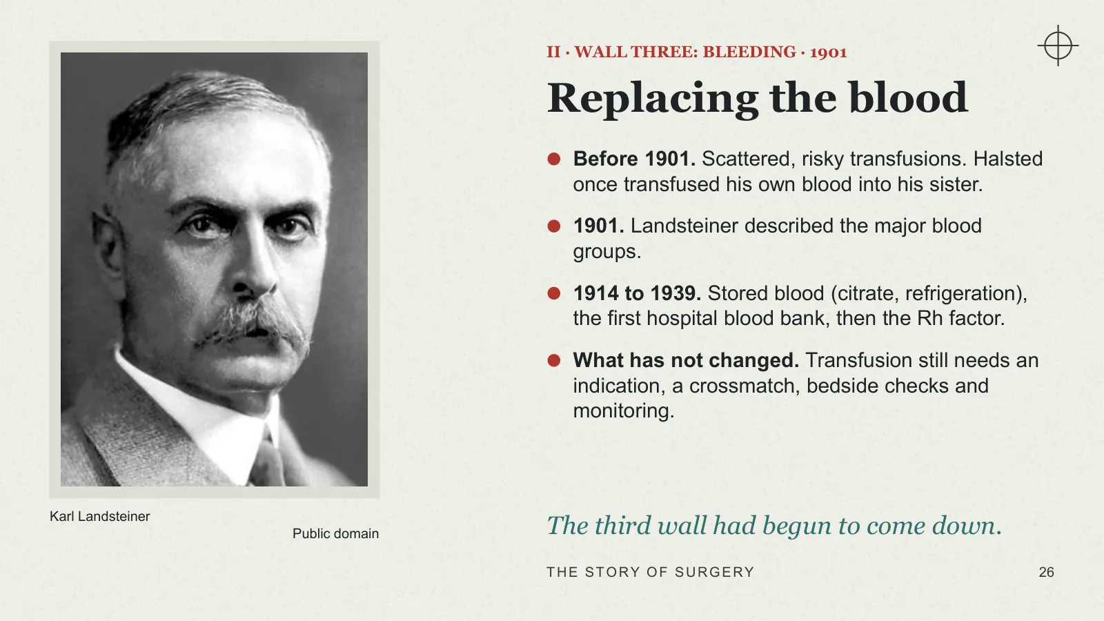
- 1901 Landsteiner described the major blood groups.
- 1914 to 1939 Stored blood (citrate, refrigeration), the first hospital blood bank, then the Rh factor.
- What has not changed Transfusion still needs an indication, a crossmatch, bedside checks and monitoring.
The third wall had begun to come down.
Karl Landsteiner · Public domain
II · THE WALLS COME DOWN
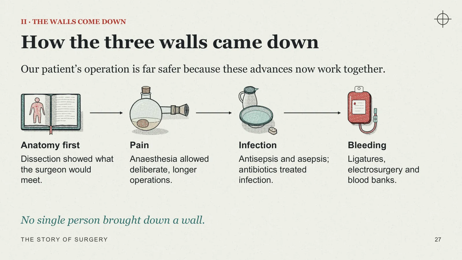
How the three walls came down
Our patient’s operation is far safer because these advances now work together.
- Anatomy first Dissection showed what the surgeon would meet.
- Pain Anaesthesia allowed deliberate, longer operations.
- Infection Antisepsis and asepsis; antibiotics treated infection.
- Bleeding Ligatures, electrosurgery and blood banks.
No single person brought down a wall.
II · 1895
Meanwhile, a discovery changed what doctors could see.
Seeing inside without cutting
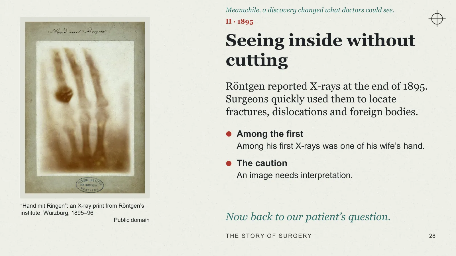
Röntgen reported X-rays at the end of 1895. Surgeons quickly used them to locate fractures, dislocations and foreign bodies.
- Among the first Among his first X-rays was one of his wife’s hand.
- The caution An image needs interpretation.
Now back to our patient’s question.
“Hand mit Ringen”: an X-ray print from Röntgen’s institute, Würzburg, 1895–96 · Public domain
Act III · Our patient, today

ACT III
Our patient, today
Our patient’s journey, from the ward to home: before, during, after and beyond.
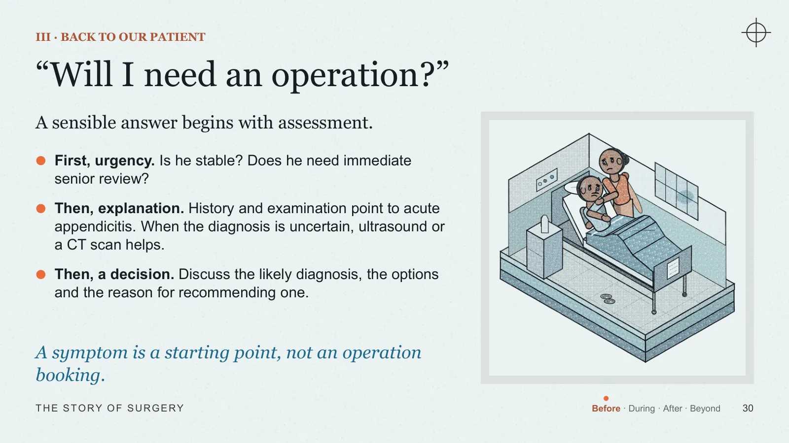
III · BACK TO OUR PATIENT
“Will I need an operation?”
A sensible answer begins with assessment.
- First, urgency Is he stable? Does he need immediate senior review?
- Then, explanation History and examination point to acute appendicitis. When the diagnosis is uncertain, ultrasound or a CT scan helps.
- Then, a decision Discuss the likely diagnosis, the options and the reason for recommending one.
A symptom is a starting point, not an operation booking.
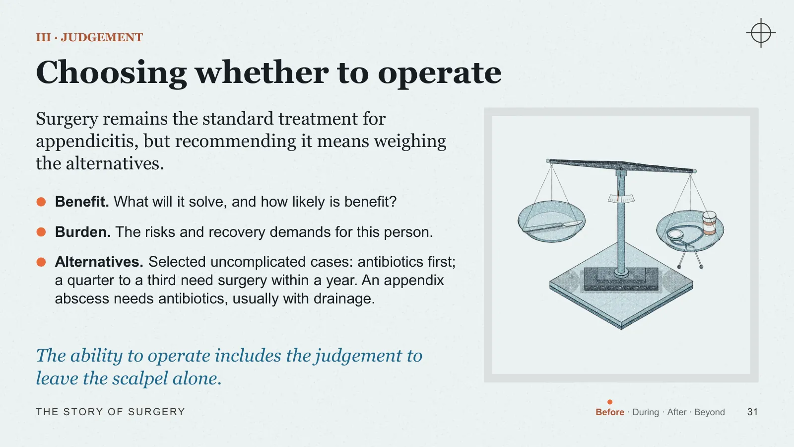
III · JUDGEMENT
Choosing whether to operate
Surgery remains the standard treatment for appendicitis, but recommending it means weighing the alternatives.
- Benefit What will it solve, and how likely is benefit?
- Burden The risks and recovery demands for this person.
- Alternatives Selected uncomplicated cases: antibiotics first; a quarter to a third need surgery within a year. An appendix abscess needs antibiotics, usually with drainage.
The ability to operate includes the judgement to leave the scalpel alone.
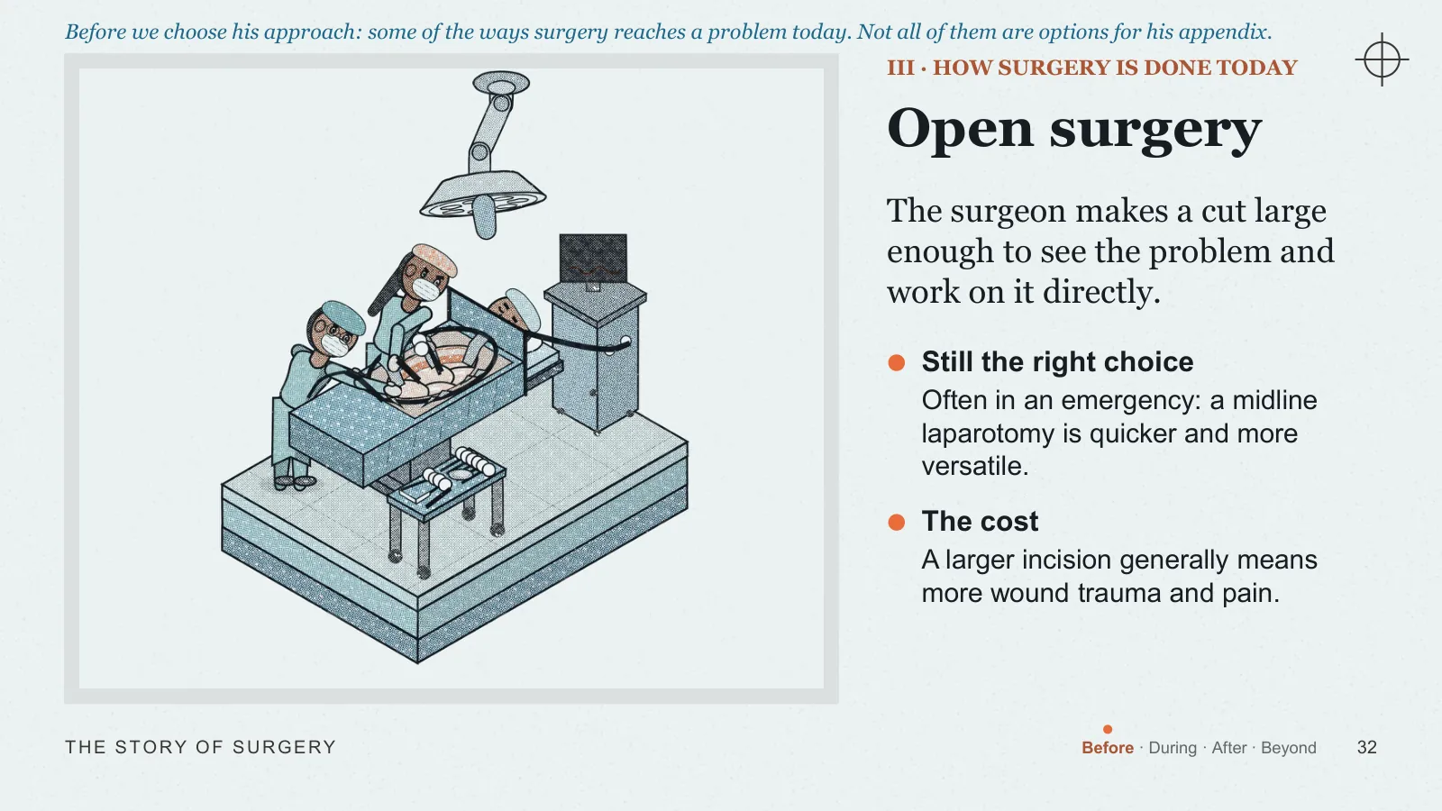
III · HOW SURGERY IS DONE TODAY
Before we choose his approach: some of the ways surgery reaches a problem today. Not all of them are options for his appendix.
Open surgery
The surgeon makes a cut large enough to see the problem and work on it directly.
- Still the right choice Often in an emergency: a midline laparotomy is quicker and more versatile.
- The cost A larger incision generally means more wound trauma and pain.
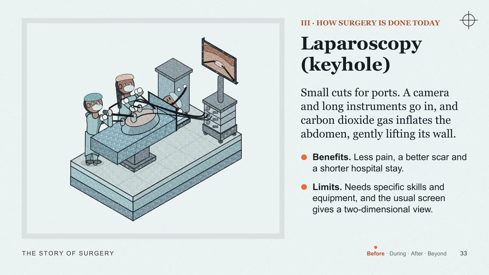
III · HOW SURGERY IS DONE TODAY
Laparoscopy (keyhole)
Small cuts for ports. A camera and long instruments go in, and carbon dioxide gas inflates the abdomen, gently lifting its wall.
- Benefits Less pain, a better scar and a shorter hospital stay.
- Limits Needs specific skills and equipment, and the usual screen gives a two-dimensional view.
III · HOW SURGERY IS DONE TODAY
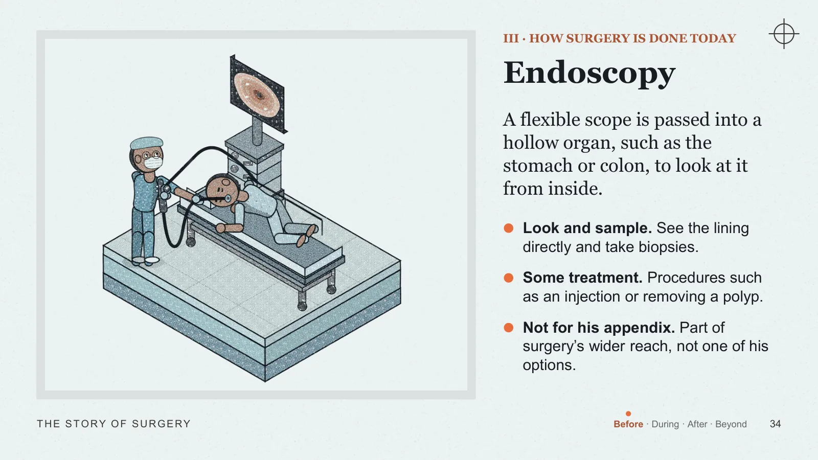
Endoscopy
A flexible scope is passed into a hollow organ, such as the stomach or colon, to look at it from inside.
- Look and sample See the lining directly and take biopsies.
- Some treatment Procedures such as an injection or removing a polyp.
- Not for his appendix Part of surgery’s wider reach, not one of his options.
III · HOW SURGERY IS DONE TODAY
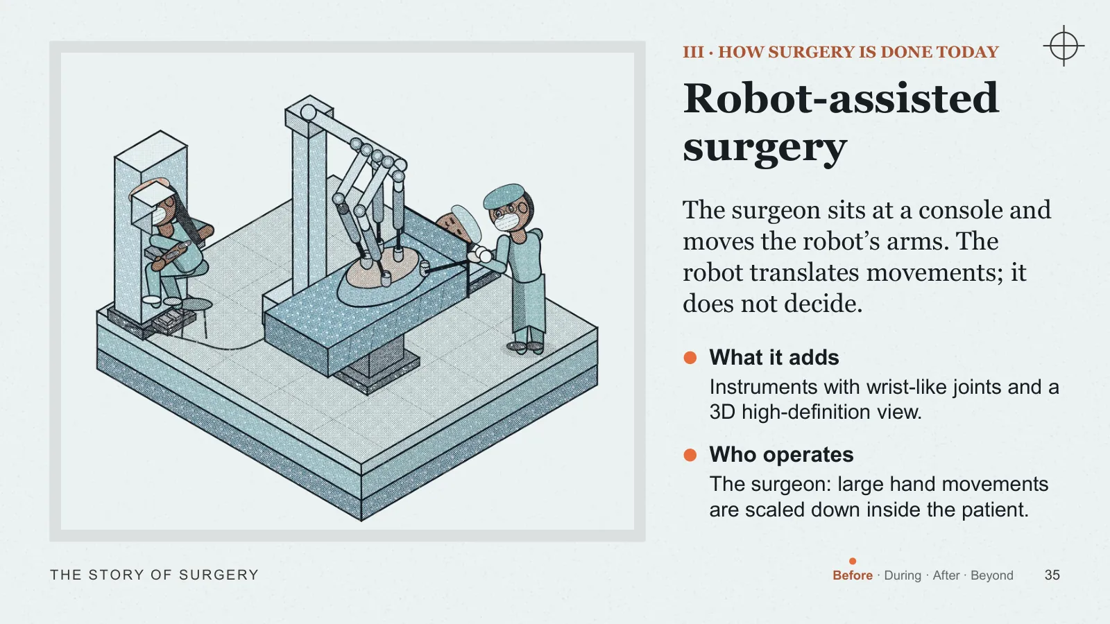
Robot-assisted surgery
The surgeon sits at a console and moves the robot’s arms. The robot translates movements; it does not decide.
- What it adds Instruments with wrist-like joints and a 3D high-definition view.
- Who operates The surgeon: large hand movements are scaled down inside the patient.
III · HOW SURGERY IS DONE TODAY
So, for him?
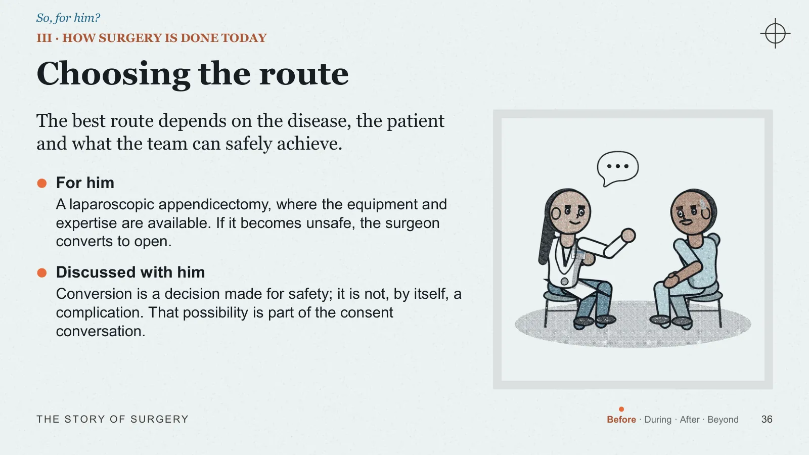
Choosing the route
The best route depends on the disease, the patient and what the team can safely achieve.
- For him A laparoscopic appendicectomy, where the equipment and expertise are available. If it becomes unsafe, the surgeon converts to open.
- Discussed with him Conversion is a decision made for safety; it is not, by itself, a complication. That possibility is part of the consent conversation.
III · EMPATHY IS A CLINICAL SKILL
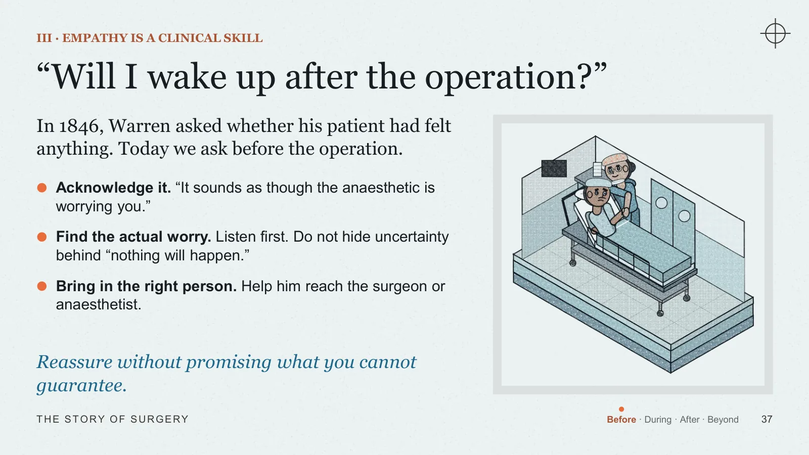
“Will I wake up after the operation?”
In 1846, Warren asked whether his patient had felt anything. Today we ask before the operation.
- Acknowledge it “It sounds as though the anaesthetic is worrying you.”
- Find the actual worry Listen first. Do not hide uncertainty behind “nothing will happen.”
- Bring in the right person Help him reach the surgeon or anaesthetist.
Reassure without promising what you cannot guarantee.
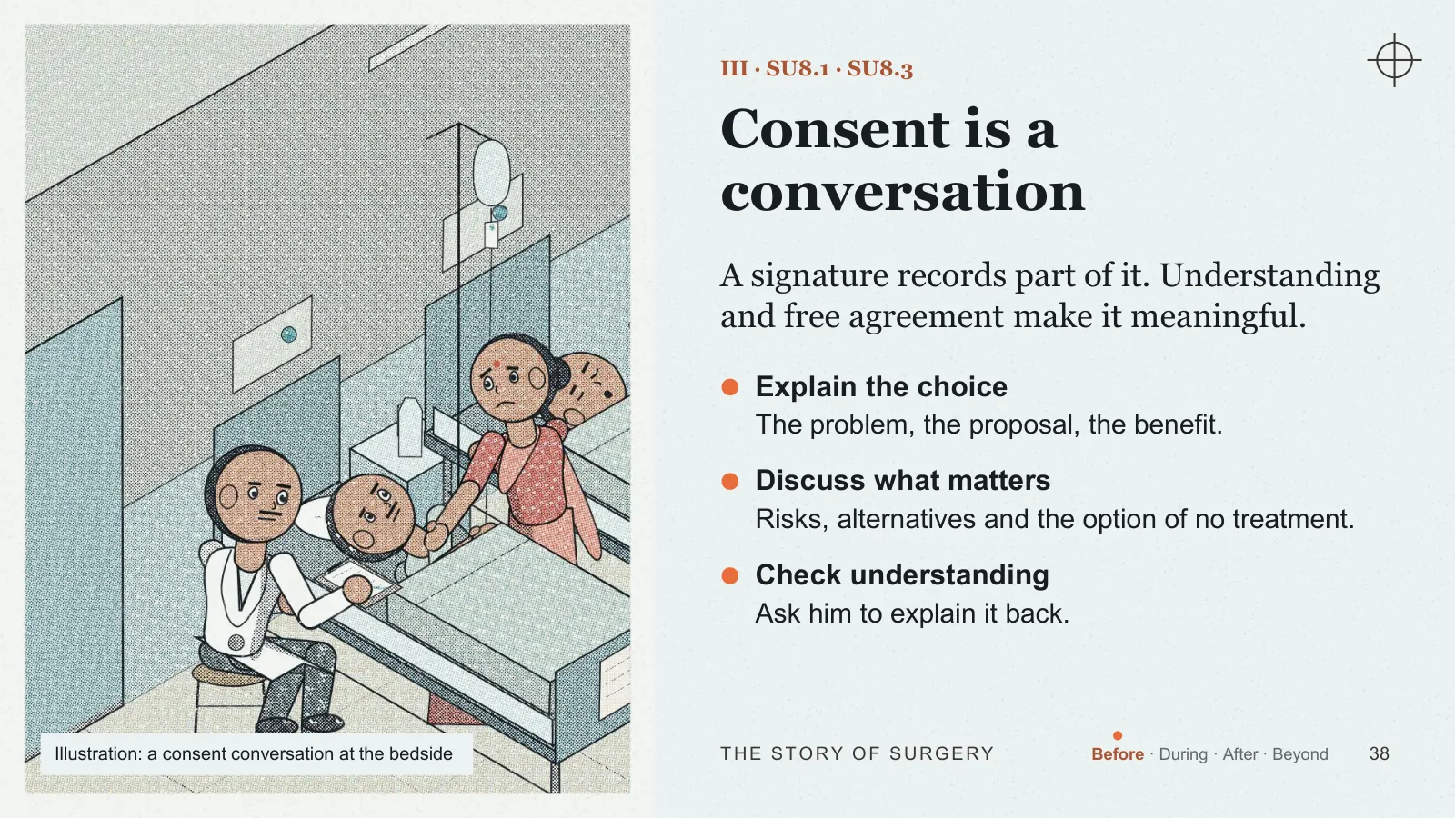
III · SU8.1 · SU8.3
Consent is a conversation
A signature records part of it. Understanding and free agreement make it meaningful.
- Explain the choice The problem, the proposal, the benefit.
- Discuss what matters Risks, alternatives and the option of no treatment.
- Check understanding Ask him to explain it back.
Illustration: a consent conversation at the bedside
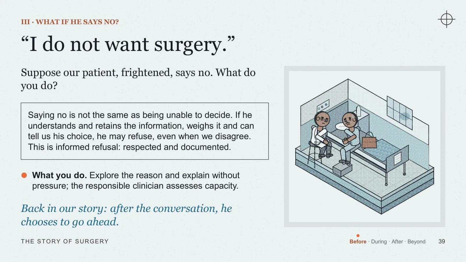
III · WHAT IF HE SAYS NO?
“I do not want surgery.”
Suppose our patient, frightened, says no. What do you do?
- What you do Explore the reason and explain without pressure; the responsible clinician assesses capacity.
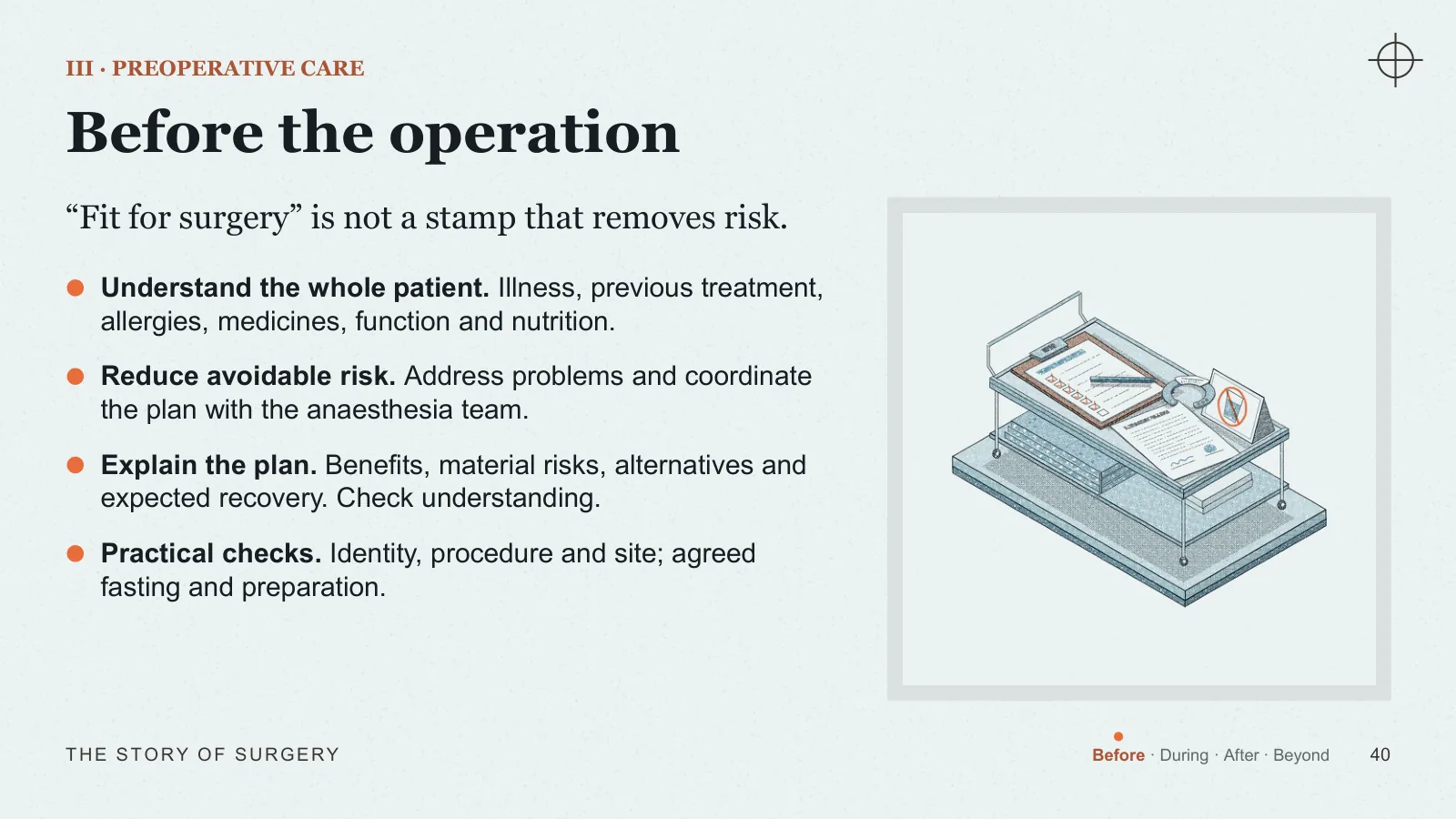
III · PREOPERATIVE CARE
Before the operation
“Fit for surgery” is not a stamp that removes risk.
- Understand the whole patient Illness, previous treatment, allergies, medicines, function and nutrition.
- Reduce avoidable risk Address problems and coordinate the plan with the anaesthesia team.
- Explain the plan Benefits, material risks, alternatives and expected recovery. Check understanding.
- Practical checks Identity, procedure and site; agreed fasting and preparation.
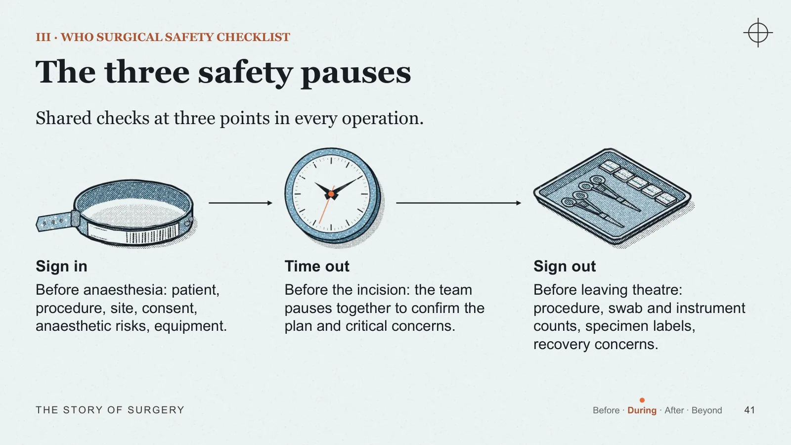
III · WHO SURGICAL SAFETY CHECKLIST
The three safety pauses
Shared checks at three points in every operation.
- Sign in Before anaesthesia: patient, procedure, site, consent, anaesthetic risks, equipment.
- Time out Before the incision: the team pauses together to confirm the plan and critical concerns.
- Sign out Before leaving theatre: procedure, swab and instrument counts, specimen labels, recovery concerns.
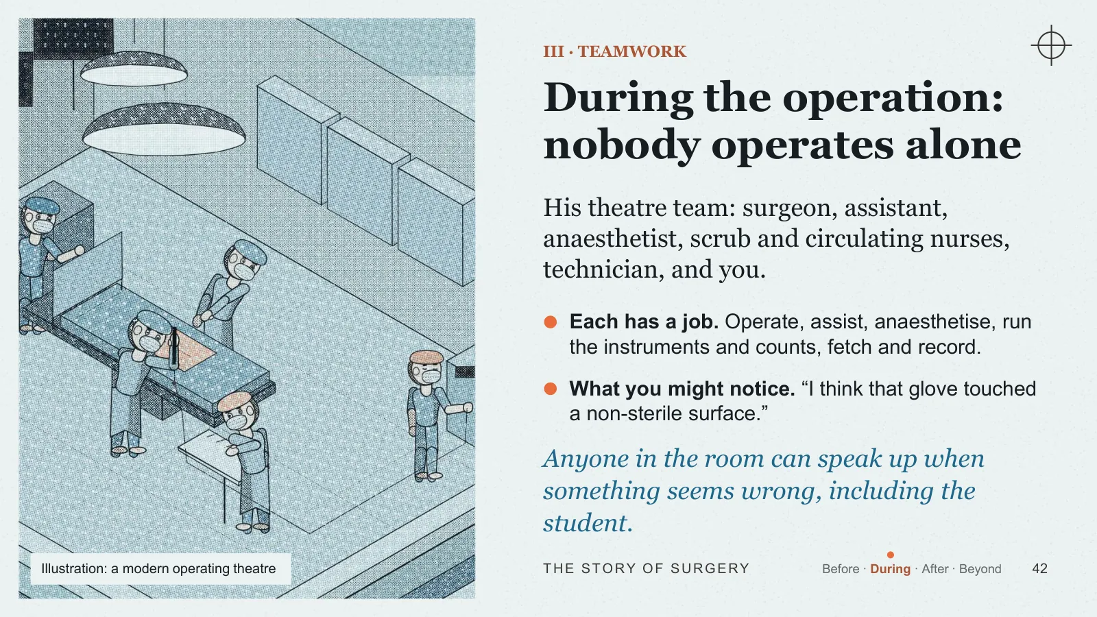
III · TEAMWORK
During the operation: nobody operates alone
His theatre team: surgeon, assistant, anaesthetist, scrub and circulating nurses, technician, and you.
- Each has a job Operate, assist, anaesthetise, run the instruments and counts, fetch and record.
- What you might notice “I think that glove touched a non-sterile surface.”
Anyone in the room can speak up when something seems wrong, including the student.
Illustration: a modern operating theatre
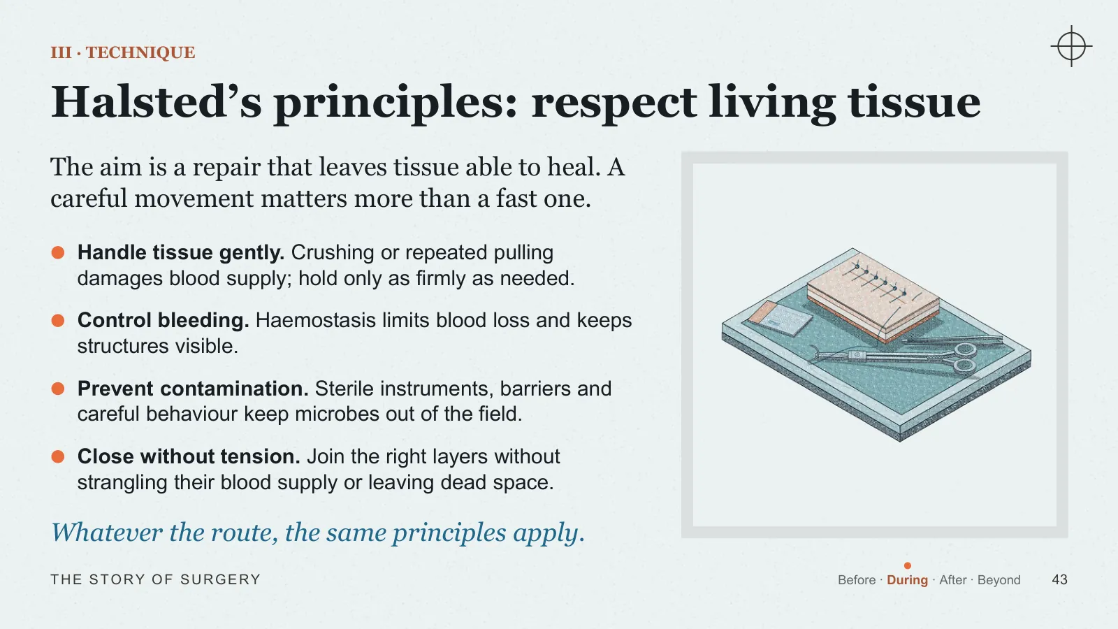
III · TECHNIQUE
Halsted’s principles: respect living tissue
The aim is a repair that leaves tissue able to heal. A careful movement matters more than a fast one.
- Handle tissue gently Crushing or repeated pulling damages blood supply; hold only as firmly as needed.
- Control bleeding Haemostasis limits blood loss and keeps structures visible.
- Prevent contamination Sterile instruments, barriers and careful behaviour keep microbes out of the field.
- Close without tension Join the right layers without strangling their blood supply or leaving dead space.
Whatever the route, the same principles apply.
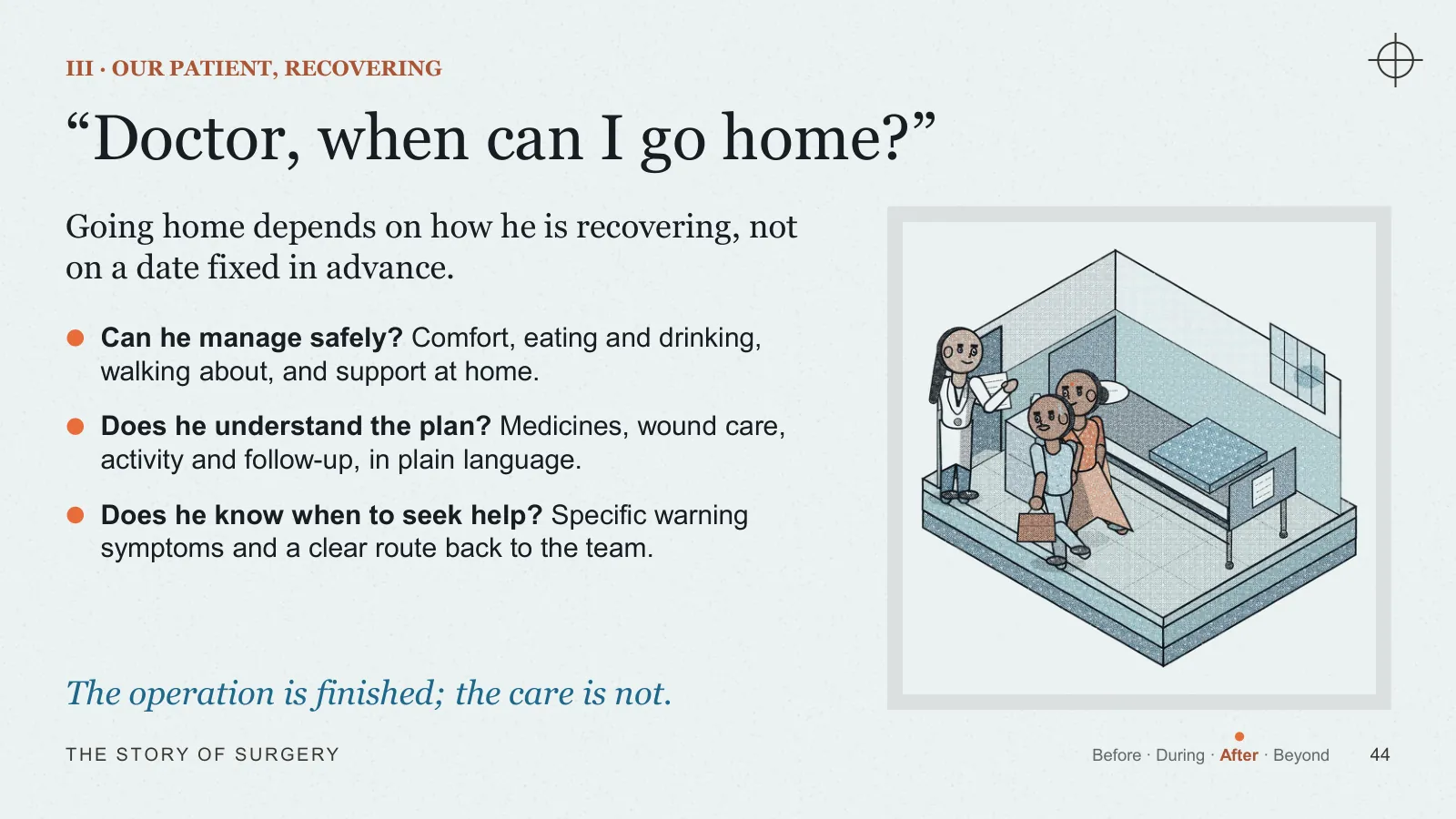
III · OUR PATIENT, RECOVERING
“Doctor, when can I go home?”
Going home depends on how he is recovering, not on a date fixed in advance.
- Can he manage safely? Comfort, eating and drinking, walking about, and support at home.
- Does he understand the plan? Medicines, wound care, activity and follow-up, in plain language.
- Does he know when to seek help? Specific warning symptoms and a clear route back to the team.
The operation is finished; the care is not.
Act IV · What no instrument can do
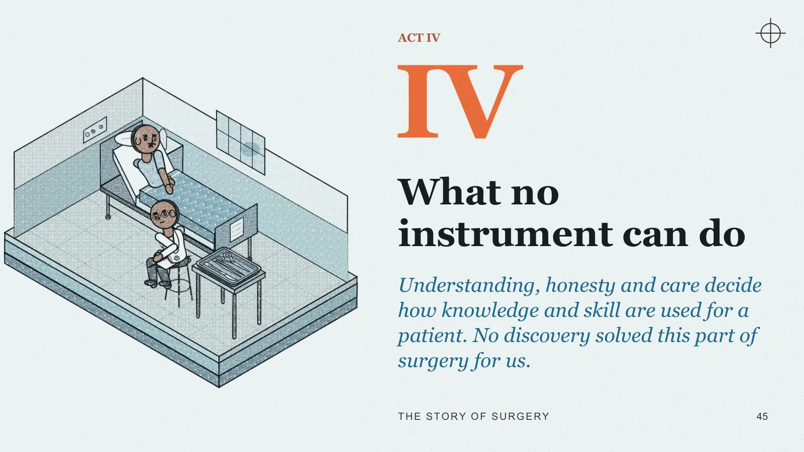
ACT IV
What no instrument can do
Understanding, honesty and care decide how knowledge and skill are used for a patient. No discovery solved this part of surgery for us.
IV · SU8.1
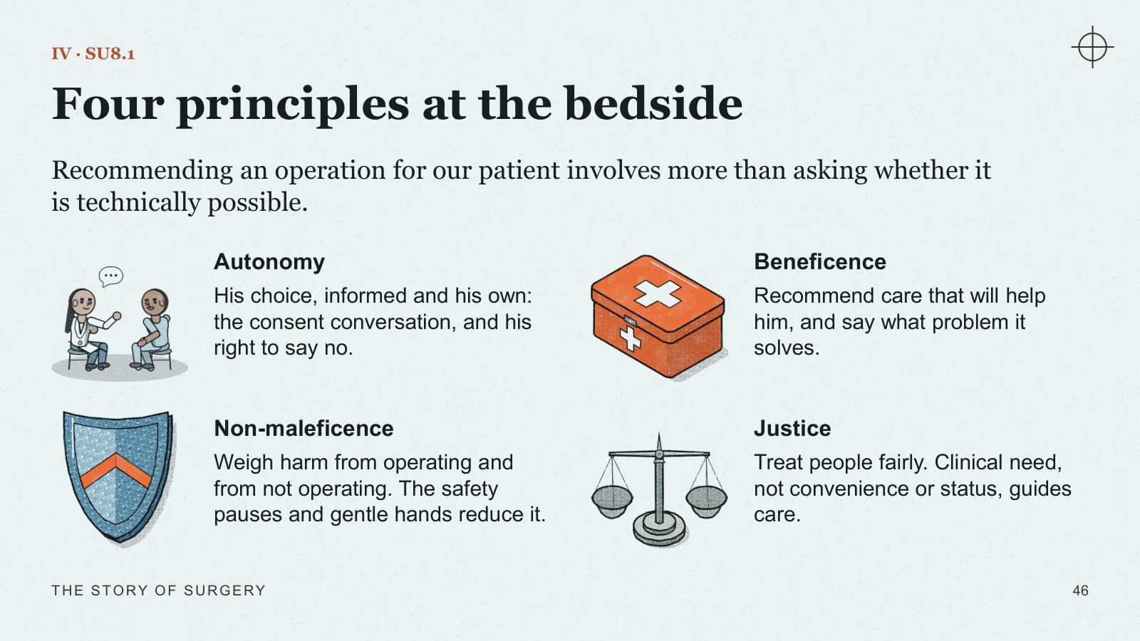
Four principles at the bedside
Recommending an operation for our patient involves more than asking whether it is technically possible.
- Autonomy His choice, informed and his own: the consent conversation, and his right to say no.
- Beneficence Recommend care that will help him, and say what problem it solves.
- Non-maleficence Weigh harm from operating and from not operating. The safety pauses and gentle hands reduce it.
- Justice Treat people fairly. Clinical need, not convenience or status, guides care.
IV · SU8.2
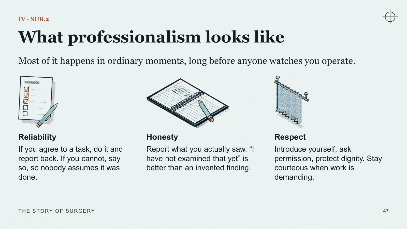
What professionalism looks like
Most of it happens in ordinary moments, long before anyone watches you operate.
- Reliability If you agree to a task, do it and report back. If you cannot, say so, so nobody assumes it was done.
- Honesty Report what you actually saw. “I have not examined that yet” is better than an invented finding.
- Respect Introduce yourself, ask permission, protect dignity. Stay courteous when work is demanding.
IV · SU8.3
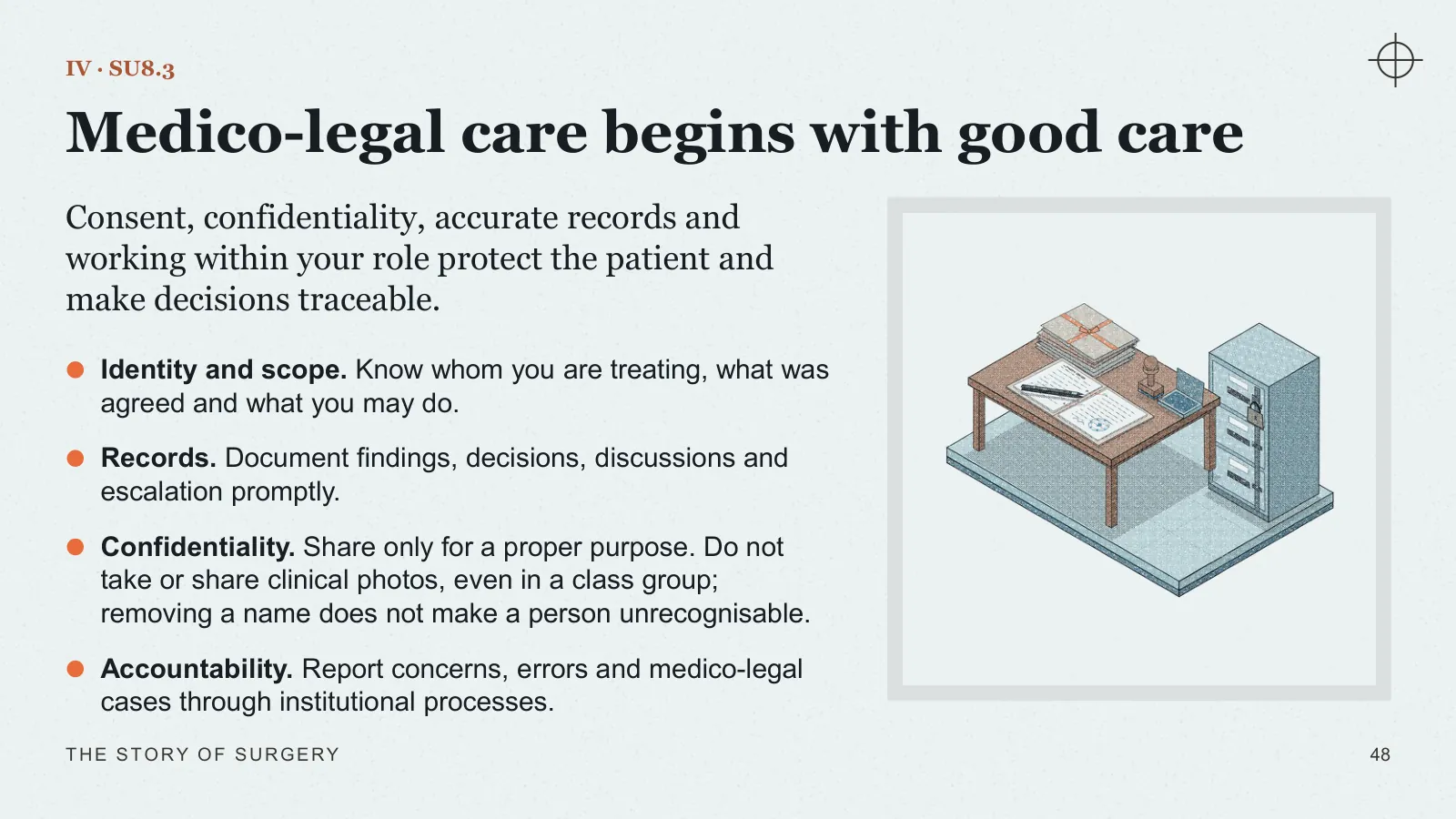
Medico-legal care begins with good care
Consent, confidentiality, accurate records and working within your role protect the patient and make decisions traceable.
- Identity and scope Know whom you are treating, what was agreed and what you may do.
- Records Document findings, decisions, discussions and escalation promptly.
- Confidentiality Share only for a proper purpose. Do not take or share clinical photos, even in a class group; removing a name does not make a person unrecognisable.
- Accountability Report concerns, errors and medico-legal cases through institutional processes.
Act V · Your chapter
ACT V
Your chapter
You begin as a supervised learner. Knowledge, participation and feedback gradually let you contribute more.
V · SCOPE
The breadth and depth of General Surgery
You will meet surgery in the clinic, the ward, emergency care and the operating theatre.
- Breadth Hernia, gallbladder and bowel, breast, thyroid, skin and soft tissue, trauma and emergencies.
- Depth The subspecialties it leads into: upper GI, hepatobiliary and pancreatic, colorectal, breast and endocrine, trauma, minimal-access surgery.
V · JOHNS HOPKINS · LATE 19TH CENTURY
Training becomes a system
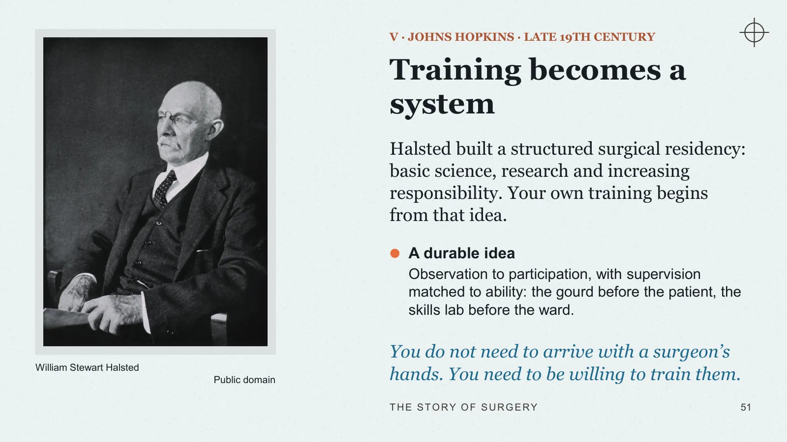
Halsted built a structured surgical residency: basic science, research and increasing responsibility. Your own training begins from that idea.
- A durable idea Observation to participation, with supervision matched to ability: the gourd before the patient, the skills lab before the ward.
You do not need to arrive with a surgeon’s hands. You need to be willing to train them.
William Stewart Halsted · Public domain
V · AETCOM
The five roles of an Indian Medical Graduate, with our patient
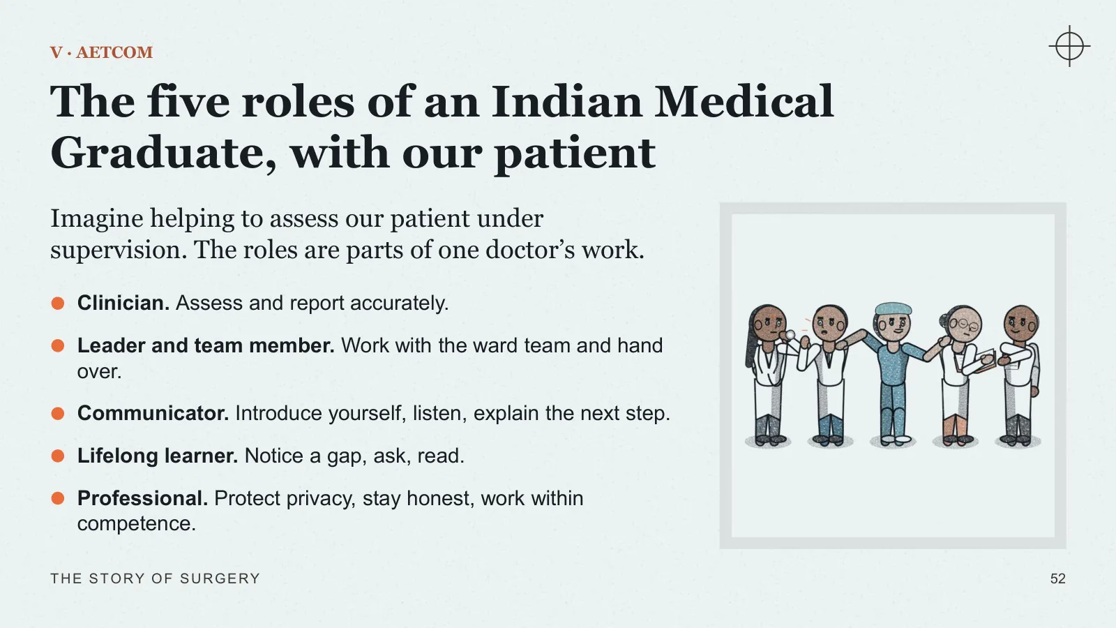
Imagine helping to assess our patient under supervision. The roles are parts of one doctor’s work.
- Clinician Assess and report accurately.
- Leader and team member Work with the ward team and hand over.
- Communicator Introduce yourself, listen, explain the next step.
- Lifelong learner Notice a gap, ask, read.
- Professional Protect privacy, stay honest, work within competence.
V · LEARNING THROUGH PATIENT CARE
The learner–doctor model
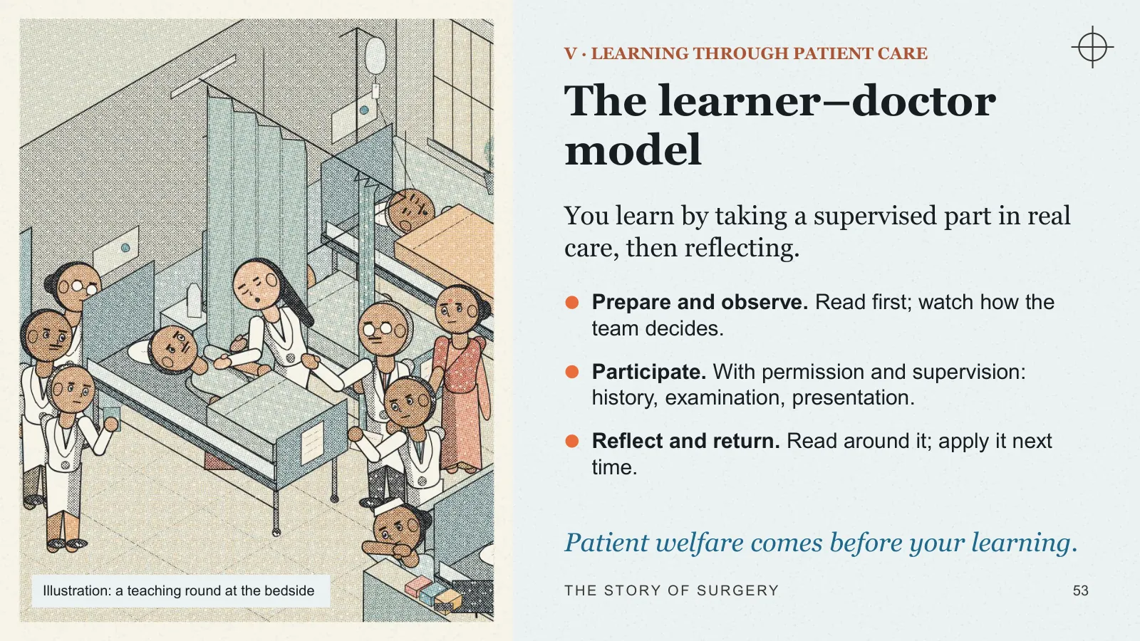
You learn by taking a supervised part in real care, then reflecting.
- Prepare and observe Read first; watch how the team decides.
- Participate With permission and supervision: history, examination, presentation.
- Reflect and return Read around it; apply it next time.
Patient welfare comes before your learning.
Illustration: a teaching round at the bedside
V · YOUR SCOPE NOW
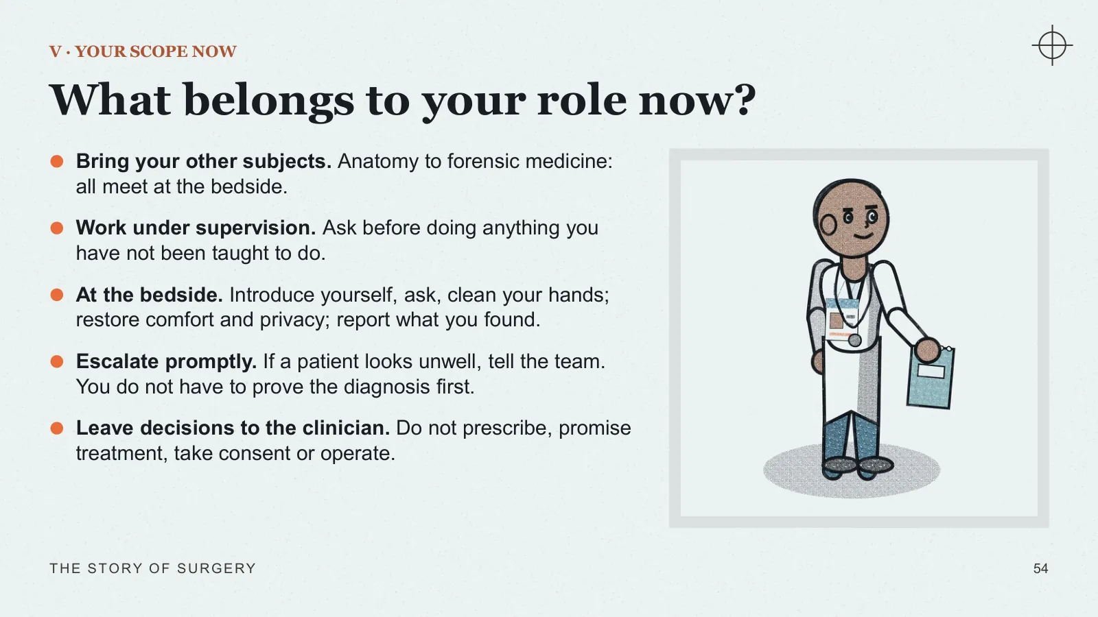
What belongs to your role now?
- Bring your other subjects Anatomy to forensic medicine: all meet at the bedside.
- Work under supervision Ask before doing anything you have not been taught to do.
- At the bedside Introduce yourself, ask, clean your hands; restore comfort and privacy; report what you found.
- Escalate promptly If a patient looks unwell, tell the team. You do not have to prove the diagnosis first.
- Leave decisions to the clinician Do not prescribe, promise treatment, take consent or operate.
Closing
TWO PATIENTS, 180 YEARS APART
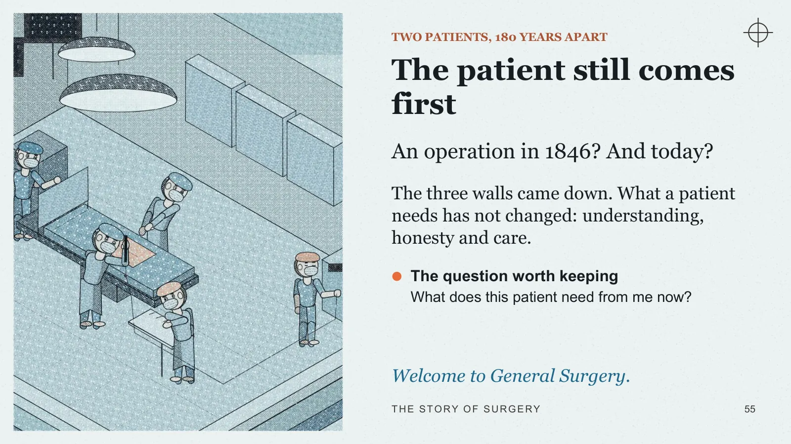
The patient still comes first
An operation in 1846? And today?
The three walls came down. What a patient needs has not changed: understanding, honesty and care.
- The question worth keeping What does this patient need from me now?
Welcome to General Surgery.
Sources and image credits
Original artwork. The animated scenes, icons and illustrated plates were made for this lecture. They contain no third-party artwork.
Images
- A modern statue of Sushruta (cropped) · Alokprasad · CC BY-SA 3.0
- Title page of the Fabrica, 1543 · Public domain
- John Hunter, portrait by John Jackson · Public domain
- A Lister carbolic spray · Wellcome Collection · CC BY 4.0
- The Gross Clinic, 1875 (the Met’s monochrome version): dark street coats, bare bloodied hands, instruments laid in an open case. · Thomas Eakins · The Met · CC0
- The Agnew Clinic, 1889: white gowns, a nurse in cap and apron, clean sheets, but still bare hands and no masks. · Thomas Eakins · Public domain
- Karl Landsteiner · Public domain
- “Hand mit Ringen”: an X-ray print from Röntgen’s institute, Würzburg, 1895–96 · Public domain
- William Stewart Halsted · Public domain


Sources
- [H] Sabiston Textbook of Surgery, 21st ed. (2021), chapter 1, The Rise of Modern Surgery: An Overview, printed pp. 2–11.
- [B] Bailey & Love’s Short Practice of Surgery, 28th ed. History, chapter 5; plastic and reconstructive surgery pp. 681–682; surgical infection pp. 50–51; surgical principles, chapter 7; minimal access and robotic surgery, chapter 10; human factors and the surgical safety checklist, chapter 15; conflict surgery, chapter 34; the vermiform appendix, chapter 76.
- [F] Fundamentals of General Surgery (Springer, 2018). Patient preparation, chapter 1; basic skills, chapter 3; incisions and closure, chapter 5; laparoscopy, endoscopy and robotics chapters.
- [A] AETCOM: Attitude, Ethics and Communication Competencies for the Indian Medical Graduate. Roles pp. 11–14; cardinal principles, module 2.2; autonomy and decision-making, modules 2.5–2.7.
- [W] WHO, New checklist to help make surgery safer, 24 June 2008, and WHO Safe Surgery resources. An introductory summary, not a substitute for the complete checklist or local protocol. Safe surgery saves lives: frequently asked questions (who.int, read 9 October 2026).
- [E] US National Library of Medicine, An Ancient Medical Treasure at Your Fingertips (2010), on the Edwin Smith Papyrus.
- [S] WHO, Unveiling of the Semmelweis statue (2019); WHO Guidelines on Hand Hygiene in Health Care (2009), historical discussion.
- [N] Nobel Assembly at Karolinska Institutet, Nobel laureates in physiology or medicine, official laureate table.
- [X] Sabiston Textbook of Surgery, 21st ed. (2021), transplantation, chapters 25 and 26.
- [J] Ambroise Paré, Journeys in Diverse Places (the Turin campaign, 1537), Harvard Classics vol. 38.
- [K] K.K.L. Bhishagratna (trans.), An English Translation of the Sushruta Samhita, Vol. I (Calcutta, 1907): introduction pp. ii–vi; Sutrasthana ch. VII–X, XVI and XXV. Internet Archive, englishtranslati01susruoft.
- [G] B. L., letter of 9 October 1794, “Curious chirurgical operation”, The Gentleman’s Magazine, October 1794, pp. 891–892; Plate I facing p. 883.
- [Q] J. C. Carpue, An Account of Two Successful Operations for Restoring a Lost Nose from the Integuments of the Forehead (London, 1816), pp. 38–39, 81–83. Wellcome Collection copy, Internet Archive b22017380.
- [Y] Shaye D.A. (2021) The history of nasal reconstruction. Curr Opin Otolaryngol Head Neck Surg 29(4). doi:10.1097/MOO.0000000000000730 (PMC8270507).
- [R] The Gentleman’s Magazine (1794), as reproduced and discussed in V. Pothula, Sushruta and Indian rhinoplasty (2021). The 1794 forehead flap is distinct from earlier cheek-flap descriptions.
- [C] ACS Surgery: Principles and Practice, surgical site infection chapter (the Halstedian principles).
- [D] Zdravković D. (2007), on Halsted’s principles of surgery. PubMed PMID 17990811 (abstract).
- [L] Spirling & Daniels (2002), doi:10.1177/146642400212200215; Hernigou (2022), doi:10.1007/s00264-022-05666-w. On Halsted’s first rubber gloves and Caroline Hampton (abstracts).
- [M] Maingot’s Abdominal Operations, 13th ed., chapter 1.
- [V] Sternbach & Varon (1993), doi:10.1016/0736-4679(93)90635-k, on Röntgen and the first X-rays (abstract).
- [Z] Schwartz’s Principles of Surgery, 11th ed. (2019), chapter 6, Surgical infections: historical background.
- [O] Oxford Handbook of Clinical Surgery, 4th ed. Gastroscopy; post-operative management.
- [T] Original teaching synthesis. Patients, dialogue and scenes are hypothetical or illustrative, not patient records or quotations of historical figures.
- [P] Dr Prateek Gupta. Department brief: Introduction to General Surgery, IMG roles and learner-doctor model; SU1.3, SU8.1, SU8.2, SU8.3, SU10.1.