Hepatobiliary surgery · Lecture 1
Biliary anatomy · 44 slides
The biliary tree.
Anatomy that explains the symptoms and guides the operation: follow bile from the liver, through the ducts and gallbladder, to the duodenum.
About the downloads
PowerPoint without speaker notes. SHA-256 checksums verify the downloaded files.
Contents · 5 parts
Introduction
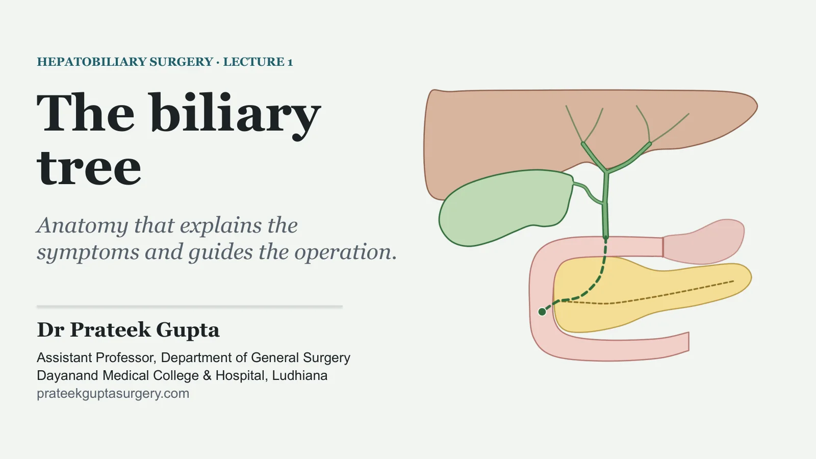
The biliary tree
HEPATOBILIARY SURGERY · LECTURE 1
Anatomy that explains the symptoms and guides the operation.
Dr Prateek Gupta
Assistant Professor, Department of General Surgery Dayanand Medical College & Hospital, Ludhiana prateekguptasurgery.com
A year of attacks of pain, and now yellow eyes
A PATIENT IN THE SURGICAL OUTPATIENT CLINIC
A 45-year-old woman has had attacks of upper abdominal pain for about a year. Each attack settled within a few hours.
This attack. The pain began three days ago, high in the middle of the abdomen, and has not fully settled.
Today. Her family has noticed that the whites of her eyes have turned yellow.
Earlier attacks. None of them made her yellow.
Questions to keep in mind
1. Which route does bile take from the liver to the duodenum?
2. Where must bile be blocked to make her yellow?
3. Why did the earlier attacks cause pain but no jaundice?
4. Which structures must a surgeon identify before removing her gallbladder?
We return to her at the end of the class.
Illustrative case: not a real patient.
THE BILIARY TREE
2
The route of bile
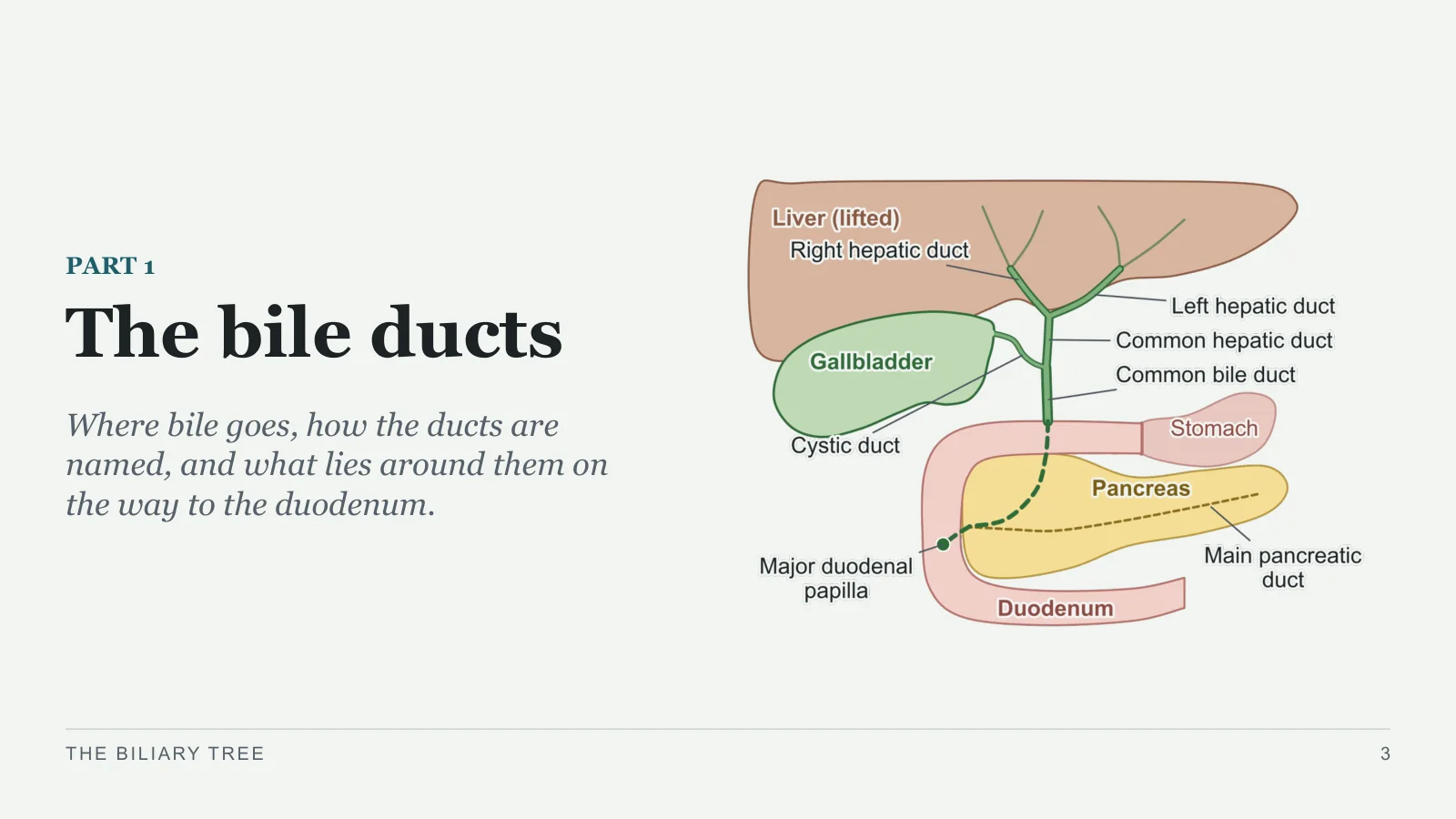
The bile ducts
PART 1
Where bile goes, how the ducts are named, and what lies around them on the way to the duodenum.
THE BILIARY TREE
3
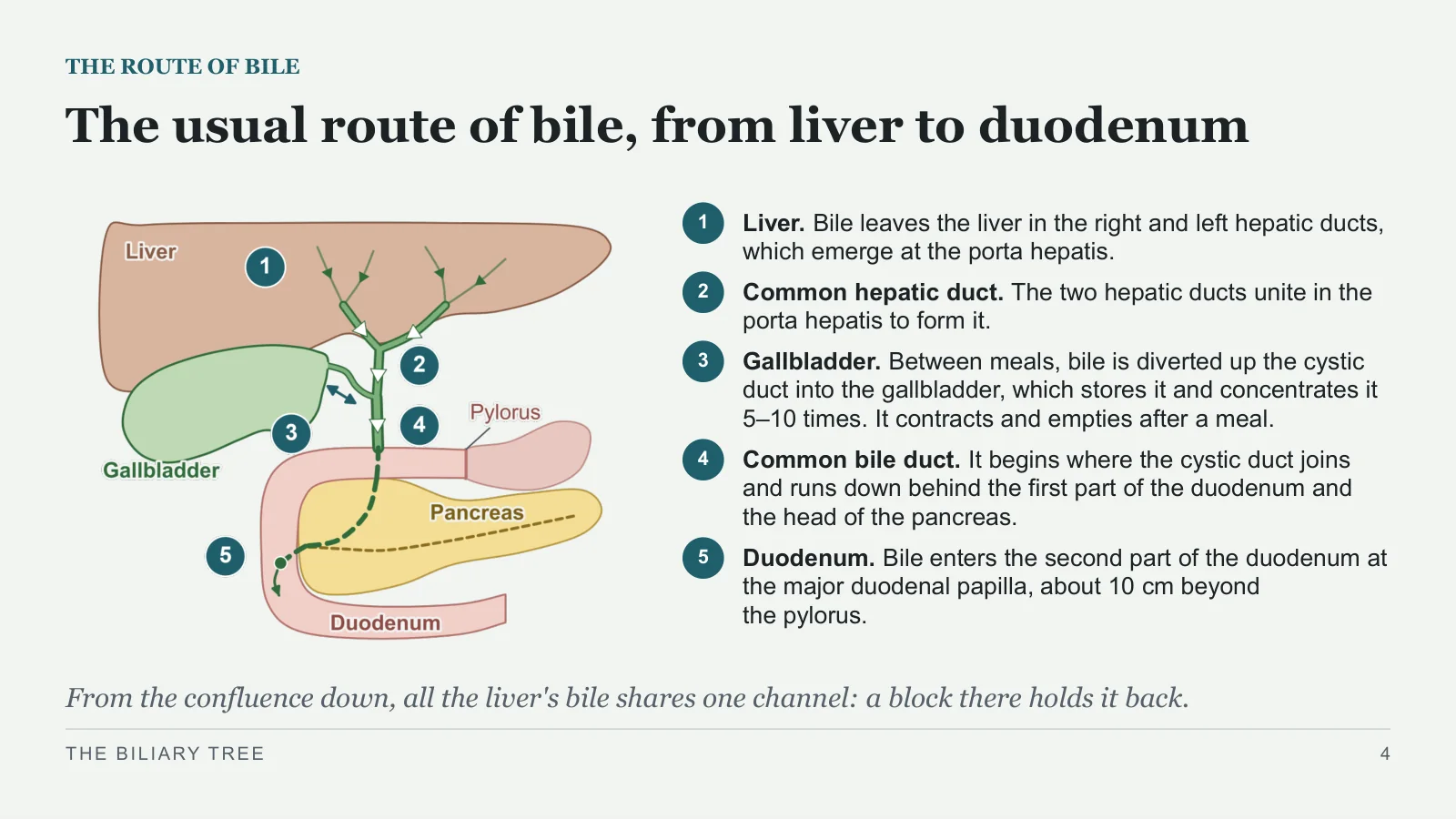
The usual route of bile, from liver to duodenum
THE ROUTE OF BILE
1
Liver. Bile leaves the liver in the right and left hepatic ducts, which emerge at the porta hepatis.
2
Common hepatic duct. The two hepatic ducts unite in the porta hepatis to form it.
3
Gallbladder. Between meals, bile is diverted up the cystic duct into the gallbladder, which stores it and concentrates it 5–10 times. It contracts and empties after a meal.
4
Common bile duct. It begins where the cystic duct joins and runs down behind the first part of the duodenum and the head of the pancreas.
5
Duodenum. Bile enters the second part of the duodenum at the major duodenal papilla, about 10 cm beyond the pylorus.
From the confluence down, all the liver's bile shares one channel: a block there holds it back.
THE BILIARY TREE
4
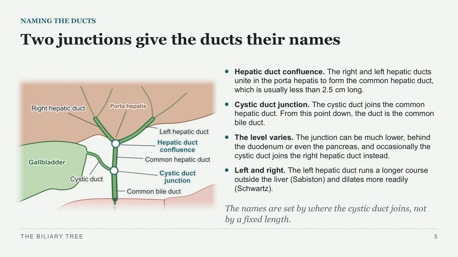
Two junctions give the ducts their names
NAMING THE DUCTS
Hepatic duct confluence. The right and left hepatic ducts unite in the porta hepatis to form the common hepatic duct, which is usually less than 2.5 cm long.
Cystic duct junction. The cystic duct joins the common hepatic duct. From this point down, the duct is the common bile duct.
The level varies. The junction can be much lower, behind the duodenum or even the pancreas, and occasionally the cystic duct joins the right hepatic duct instead.
Left and right. The left hepatic duct runs a longer course outside the liver (Sabiston) and dilates more readily (Schwartz).
The names are set by where the cystic duct joins, not by a fixed length.
THE BILIARY TREE
5
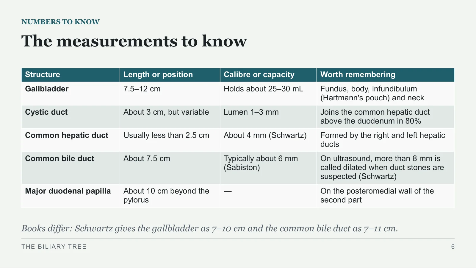
The measurements to know
NUMBERS TO KNOW
Books differ: Schwartz gives the gallbladder as 7–10 cm and the common bile duct as 7–11 cm.
THE BILIARY TREE
6
| Structure | Length or position | Calibre or capacity | Worth remembering |
| Gallbladder | 7.5–12 cm | Holds about 25–30 mL | Fundus, body, infundibulum (Hartmann's pouch) and neck |
| Cystic duct | About 3 cm, but variable | Lumen 1–3 mm | Joins the common hepatic duct above the duodenum in 80% |
| Common hepatic duct | Usually less than 2.5 cm | About 4 mm (Schwartz) | Formed by the right and left hepatic ducts |
| Common bile duct | About 7.5 cm | Typically about 6 mm (Sabiston) | On ultrasound, more than 8 mm is called dilated when duct stones are suspected (Schwartz) |
| Major duodenal papilla | About 10 cm beyond the pylorus | — | On the posteromedial wall of the second part |
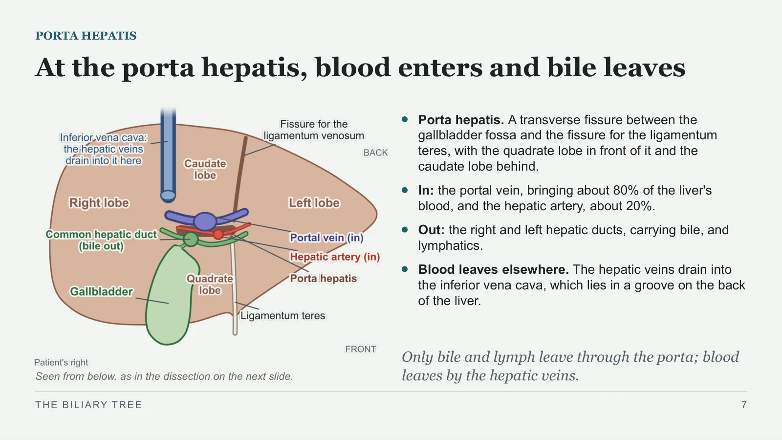
At the porta hepatis, blood enters and bile leaves
PORTA HEPATIS
Seen from below, as in the dissection on the next slide.
Porta hepatis. A transverse fissure between the gallbladder fossa and the fissure for the ligamentum teres, with the quadrate lobe in front of it and the caudate lobe behind.
In: the portal vein, bringing about 80% of the liver's blood, and the hepatic artery, about 20%.
Out: the right and left hepatic ducts, carrying bile, and lymphatics.
Blood leaves elsewhere. The hepatic veins drain into the inferior vena cava, which lies in a groove on the back of the liver.
Only bile and lymph leave through the porta; blood leaves by the hepatic veins.
THE BILIARY TREE
7
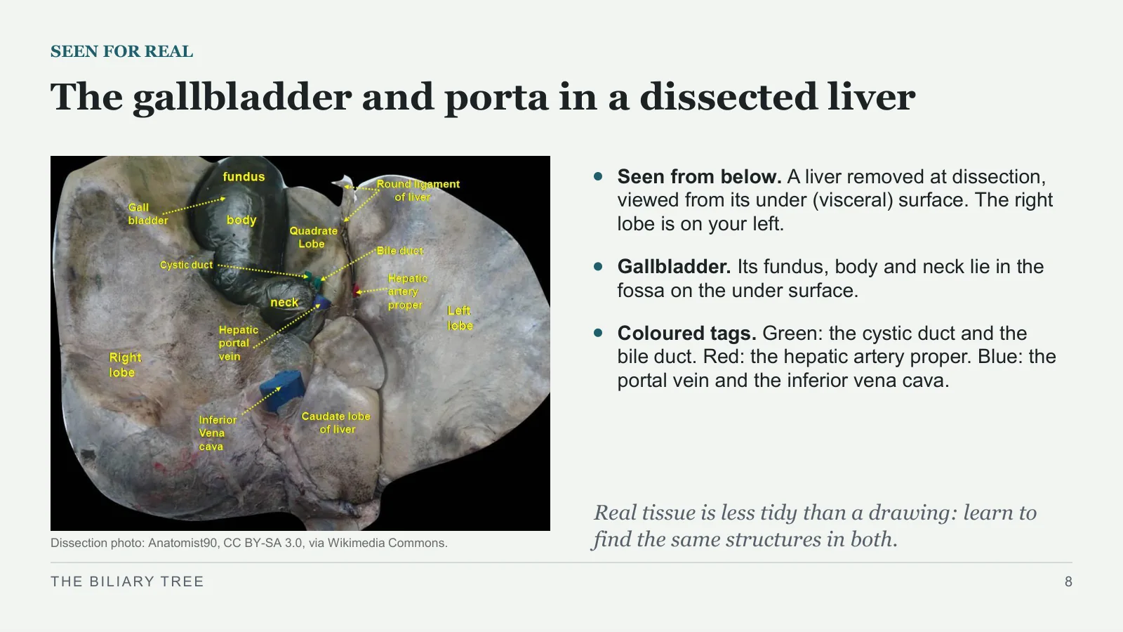
The gallbladder and porta in a dissected liver
SEEN FOR REAL
Dissection photo: Anatomist90, CC BY-SA 3.0, via Wikimedia Commons.
Seen from below. A liver removed at dissection, viewed from its under (visceral) surface. The right lobe is on your left.
Gallbladder. Its fundus, body and neck lie in the fossa on the under surface.
Coloured tags. Green: the cystic duct and the bile duct. Red: the hepatic artery proper. Blue: the portal vein and the inferior vena cava.
Real tissue is less tidy than a drawing: learn to find the same structures in both.
THE BILIARY TREE
8
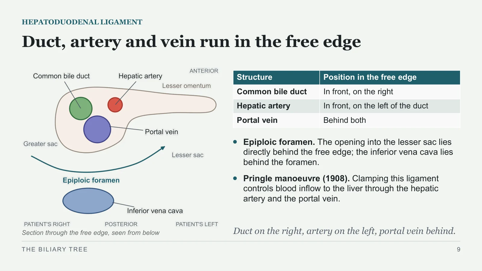
Duct, artery and vein run in the free edge
HEPATODUODENAL LIGAMENT
Section through the free edge, seen from below
Epiploic foramen. The opening into the lesser sac lies directly behind the free edge; the inferior vena cava lies behind the foramen.
Pringle manoeuvre (1908). Clamping this ligament controls blood inflow to the liver through the hepatic artery and the portal vein.
Duct on the right, artery on the left, portal vein behind.
THE BILIARY TREE
9
| Structure | Position in the free edge |
| Common bile duct | In front, on the right |
| Hepatic artery | In front, on the left of the duct |
| Portal vein | Behind both |
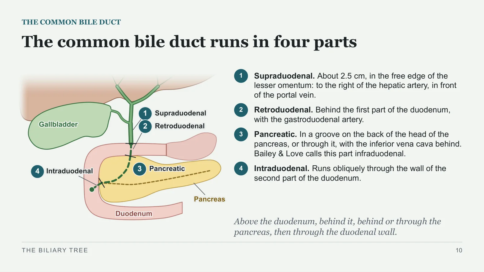
The common bile duct runs in four parts
THE COMMON BILE DUCT
1
Supraduodenal. About 2.5 cm, in the free edge of the lesser omentum: to the right of the hepatic artery, in front of the portal vein.
2
Retroduodenal. Behind the first part of the duodenum, with the gastroduodenal artery.
3
Pancreatic. In a groove on the back of the head of the pancreas, or through it, with the inferior vena cava behind. Bailey & Love calls this part infraduodenal.
4
Intraduodenal. Runs obliquely through the wall of the second part of the duodenum.
Above the duodenum, behind it, behind or through the pancreas, then through the duodenal wall.
THE BILIARY TREE
10
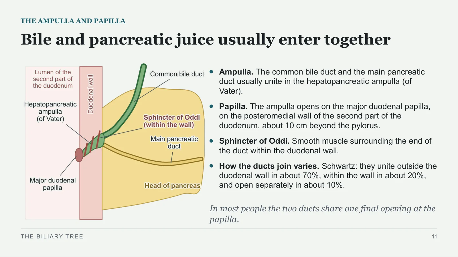
Bile and pancreatic juice usually enter together
THE AMPULLA AND PAPILLA
Ampulla. The common bile duct and the main pancreatic duct usually unite in the hepatopancreatic ampulla (of Vater).
Papilla. The ampulla opens on the major duodenal papilla, on the posteromedial wall of the second part of the duodenum, about 10 cm beyond the pylorus.
Sphincter of Oddi. Smooth muscle surrounding the end of the duct within the duodenal wall.
How the ducts join varies. Schwartz: they unite outside the duodenal wall in about 70%, within the wall in about 20%, and open separately in about 10%.
In most people the two ducts share one final opening at the papilla.
THE BILIARY TREE
11
The gallbladder
The gallbladder
PART 2
Where it lies, its parts and wall, its blood supply, lymph and nerves, and what a stone does at its outlet.
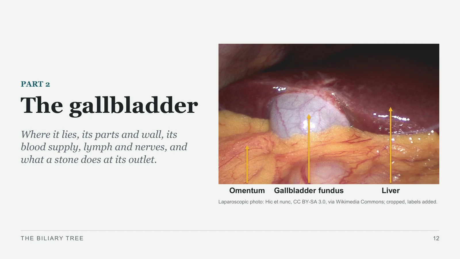
Omentum
Gallbladder fundus
Liver
Laparoscopic photo: Hic et nunc, CC BY-SA 3.0, via Wikimedia Commons; cropped, labels added.
THE BILIARY TREE
12
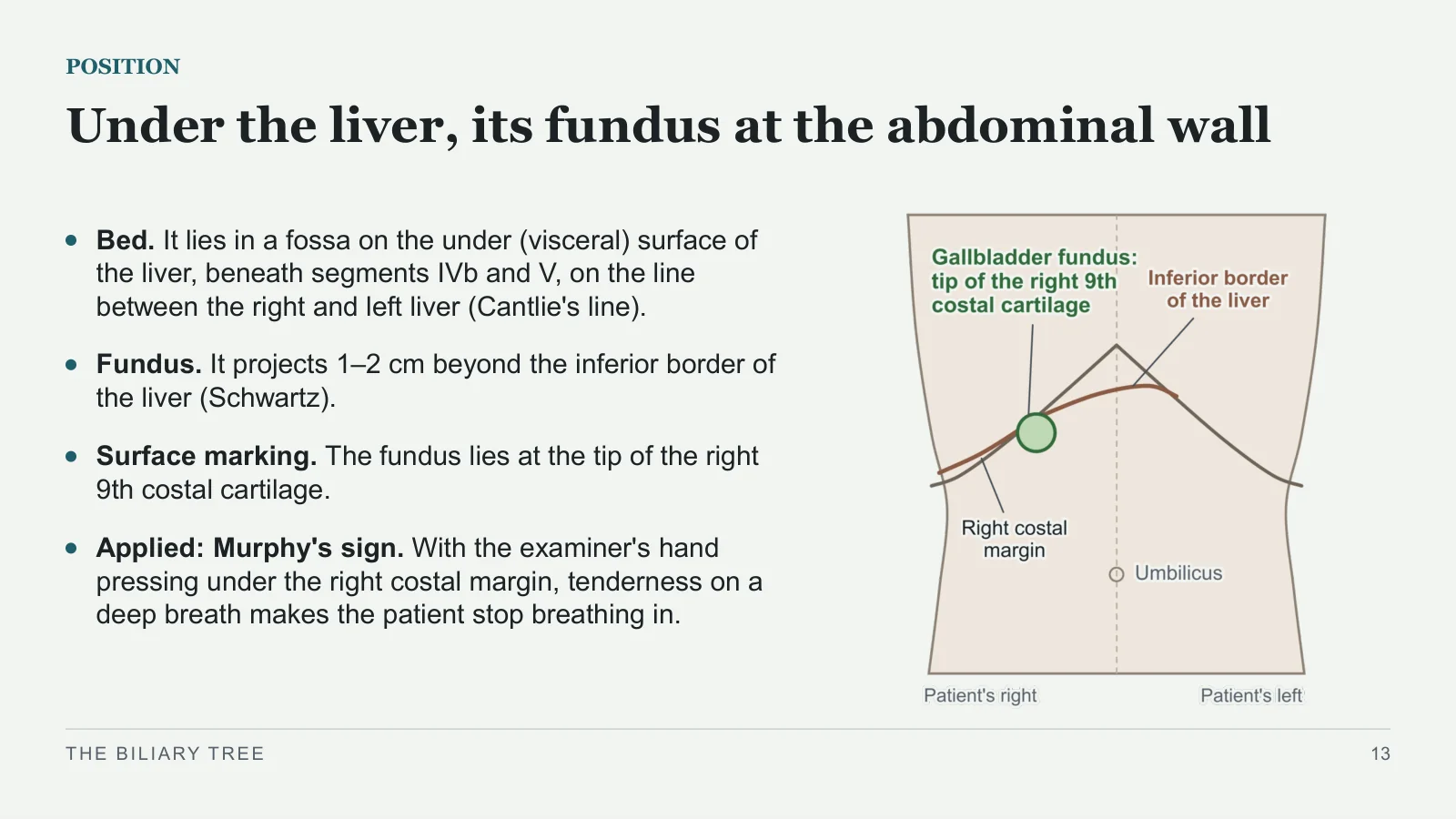
Under the liver, its fundus at the abdominal wall
POSITION
Bed. It lies in a fossa on the under (visceral) surface of the liver, beneath segments IVb and V, on the line between the right and left liver (Cantlie's line).
Fundus. It projects 1–2 cm beyond the inferior border of the liver (Schwartz).
Surface marking. The fundus lies at the tip of the right 9th costal cartilage.
Applied: Murphy's sign. With the examiner's hand pressing under the right costal margin, tenderness on a deep breath makes the patient stop breathing in.
THE BILIARY TREE
13
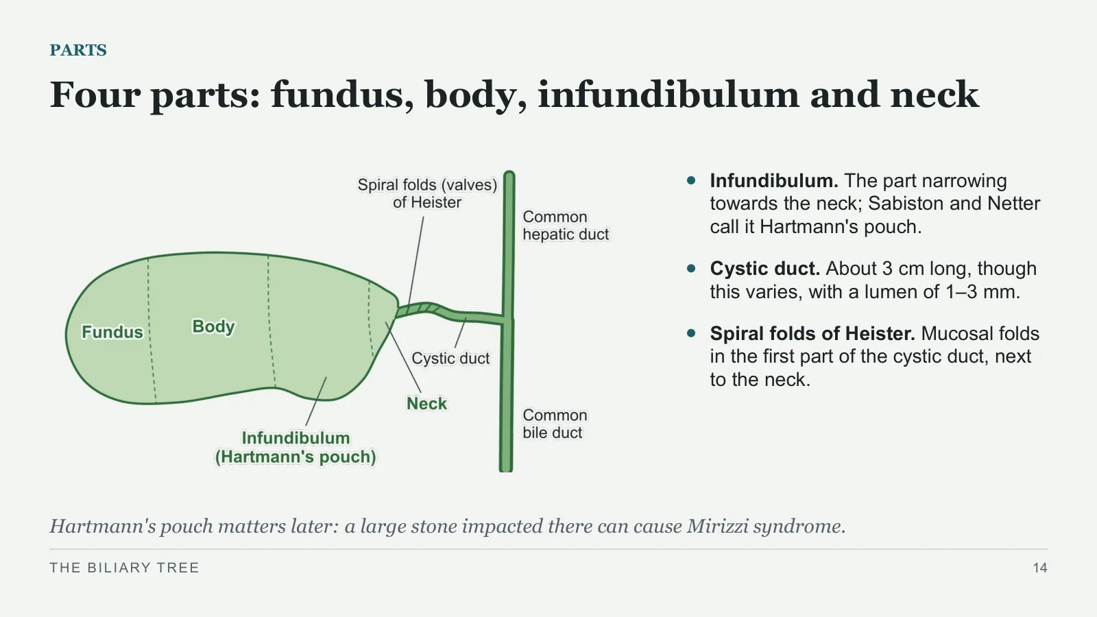
Four parts: fundus, body, infundibulum and neck
PARTS
Infundibulum. The part narrowing towards the neck; Sabiston and Netter call it Hartmann's pouch.
Cystic duct. About 3 cm long, though this varies, with a lumen of 1–3 mm.
Spiral folds of Heister. Mucosal folds in the first part of the cystic duct, next to the neck.
Hartmann's pouch matters later: a large stone impacted there can cause Mirizzi syndrome.
THE BILIARY TREE
14
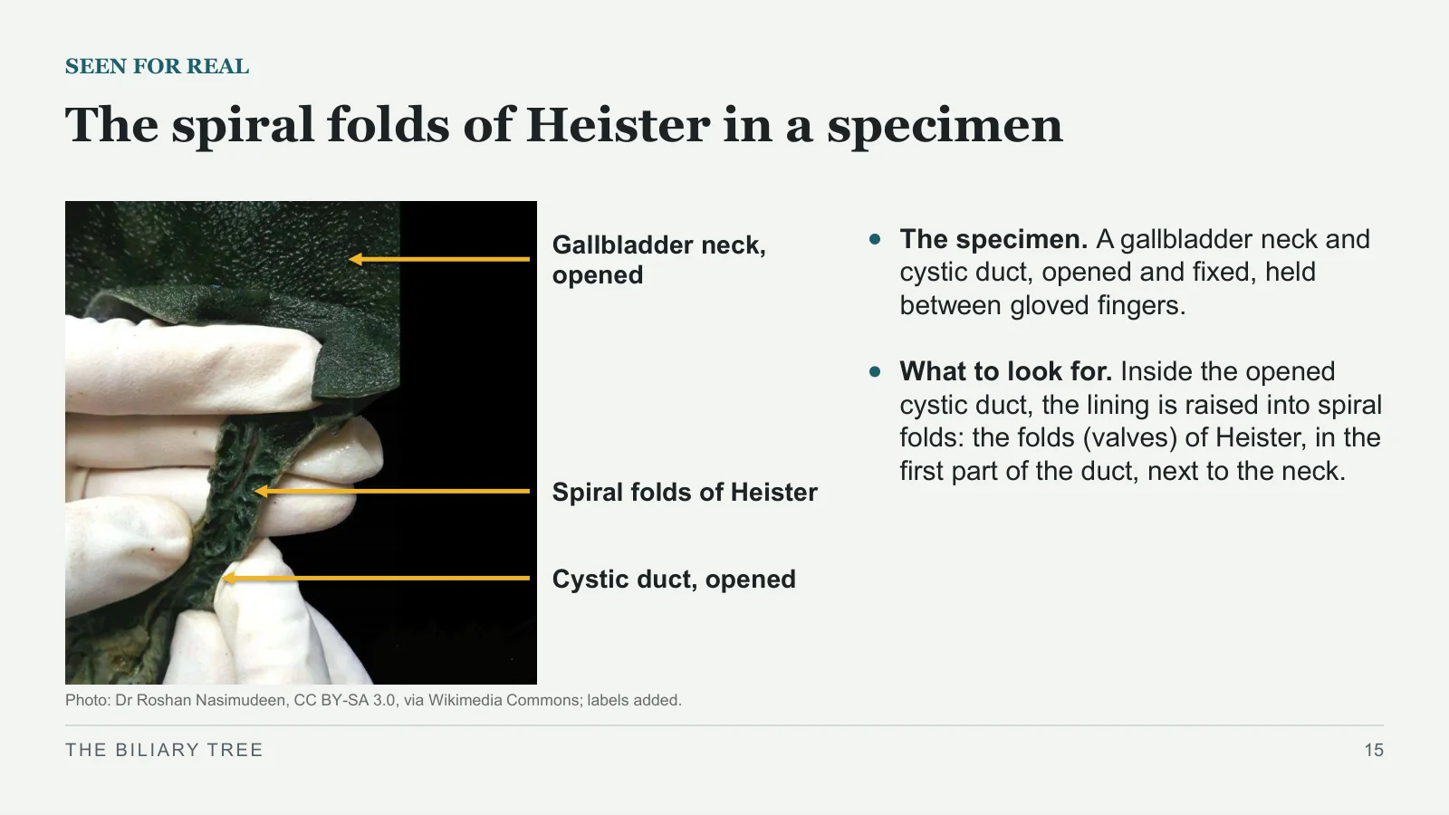
The spiral folds of Heister in a specimen
SEEN FOR REAL
Photo: Dr Roshan Nasimudeen, CC BY-SA 3.0, via Wikimedia Commons; labels added.
Gallbladder neck, opened
Spiral folds of Heister
Cystic duct, opened
The specimen. A gallbladder neck and cystic duct, opened and fixed, held between gloved fingers.
What to look for. Inside the opened cystic duct, the lining is raised into spiral folds: the folds (valves) of Heister, in the first part of the duct, next to the neck.
THE BILIARY TREE
15
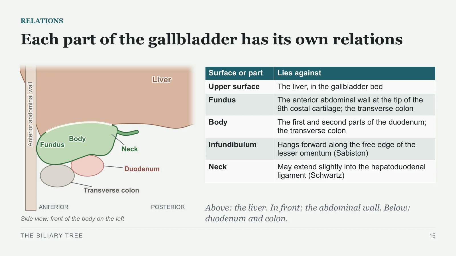
Each part of the gallbladder has its own relations
RELATIONS
Side view: front of the body on the left
Above: the liver. In front: the abdominal wall. Below: duodenum and colon.
THE BILIARY TREE
16
| Surface or part | Lies against |
| Upper surface | The liver, in the gallbladder bed |
| Fundus | The anterior abdominal wall at the tip of the 9th costal cartilage; the transverse colon |
| Body | The first and second parts of the duodenum; the transverse colon |
| Infundibulum | Hangs forward along the free edge of the lesser omentum (Sabiston) |
| Neck | May extend slightly into the hepatoduodenal ligament (Schwartz) |
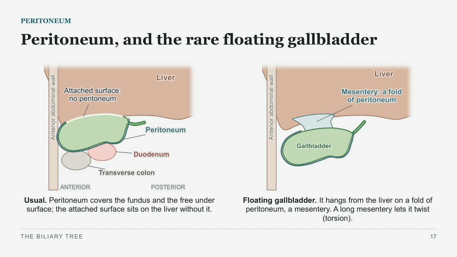
Peritoneum, and the rare floating gallbladder
PERITONEUM
Usual. Peritoneum covers the fundus and the free under surface; the attached surface sits on the liver without it.
Floating gallbladder. It hangs from the liver on a fold of peritoneum, a mesentery. A long mesentery lets it twist (torsion).
THE BILIARY TREE
17
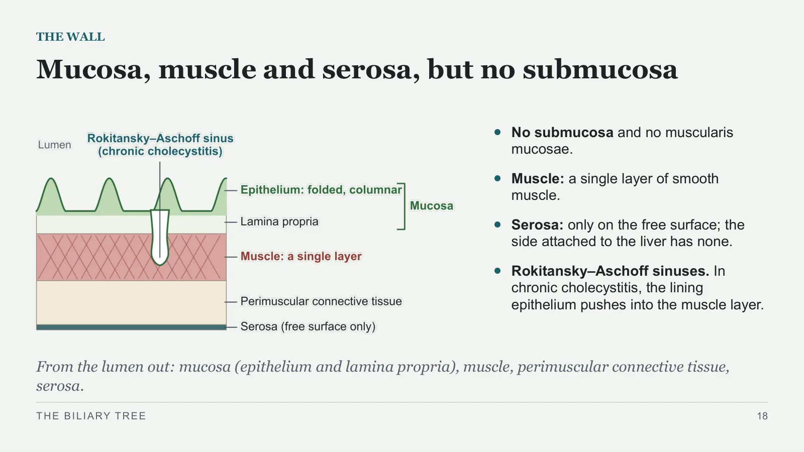
Mucosa, muscle and serosa, but no submucosa
THE WALL
No submucosa and no muscularis mucosae.
Muscle: a single layer of smooth muscle.
Serosa: only on the free surface; the side attached to the liver has none.
Rokitansky–Aschoff sinuses. In chronic cholecystitis, the lining epithelium pushes into the muscle layer.
From the lumen out: mucosa (epithelium and lamina propria), muscle, perimuscular connective tissue, serosa.
THE BILIARY TREE
18
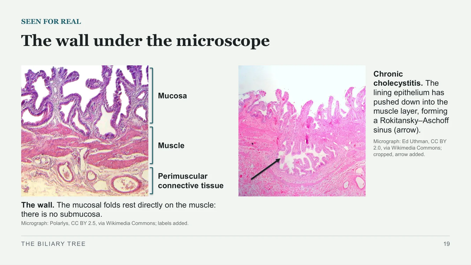
The wall under the microscope
SEEN FOR REAL
Mucosa
Muscle
Perimuscular connective tissue
The wall. The mucosal folds rest directly on the muscle: there is no submucosa.
Micrograph: Polarlys, CC BY 2.5, via Wikimedia Commons; labels added.
Chronic cholecystitis. The lining epithelium has pushed down into the muscle layer, forming a Rokitansky–Aschoff sinus (arrow).
Micrograph: Ed Uthman, CC BY 2.0, via Wikimedia Commons; cropped, arrow added.
THE BILIARY TREE
19
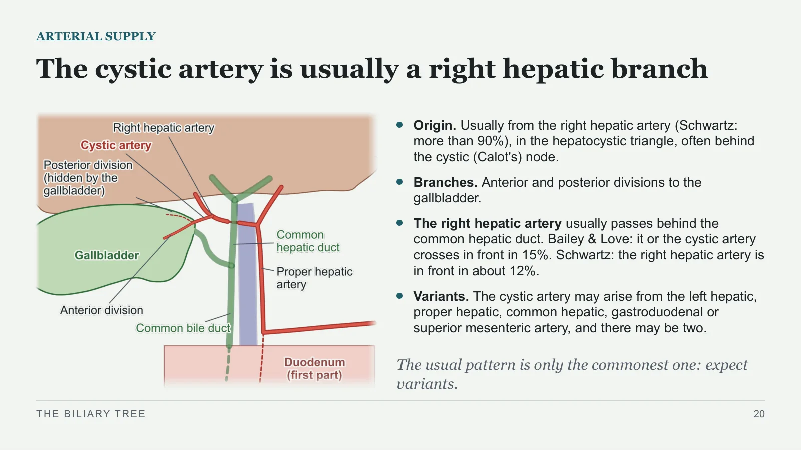
The cystic artery is usually a right hepatic branch
ARTERIAL SUPPLY
Origin. Usually from the right hepatic artery (Schwartz: more than 90%), in the hepatocystic triangle, often behind the cystic (Calot's) node.
Branches. Anterior and posterior divisions to the gallbladder.
The right hepatic artery usually passes behind the common hepatic duct. Bailey & Love: it or the cystic artery crosses in front in 15%. Schwartz: the right hepatic artery is in front in about 12%.
Variants. The cystic artery may arise from the left hepatic, proper hepatic, common hepatic, gastroduodenal or superior mesenteric artery, and there may be two.
The usual pattern is only the commonest one: expect variants.
THE BILIARY TREE
20
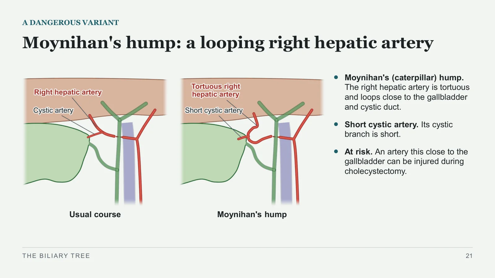
Moynihan's hump: a looping right hepatic artery
A DANGEROUS VARIANT
Usual course
Moynihan's hump
Moynihan's (caterpillar) hump. The right hepatic artery is tortuous and loops close to the gallbladder and cystic duct.
Short cystic artery. Its cystic branch is short.
At risk. An artery this close to the gallbladder can be injured during cholecystectomy.
THE BILIARY TREE
21
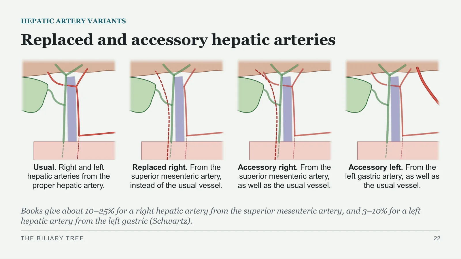
Replaced and accessory hepatic arteries
HEPATIC ARTERY VARIANTS
Usual. Right and left hepatic arteries from the proper hepatic artery.
Replaced right. From the superior mesenteric artery, instead of the usual vessel.
Accessory right. From the superior mesenteric artery, as well as the usual vessel.
Accessory left. From the left gastric artery, as well as the usual vessel.
Books give about 10–25% for a right hepatic artery from the superior mesenteric artery, and 3–10% for a left hepatic artery from the left gastric (Schwartz).
THE BILIARY TREE
22
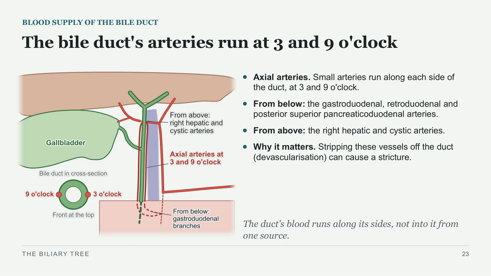
The bile duct's arteries run at 3 and 9 o'clock
BLOOD SUPPLY OF THE BILE DUCT
Axial arteries. Small arteries run along each side of the duct, at 3 and 9 o'clock.
From below: the gastroduodenal, retroduodenal and posterior superior pancreaticoduodenal arteries.
From above: the right hepatic and cystic arteries.
Why it matters. Stripping these vessels off the duct (devascularisation) can cause a stricture.
The duct's blood runs along its sides, not into it from one source.
THE BILIARY TREE
23
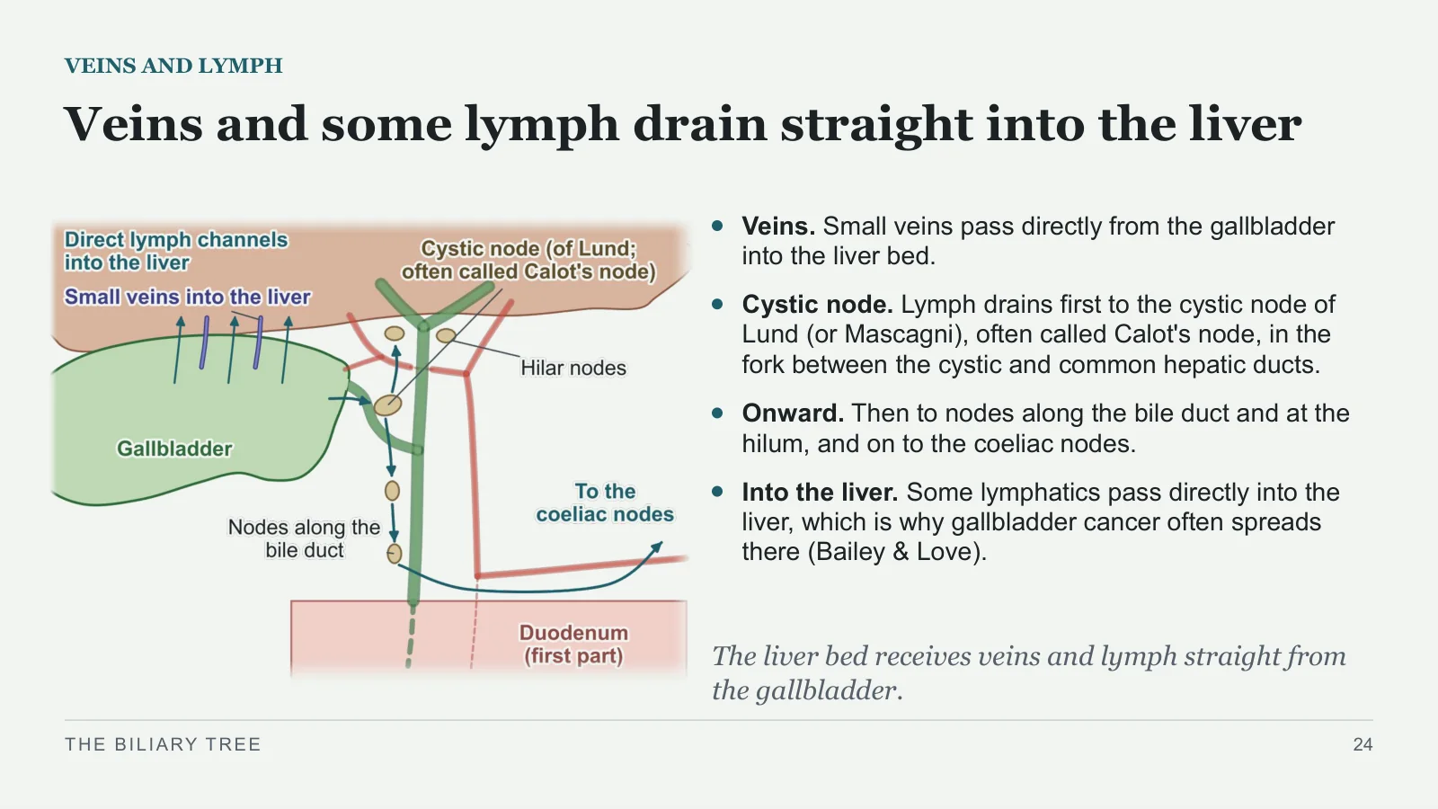
Veins and some lymph drain straight into the liver
VEINS AND LYMPH
Veins. Small veins pass directly from the gallbladder into the liver bed.
Cystic node. Lymph drains first to the cystic node of Lund (or Mascagni), often called Calot's node, in the fork between the cystic and common hepatic ducts.
Onward. Then to nodes along the bile duct and at the hilum, and on to the coeliac nodes.
Into the liver. Some lymphatics pass directly into the liver, which is why gallbladder cancer often spreads there (Bailey & Love).
The liver bed receives veins and lymph straight from the gallbladder.
THE BILIARY TREE
24
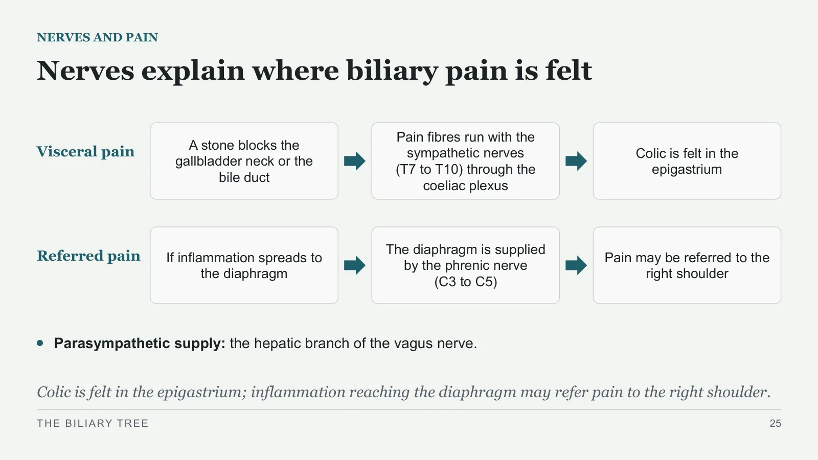
Nerves explain where biliary pain is felt
NERVES AND PAIN
Visceral pain
A stone blocks the gallbladder neck or the bile duct
Pain fibres run with the sympathetic nerves (T7 to T10) through the coeliac plexus
Colic is felt in the epigastrium
Referred pain
If inflammation spreads to the diaphragm
The diaphragm is supplied by the phrenic nerve (C3 to C5)
Pain may be referred to the right shoulder
Parasympathetic supply: the hepatic branch of the vagus nerve.
Colic is felt in the epigastrium; inflammation reaching the diaphragm may refer pain to the right shoulder.
THE BILIARY TREE
25
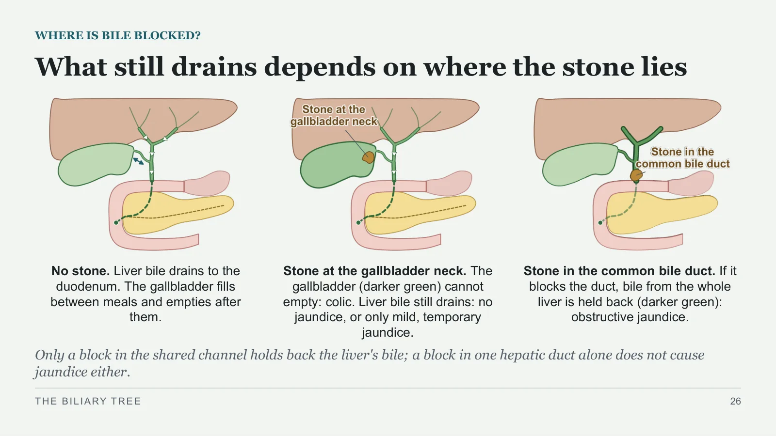
What still drains depends on where the stone lies
WHERE IS BILE BLOCKED?
No stone. Liver bile drains to the duodenum. The gallbladder fills between meals and empties after them.
Stone at the gallbladder neck. The gallbladder (darker green) cannot empty: colic. Liver bile still drains: no jaundice, or only mild, temporary jaundice.
Stone in the common bile duct. If it blocks the duct, bile from the whole liver is held back (darker green): obstructive jaundice.
Only a block in the shared channel holds back the liver's bile; a block in one hepatic duct alone does not cause jaundice either.
THE BILIARY TREE
26
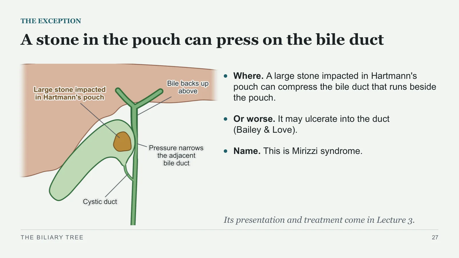
A stone in the pouch can press on the bile duct
THE EXCEPTION
Where. A large stone impacted in Hartmann's pouch can compress the bile duct that runs beside the pouch.
Or worse. It may ulcerate into the duct (Bailey & Love).
Name. This is Mirizzi syndrome.
Its presentation and treatment come in Lecture 3.
THE BILIARY TREE
27
Anatomy at operation
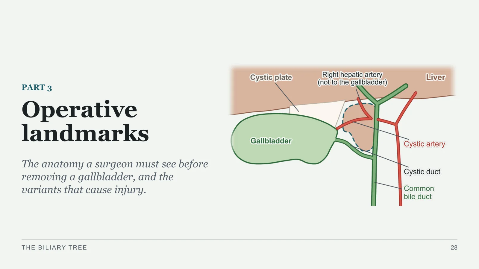
Operative landmarks
PART 3
The anatomy a surgeon must see before removing a gallbladder, and the variants that cause injury.
THE BILIARY TREE
28
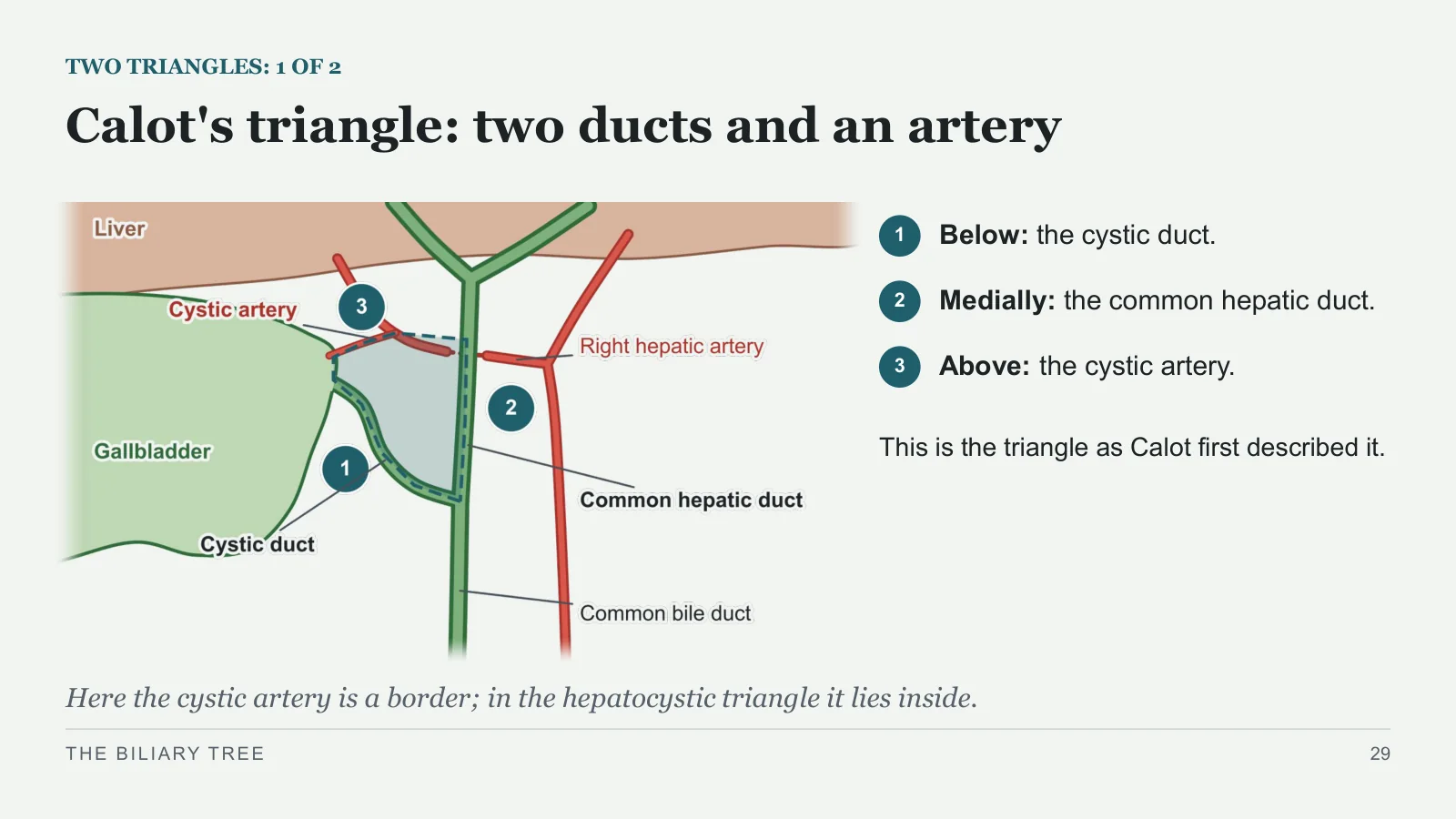
Calot's triangle: two ducts and an artery
TWO TRIANGLES: 1 OF 2
1
Below: the cystic duct.
2
Medially: the common hepatic duct.
3
Above: the cystic artery.
This is the triangle as Calot first described it.
Here the cystic artery is a border; in the hepatocystic triangle it lies inside.
THE BILIARY TREE
29
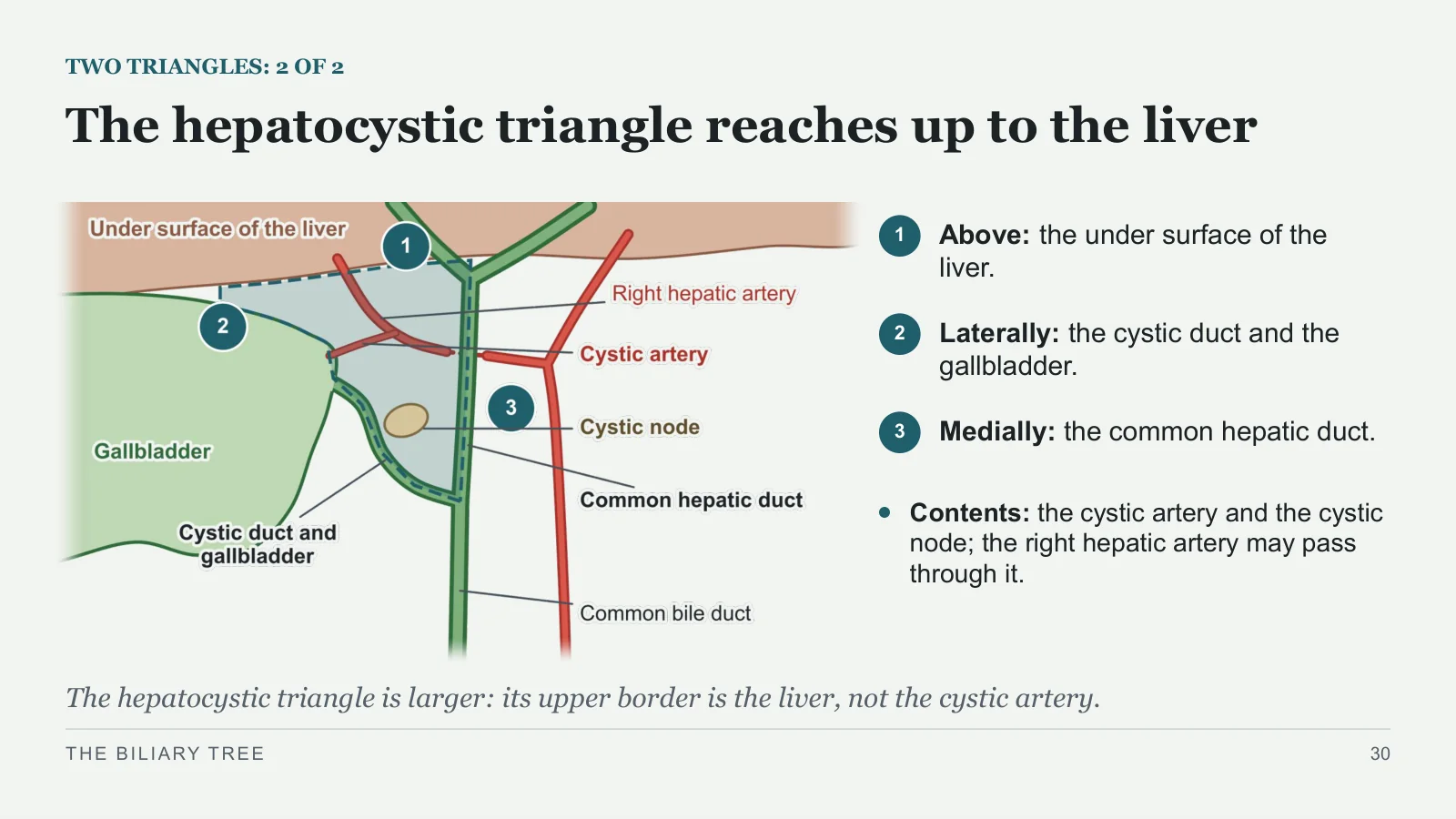
The hepatocystic triangle reaches up to the liver
TWO TRIANGLES: 2 OF 2
1
Above: the under surface of the liver.
2
Laterally: the cystic duct and the gallbladder.
3
Medially: the common hepatic duct.
Contents: the cystic artery and the cystic node; the right hepatic artery may pass through it.
The hepatocystic triangle is larger: its upper border is the liver, not the cystic artery.
THE BILIARY TREE
30
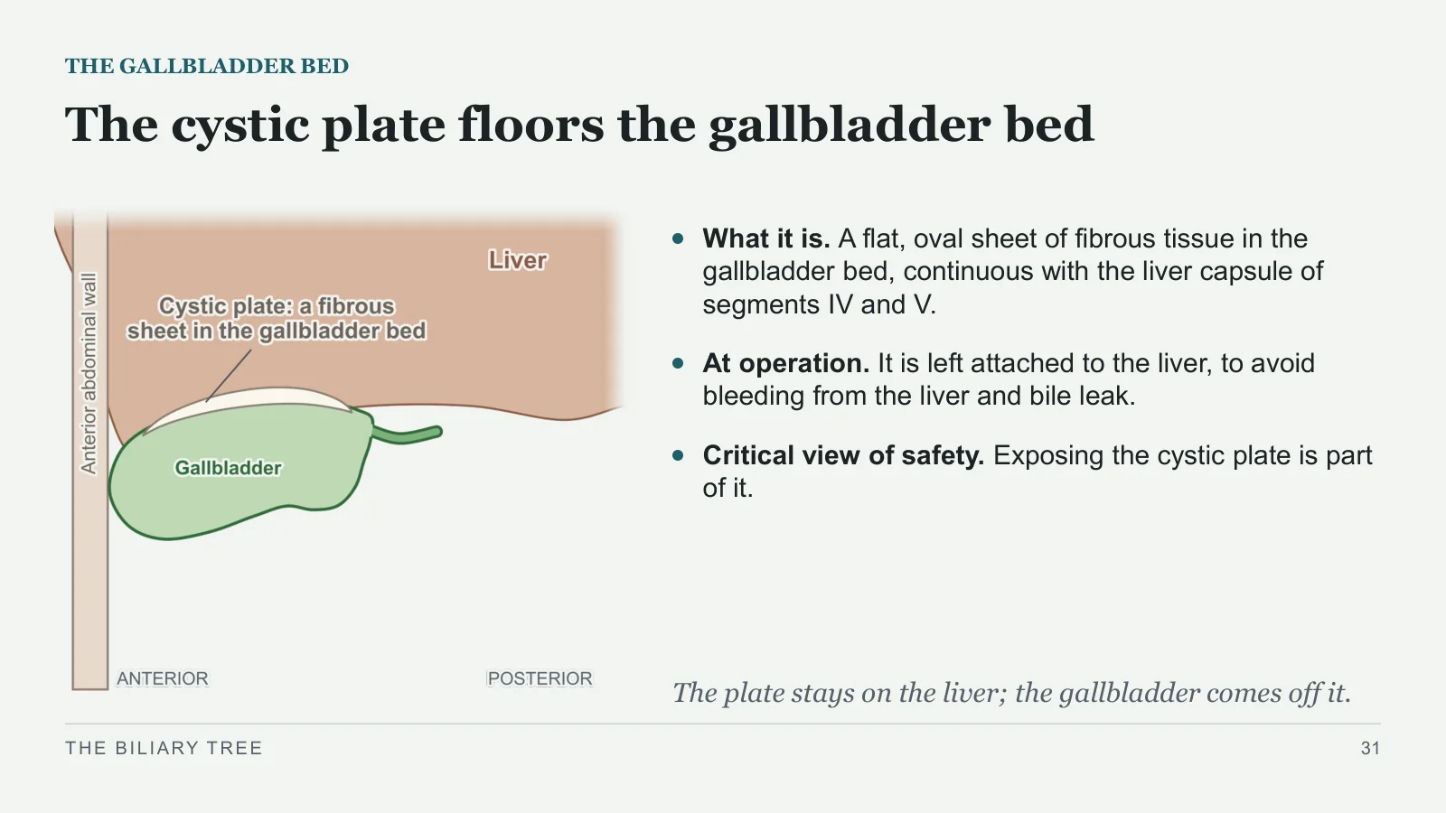
The cystic plate floors the gallbladder bed
THE GALLBLADDER BED
What it is. A flat, oval sheet of fibrous tissue in the gallbladder bed, continuous with the liver capsule of segments IV and V.
At operation. It is left attached to the liver, to avoid bleeding from the liver and bile leak.
Critical view of safety. Exposing the cystic plate is part of it.
The plate stays on the liver; the gallbladder comes off it.
THE BILIARY TREE
31
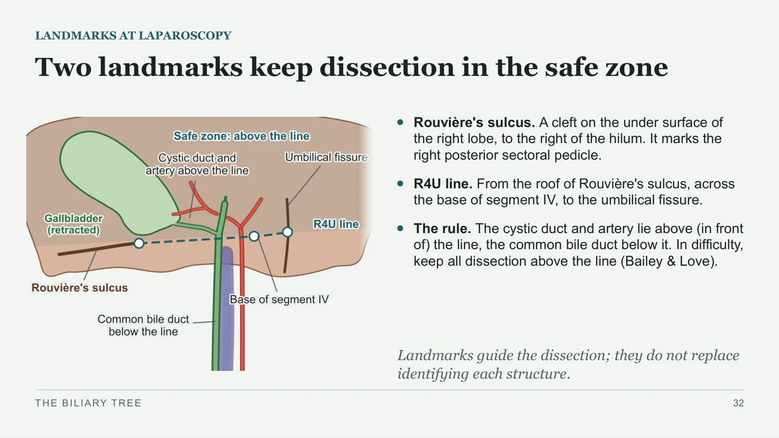
Two landmarks keep dissection in the safe zone
LANDMARKS AT LAPAROSCOPY
Rouvière's sulcus. A cleft on the under surface of the right lobe, to the right of the hilum. It marks the right posterior sectoral pedicle.
R4U line. From the roof of Rouvière's sulcus, across the base of segment IV, to the umbilical fissure.
The rule. The cystic duct and artery lie above (in front of) the line, the common bile duct below it. In difficulty, keep all dissection above the line (Bailey & Love).
Landmarks guide the dissection; they do not replace identifying each structure.
THE BILIARY TREE
32
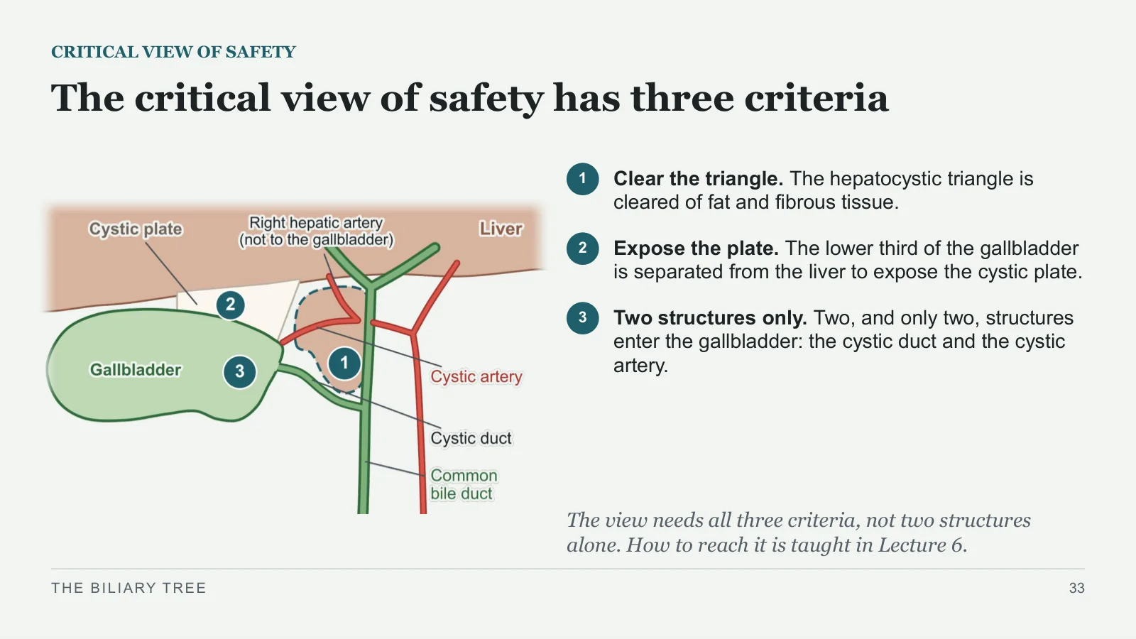
The critical view of safety has three criteria
CRITICAL VIEW OF SAFETY
1
Clear the triangle. The hepatocystic triangle is cleared of fat and fibrous tissue.
2
Expose the plate. The lower third of the gallbladder is separated from the liver to expose the cystic plate.
3
Two structures only. Two, and only two, structures enter the gallbladder: the cystic duct and the cystic artery.
The view needs all three criteria, not two structures alone. How to reach it is taught in Lecture 6.
THE BILIARY TREE
33
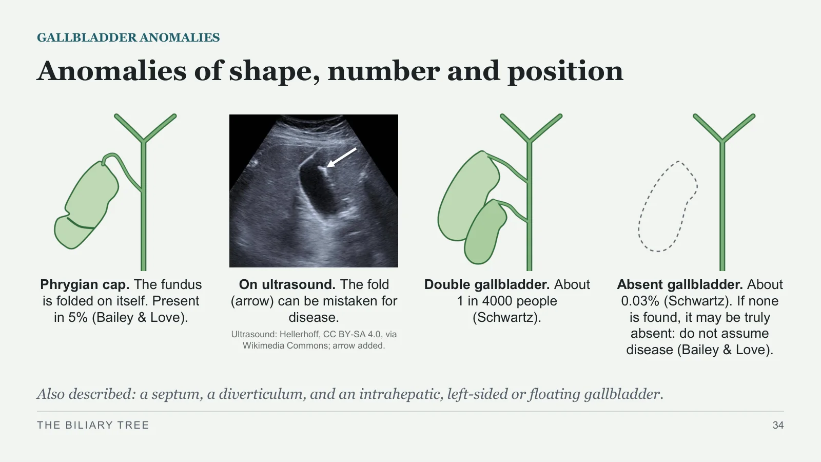
Anomalies of shape, number and position
GALLBLADDER ANOMALIES
Phrygian cap. The fundus is folded on itself. Present in 5% (Bailey & Love).
On ultrasound. The fold (arrow) can be mistaken for disease.
Double gallbladder. About 1 in 4000 people (Schwartz).
Absent gallbladder. About 0.03% (Schwartz). If none is found, it may be truly absent: do not assume disease (Bailey & Love).
Ultrasound: Hellerhoff, CC BY-SA 4.0, via Wikimedia Commons; arrow added.
Also described: a septum, a diverticulum, and an intrahepatic, left-sided or floating gallbladder.
THE BILIARY TREE
34
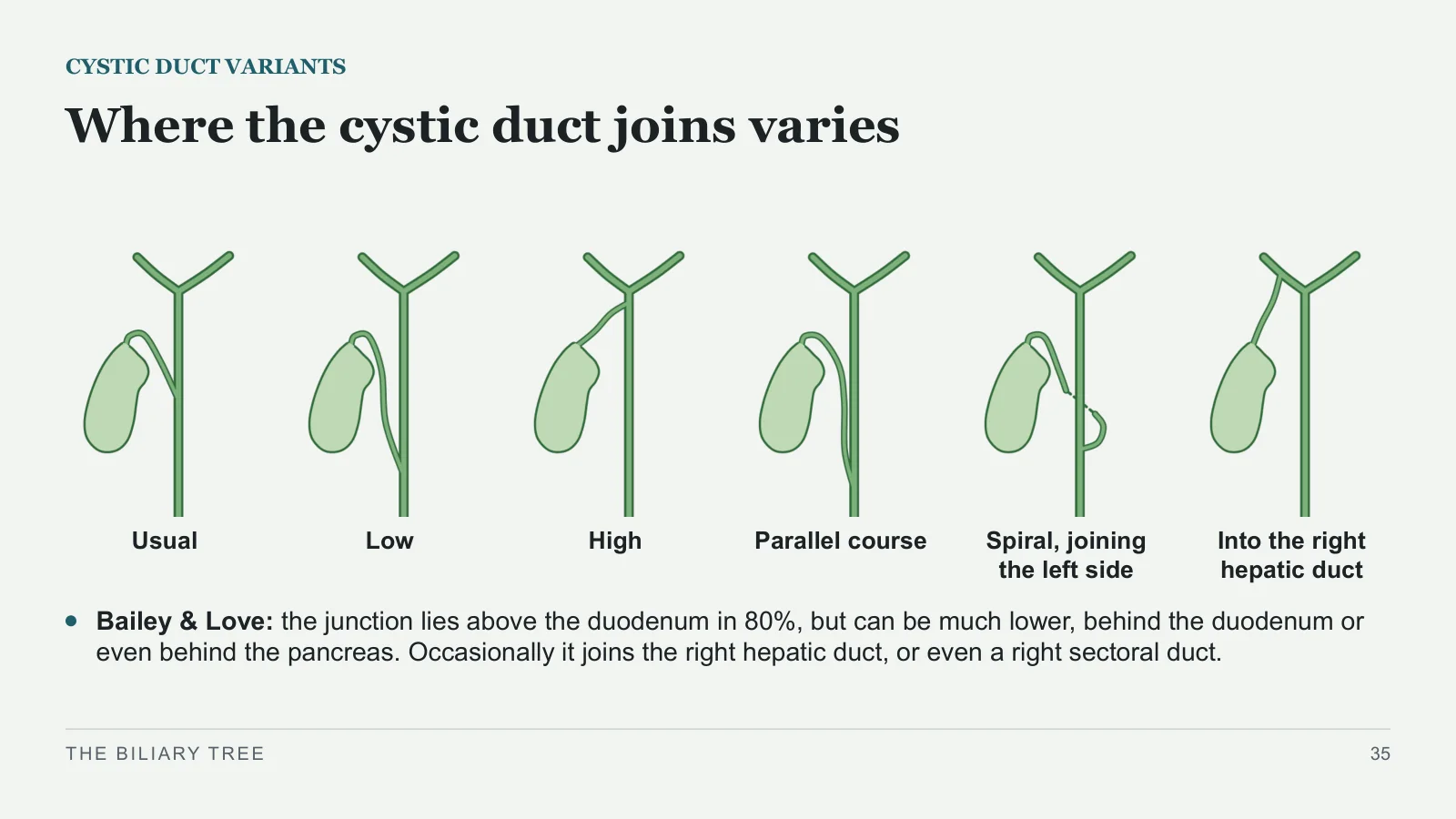
Where the cystic duct joins varies
CYSTIC DUCT VARIANTS
Usual
Low
High
Parallel course
Spiral, joining the left side
Into the right hepatic duct
Bailey & Love: the junction lies above the duodenum in 80%, but can be much lower, behind the duodenum or even behind the pancreas. Occasionally it joins the right hepatic duct, or even a right sectoral duct.
THE BILIARY TREE
35
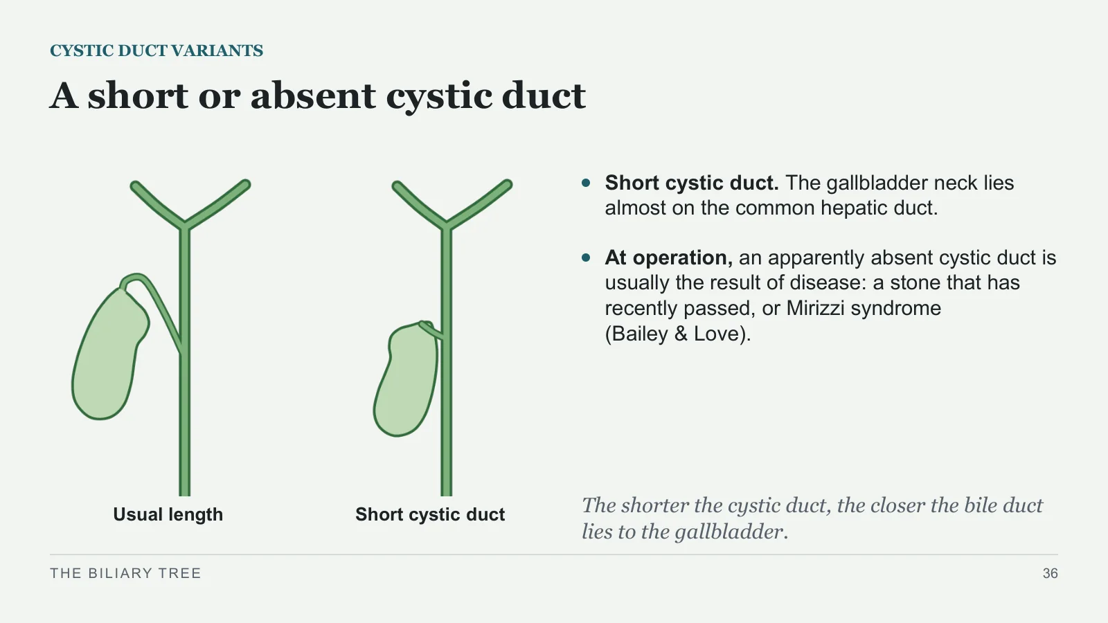
A short or absent cystic duct
CYSTIC DUCT VARIANTS
Usual length
Short cystic duct
Short cystic duct. The gallbladder neck lies almost on the common hepatic duct.
At operation, an apparently absent cystic duct is usually the result of disease: a stone that has recently passed, or Mirizzi syndrome (Bailey & Love).
The shorter the cystic duct, the closer the bile duct lies to the gallbladder.
THE BILIARY TREE
36
Where a right sectoral duct drains varies
DUCT VARIANTS
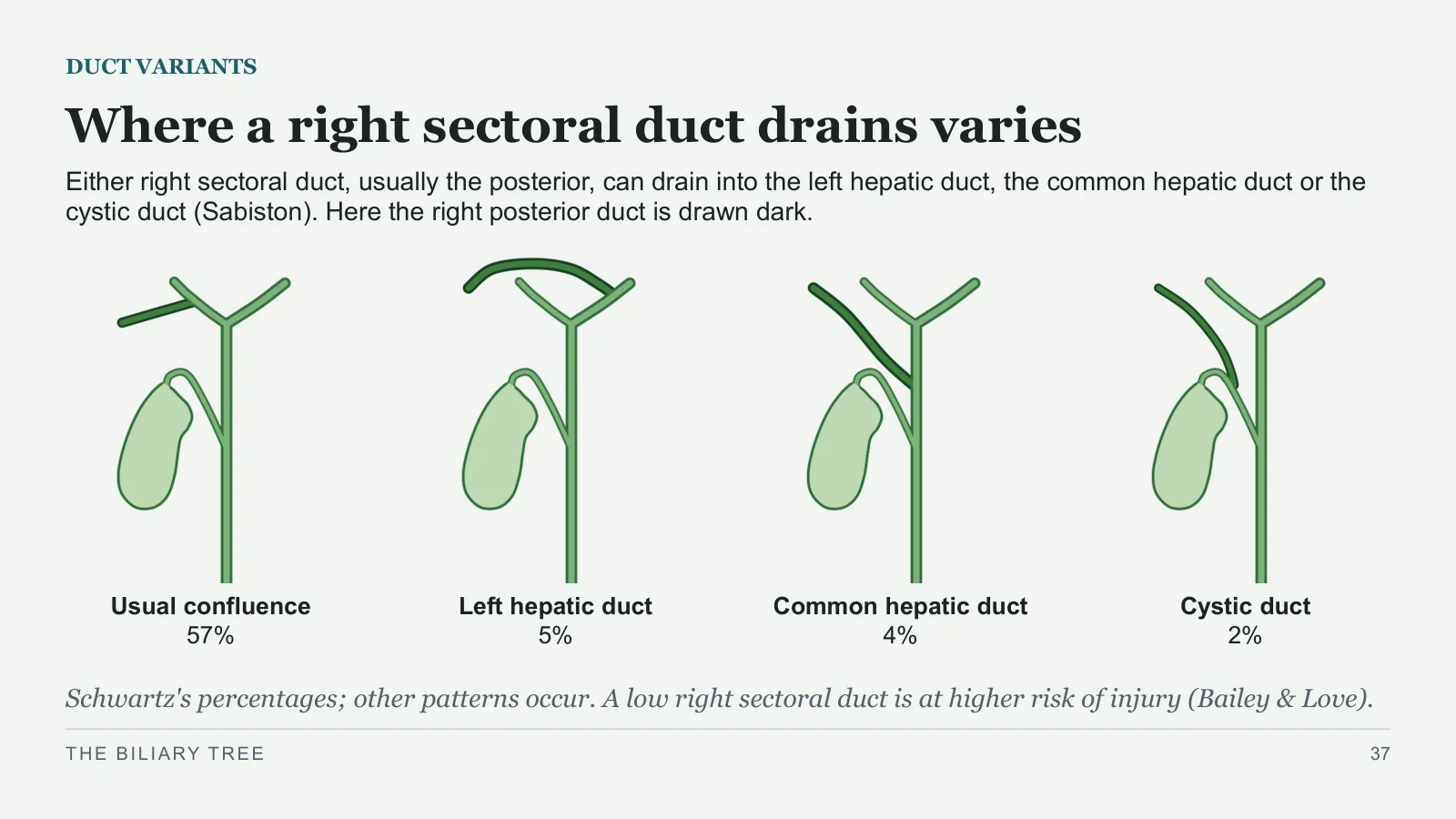
Either right sectoral duct, usually the posterior, can drain into the left hepatic duct, the common hepatic duct or the cystic duct (Sabiston). Here the right posterior duct is drawn dark.
Usual confluence
57%
Left hepatic duct
5%
Common hepatic duct
4%
Cystic duct
2%
Schwartz's percentages; other patterns occur. A low right sectoral duct is at higher risk of injury (Bailey & Love).
THE BILIARY TREE
37
Ducts of Luschka drain into the gallbladder
ACCESSORY DUCTS
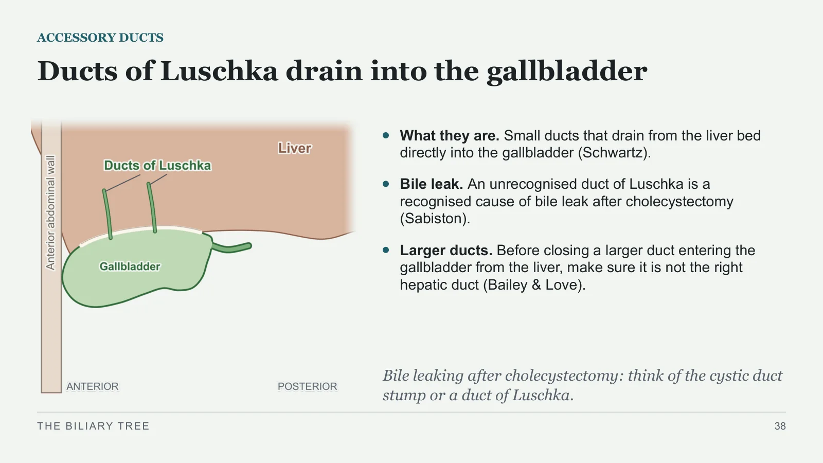
What they are. Small ducts that drain from the liver bed directly into the gallbladder (Schwartz).
Bile leak. An unrecognised duct of Luschka is a recognised cause of bile leak after cholecystectomy (Sabiston).
Larger ducts. Before closing a larger duct entering the gallbladder from the liver, make sure it is not the right hepatic duct (Bailey & Love).
Bile leaking after cholecystectomy: think of the cystic duct stump or a duct of Luschka.
THE BILIARY TREE
38
Two classic hazards and their anatomy
HAZARDS AT OPERATION
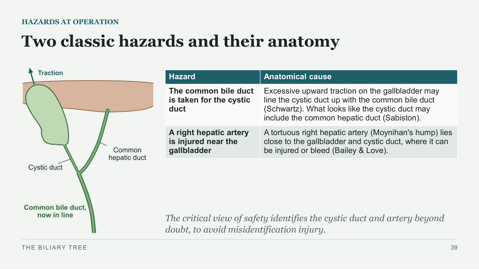
The critical view of safety identifies the cystic duct and artery beyond doubt, to avoid misidentification injury.
THE BILIARY TREE
39
| Hazard | Anatomical cause |
| The common bile duct is taken for the cystic duct | Excessive upward traction on the gallbladder may line the cystic duct up with the common bile duct (Schwartz). What looks like the cystic duct may include the common hepatic duct (Sabiston). |
| A right hepatic artery is injured near the gallbladder | A tortuous right hepatic artery (Moynihan's hump) lies close to the gallbladder and cystic duct, where it can be injured or bleed (Bailey & Love). |
A stone in the common bile duct made her yellow
BACK TO THE PATIENT
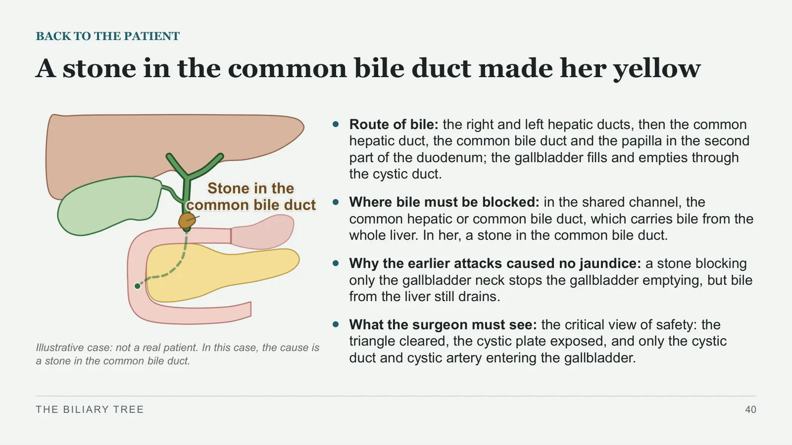
Illustrative case: not a real patient. In this case, the cause is a stone in the common bile duct.
Route of bile: the right and left hepatic ducts, then the common hepatic duct, the common bile duct and the papilla in the second part of the duodenum; the gallbladder fills and empties through the cystic duct.
Where bile must be blocked: in the shared channel, the common hepatic or common bile duct, which carries bile from the whole liver. In her, a stone in the common bile duct.
Why the earlier attacks caused no jaundice: a stone blocking only the gallbladder neck stops the gallbladder emptying, but bile from the liver still drains.
What the surgeon must see: the critical view of safety: the triangle cleared, the cystic plate exposed, and only the cystic duct and cystic artery entering the gallbladder.
THE BILIARY TREE
40
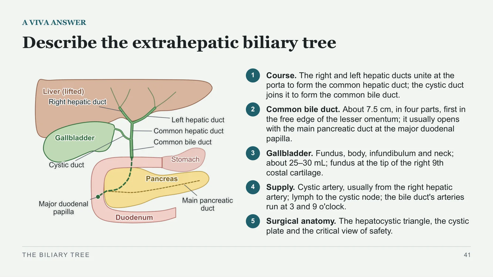
Describe the extrahepatic biliary tree
A VIVA ANSWER
1
Course. The right and left hepatic ducts unite at the porta to form the common hepatic duct; the cystic duct joins it to form the common bile duct.
2
Common bile duct. About 7.5 cm, in four parts, first in the free edge of the lesser omentum; it usually opens with the main pancreatic duct at the major duodenal papilla.
3
Gallbladder. Fundus, body, infundibulum and neck; about 25–30 mL; fundus at the tip of the right 9th costal cartilage.
4
Supply. Cystic artery, usually from the right hepatic artery; lymph to the cystic node; the bile duct's arteries run at 3 and 9 o'clock.
5
Surgical anatomy. The hepatocystic triangle, the cystic plate and the critical view of safety.
THE BILIARY TREE
41
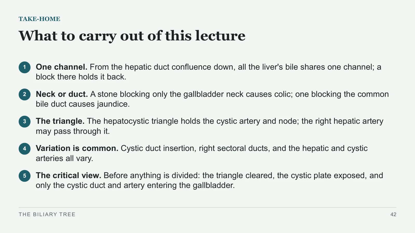
What to carry out of this lecture
TAKE-HOME
1
One channel. From the hepatic duct confluence down, all the liver's bile shares one channel; a block there holds it back.
2
Neck or duct. A stone blocking only the gallbladder neck causes colic; one blocking the common bile duct causes jaundice.
3
The triangle. The hepatocystic triangle holds the cystic artery and node; the right hepatic artery may pass through it.
4
Variation is common. Cystic duct insertion, right sectoral ducts, and the hepatic and cystic arteries all vary.
5
The critical view. Before anything is divided: the triangle cleared, the cystic plate exposed, and only the cystic duct and artery entering the gallbladder.
THE BILIARY TREE
42
References
Books used for this lecture
REFERENCES
Bailey & Love's Short Practice of Surgery, 28th edition. Liver pp. 1191–1193; gallbladder and bile ducts, chapter 71, pp. 1232–1252.
Bailey & Love's Essential Clinical Anatomy (2019). Pp. 57, 89–91, 97, 107, 113–122.
Sabiston Textbook of Surgery, 21st edition. Chapters 54 and 55.
Schwartz's Principles of Surgery, 11th edition. Chapters 31 and 32.
Netter's Atlas of Human Anatomy, 7th edition. Plates 274, 287, 309, BP63, BP68, BP69, BP70.
THE BILIARY TREE
43
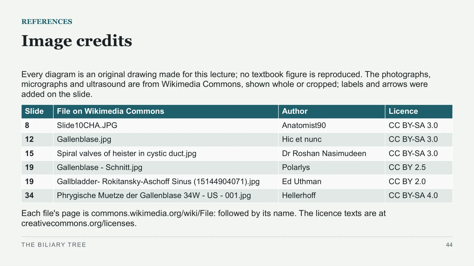
Image credits
REFERENCES
Every diagram is an original drawing made for this lecture; no textbook figure is reproduced. The photographs, micrographs and ultrasound are from Wikimedia Commons, shown whole or cropped; labels and arrows were added on the slide.
Each file's page is commons.wikimedia.org/wiki/File: followed by its name. The licence texts are at creativecommons.org/licenses.
THE BILIARY TREE
44
| Slide | File on Wikimedia Commons | Author | Licence |
| 8 | Slide10CHA.JPG | Anatomist90 | CC BY-SA 3.0 |
| 12 | Gallenblase.jpg | Hic et nunc | CC BY-SA 3.0 |
| 15 | Spiral valves of heister in cystic duct.jpg | Dr Roshan Nasimudeen | CC BY-SA 3.0 |
| 19 | Gallenblase - Schnitt.jpg | Polarlys | CC BY 2.5 |
| 19 | Gallbladder- Rokitansky-Aschoff Sinus (15144904071).jpg | Ed Uthman | CC BY 2.0 |
| 34 | Phrygische Muetze der Gallenblase 34W - US - 001.jpg | Hellerhoff | CC BY-SA 4.0 |
Sources and image credits
Every diagram is an original drawing made for this lecture; no textbook figure is reproduced. The photographs, micrographs and ultrasound are from Wikimedia Commons, shown whole or cropped; labels and arrows were added on the slide. The source licences below apply to the credited images; crops and annotated versions of CC BY-SA images are shared under the same licence as their source. Slide captions identify the crops and added markers.
Images
- Slide 8 · Slide10CHA.JPG · Anatomist90 · CC BY-SA 3.0
- Slide 12 · Gallenblase.jpg · Hic et nunc · CC BY-SA 3.0
- Slide 15 · Spiral valves of heister in cystic duct.jpg · Dr Roshan Nasimudeen · CC BY-SA 3.0
- Slide 19 · Gallenblase - Schnitt.jpg · Polarlys · CC BY 2.5
- Slide 19 · Gallbladder- Rokitansky-Aschoff Sinus (15144904071).jpg · Ed Uthman · CC BY 2.0
- Slide 34 · Phrygische Muetze der Gallenblase 34W - US - 001.jpg · Hellerhoff · CC BY-SA 4.0


Sources
- [1] Bailey & Love's Short Practice of Surgery, 28th edition. Liver pp. 1191–1193; gallbladder and bile ducts, chapter 71, pp. 1232–1252.
- [2] Bailey & Love's Essential Clinical Anatomy (2019). Pp. 57, 89–91, 97, 107, 113–122.
- [3] Sabiston Textbook of Surgery, 21st edition. Chapters 54 and 55.
- [4] Schwartz's Principles of Surgery, 11th edition. Chapters 31 and 32.
- [5] Netter's Atlas of Human Anatomy, 7th edition. Plates 274, 287, 309, BP63, BP68, BP69, BP70.