Hepatobiliary surgery · Lecture 2
Physiology · Jaundice · Investigation · 49 slides
Bile, jaundice and investigation.
Why a patient turns yellow, and how to find where the fault lies: bile flow, bilirubin, the pattern of liver tests and the choice of investigation.
About the downloads
PowerPoint without speaker notes. SHA-256 checksums verify the downloaded files.
Contents · 7 parts
Introduction
Bile, jaundice and investigation
HEPATOBILIARY SURGERY · LECTURE 2
Bile, jaundice and investigation
Why a patient turns yellow, and how to find where the fault lies.
Dr Prateek Gupta
Assistant Professor, Department of General Surgery Dayanand Medical College & Hospital, Ludhiana prateekguptasurgery.com
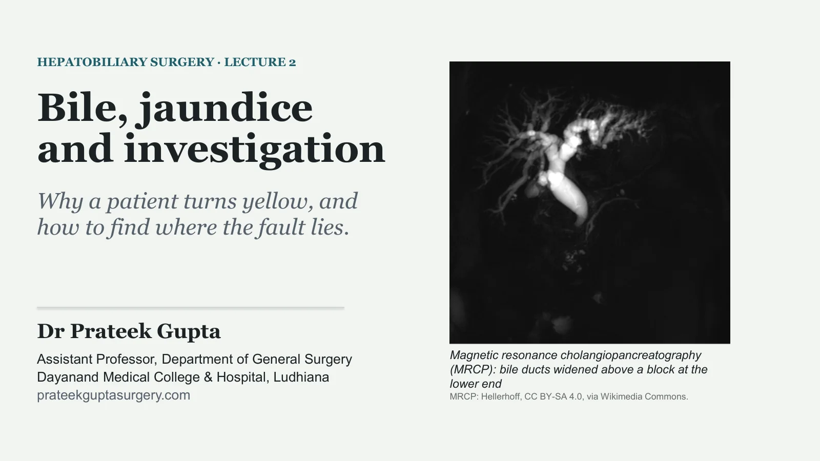
Magnetic resonance cholangiopancreatography (MRCP): bile ducts widened above a block at the lower end
MRCP: Hellerhoff, CC BY-SA 4.0, via Wikimedia Commons.
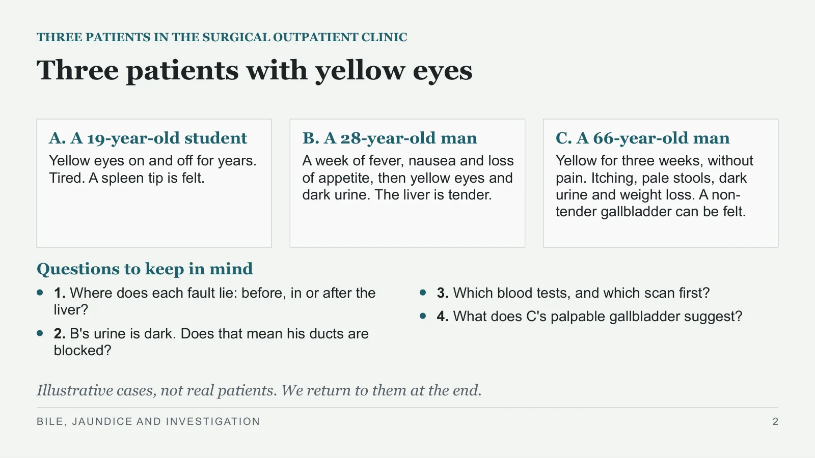
Three patients with yellow eyes
THREE PATIENTS IN THE SURGICAL OUTPATIENT CLINIC
A. A 19-year-old student
Yellow eyes on and off for years. Tired. A spleen tip is felt.
B. A 28-year-old man
A week of fever, nausea and loss of appetite, then yellow eyes and dark urine. The liver is tender.
C. A 66-year-old man
Yellow for three weeks, without pain. Itching, pale stools, dark urine and weight loss. A non-tender gallbladder can be felt.
Questions to keep in mind
1. Where does each fault lie: before, in or after the liver?
2. B's urine is dark. Does that mean his ducts are blocked?
3. Which blood tests, and which scan first?
4. What does C's palpable gallbladder suggest?
Illustrative cases, not real patients. We return to them at the end.
BILE, JAUNDICE AND INVESTIGATION
2
Bile formation and flow
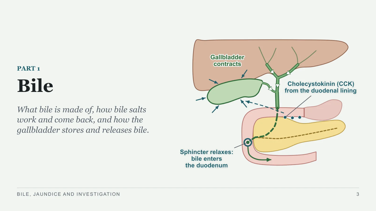
Bile
PART 1
What bile is made of, how bile salts work and come back, and how the gallbladder stores and releases bile.
BILE, JAUNDICE AND INVESTIGATION
3
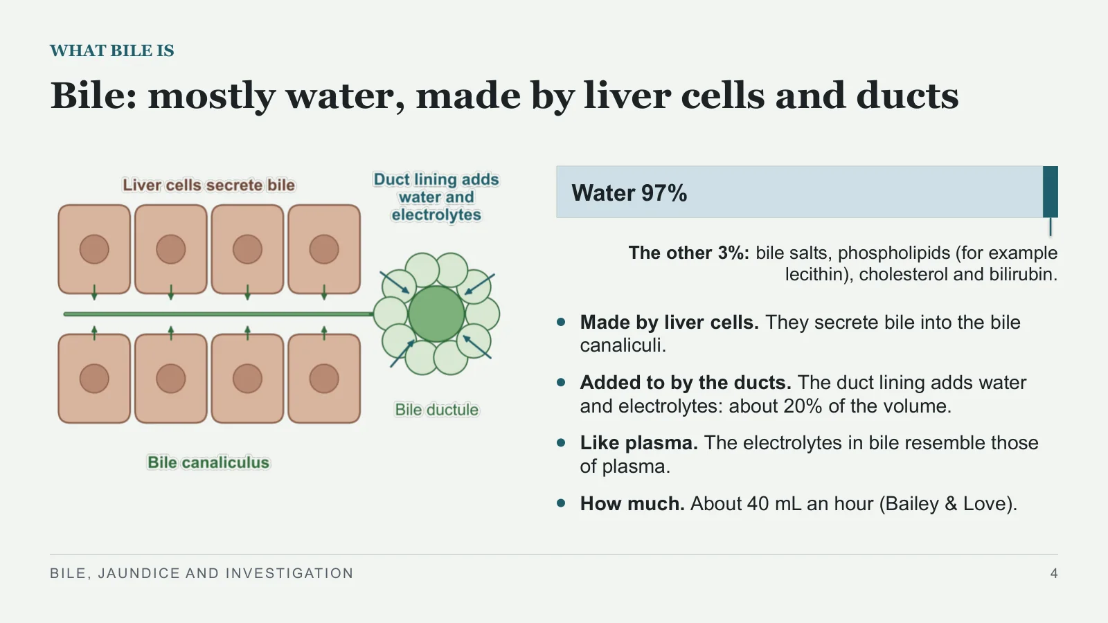
Bile: mostly water, made by liver cells and ducts
WHAT BILE IS
Water 97%
The other 3%: bile salts, phospholipids (for example lecithin), cholesterol and bilirubin.
Made by liver cells. They secrete bile into the bile canaliculi.
Added to by the ducts. The duct lining adds water and electrolytes: about 20% of the volume.
Like plasma. The electrolytes in bile resemble those of plasma.
How much. About 40 mL an hour (Bailey & Love).
BILE, JAUNDICE AND INVESTIGATION
4
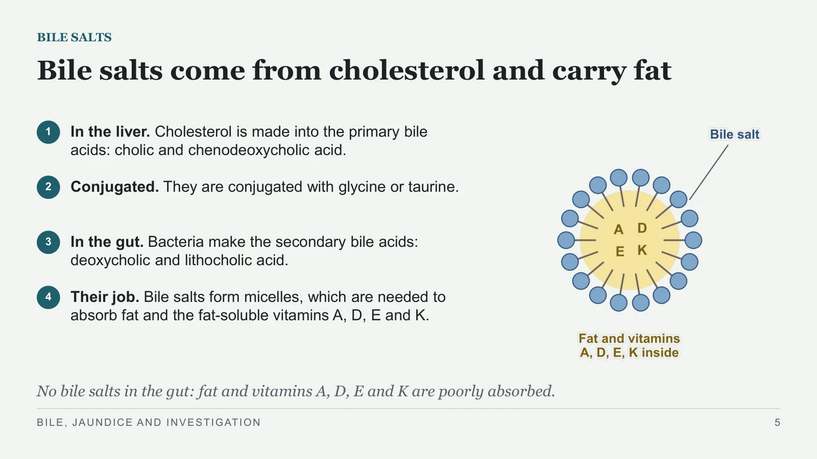
Bile salts come from cholesterol and carry fat
BILE SALTS
1
In the liver. Cholesterol is made into the primary bile acids: cholic and chenodeoxycholic acid.
2
Conjugated. They are conjugated with glycine or taurine.
3
In the gut. Bacteria make the secondary bile acids: deoxycholic and lithocholic acid.
4
Their job. Bile salts form micelles, which are needed to absorb fat and the fat-soluble vitamins A, D, E and K.
No bile salts in the gut: fat and vitamins A, D, E and K are poorly absorbed.
BILE, JAUNDICE AND INVESTIGATION
5
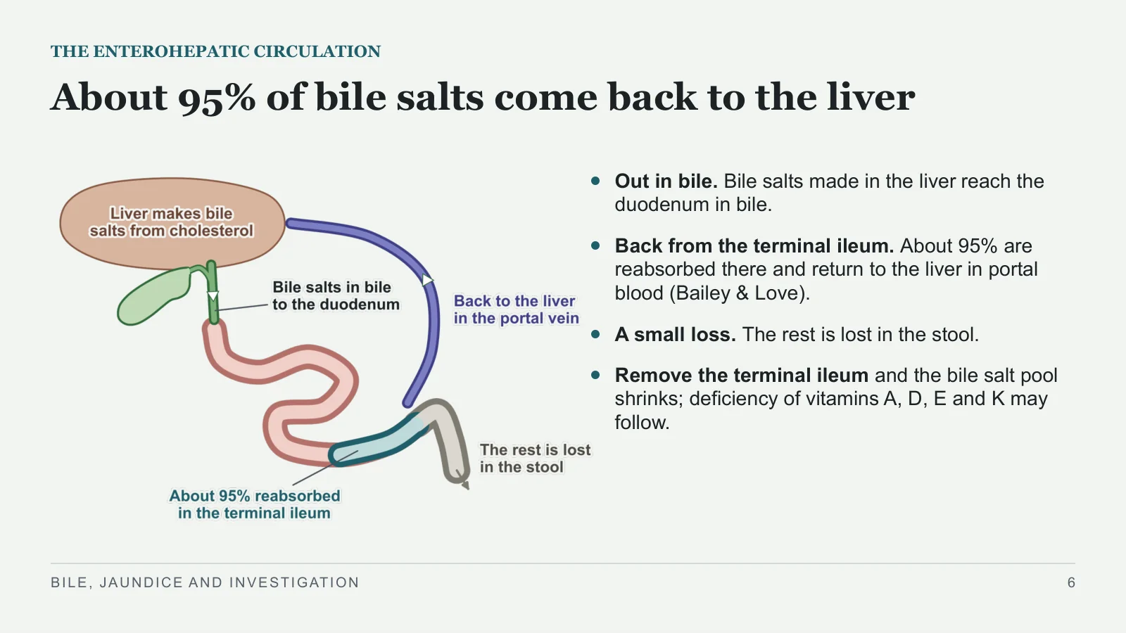
About 95% of bile salts come back to the liver
THE ENTEROHEPATIC CIRCULATION
Out in bile. Bile salts made in the liver reach the duodenum in bile.
Back from the terminal ileum. About 95% are reabsorbed there and return to the liver in portal blood (Bailey & Love).
A small loss. The rest is lost in the stool.
Remove the terminal ileum and the bile salt pool shrinks; deficiency of vitamins A, D, E and K may follow.
BILE, JAUNDICE AND INVESTIGATION
6
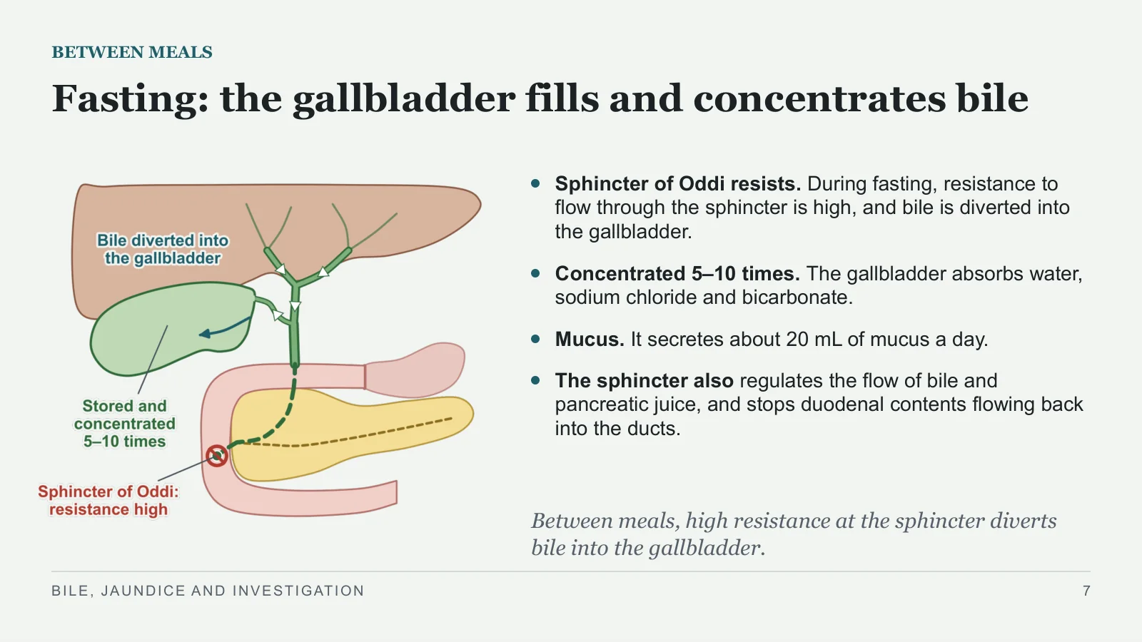
Fasting: the gallbladder fills and concentrates bile
BETWEEN MEALS
Sphincter of Oddi resists. During fasting, resistance to flow through the sphincter is high, and bile is diverted into the gallbladder.
Concentrated 5–10 times. The gallbladder absorbs water, sodium chloride and bicarbonate.
Mucus. It secretes about 20 mL of mucus a day.
The sphincter also regulates the flow of bile and pancreatic juice, and stops duodenal contents flowing back into the ducts.
Between meals, high resistance at the sphincter diverts bile into the gallbladder.
BILE, JAUNDICE AND INVESTIGATION
7
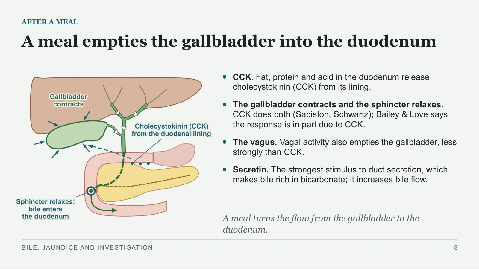
A meal empties the gallbladder into the duodenum
AFTER A MEAL
CCK. Fat, protein and acid in the duodenum release cholecystokinin (CCK) from its lining.
The gallbladder contracts and the sphincter relaxes. CCK does both (Sabiston, Schwartz); Bailey & Love says the response is in part due to CCK.
The vagus. Vagal activity also empties the gallbladder, less strongly than CCK.
Secretin. The strongest stimulus to duct secretion, which makes bile rich in bicarbonate; it increases bile flow.
A meal turns the flow from the gallbladder to the duodenum.
BILE, JAUNDICE AND INVESTIGATION
8
Bilirubin
Bilirubin
PART 2
Where bilirubin comes from, how the liver changes it, and how it leaves the body.
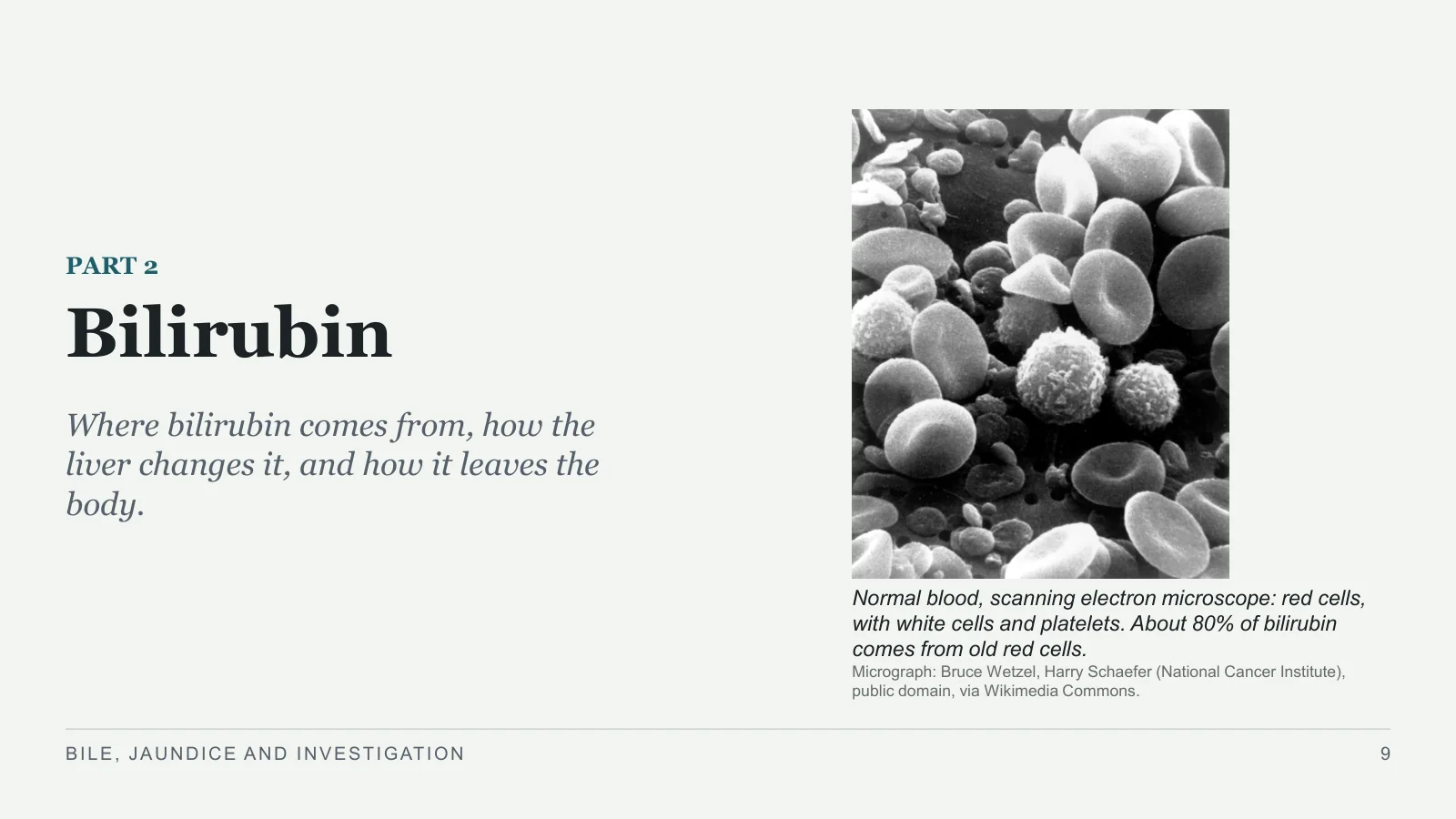
Normal blood, scanning electron microscope: red cells, with white cells and platelets. About 80% of bilirubin comes from old red cells.
Micrograph: Bruce Wetzel, Harry Schaefer (National Cancer Institute), public domain, via Wikimedia Commons.
BILE, JAUNDICE AND INVESTIGATION
9
Bilirubin comes mostly from old red cells
THE PATH OF BILIRUBIN, 1
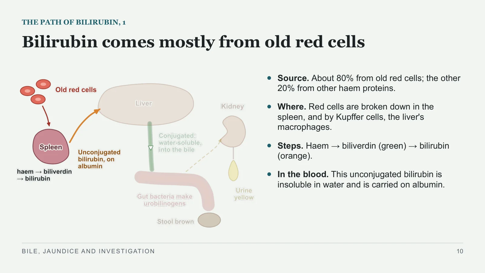
Source. About 80% from old red cells; the other 20% from other haem proteins.
Where. Red cells are broken down in the spleen, and by Kupffer cells, the liver's macrophages.
Steps. Haem → biliverdin (green) → bilirubin (orange).
In the blood. This unconjugated bilirubin is insoluble in water and is carried on albumin.
BILE, JAUNDICE AND INVESTIGATION
10
The liver makes bilirubin water-soluble
THE PATH OF BILIRUBIN, 2
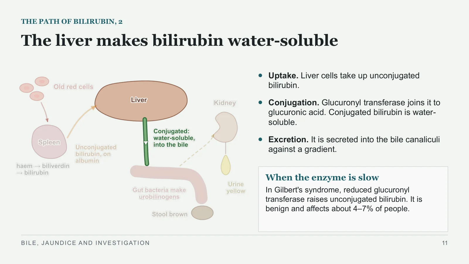
Uptake. Liver cells take up unconjugated bilirubin.
Conjugation. Glucuronyl transferase joins it to glucuronic acid. Conjugated bilirubin is water-soluble.
Excretion. It is secreted into the bile canaliculi against a gradient.
When the enzyme is slow
In Gilbert's syndrome, reduced glucuronyl transferase raises unconjugated bilirubin. It is benign and affects about 4–7% of people.
BILE, JAUNDICE AND INVESTIGATION
11
Oxidised urobilinogens colour stool and urine
THE PATH OF BILIRUBIN, 3
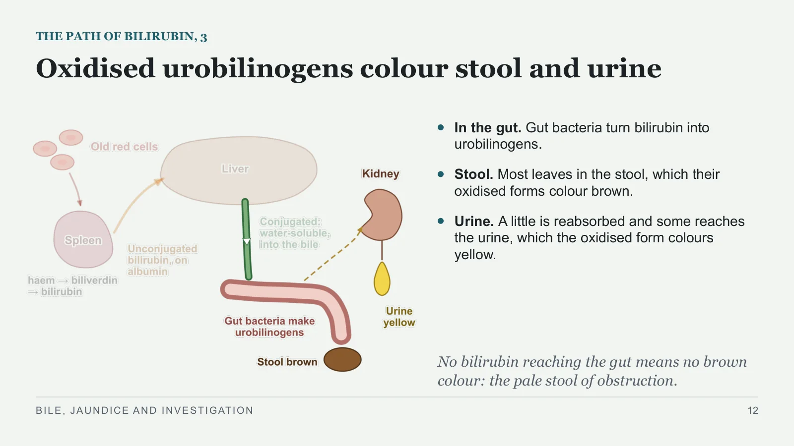
In the gut. Gut bacteria turn bilirubin into urobilinogens.
Stool. Most leaves in the stool, which their oxidised forms colour brown.
Urine. A little is reabsorbed and some reaches the urine, which the oxidised form colours yellow.
No bilirubin reaching the gut means no brown colour: the pale stool of obstruction.
BILE, JAUNDICE AND INVESTIGATION
12
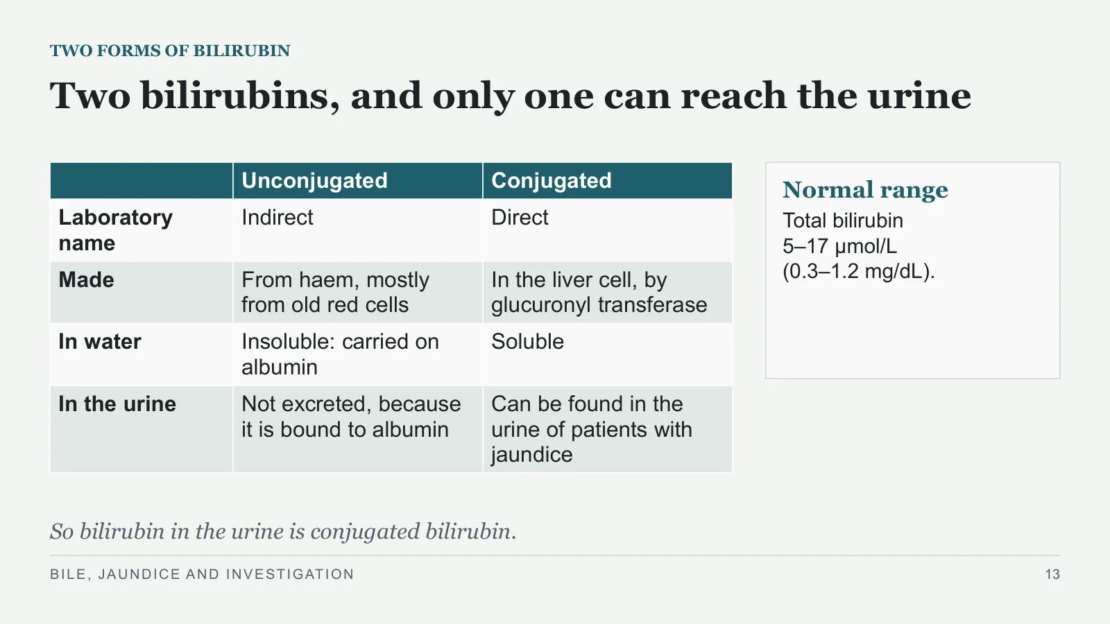
Two bilirubins, and only one can reach the urine
TWO FORMS OF BILIRUBIN
Normal range
Total bilirubin 5–17 μmol/L (0.3–1.2 mg/dL).
So bilirubin in the urine is conjugated bilirubin.
BILE, JAUNDICE AND INVESTIGATION
13
| Unconjugated | Conjugated | |
| Laboratory name | Indirect | Direct |
| Made | From haem, mostly from old red cells | In the liver cell, by glucuronyl transferase |
| In water | Insoluble: carried on albumin | Soluble |
| In the urine | Not excreted, because it is bound to albumin | Can be found in the urine of patients with jaundice |
Jaundice
Jaundice
PART 3
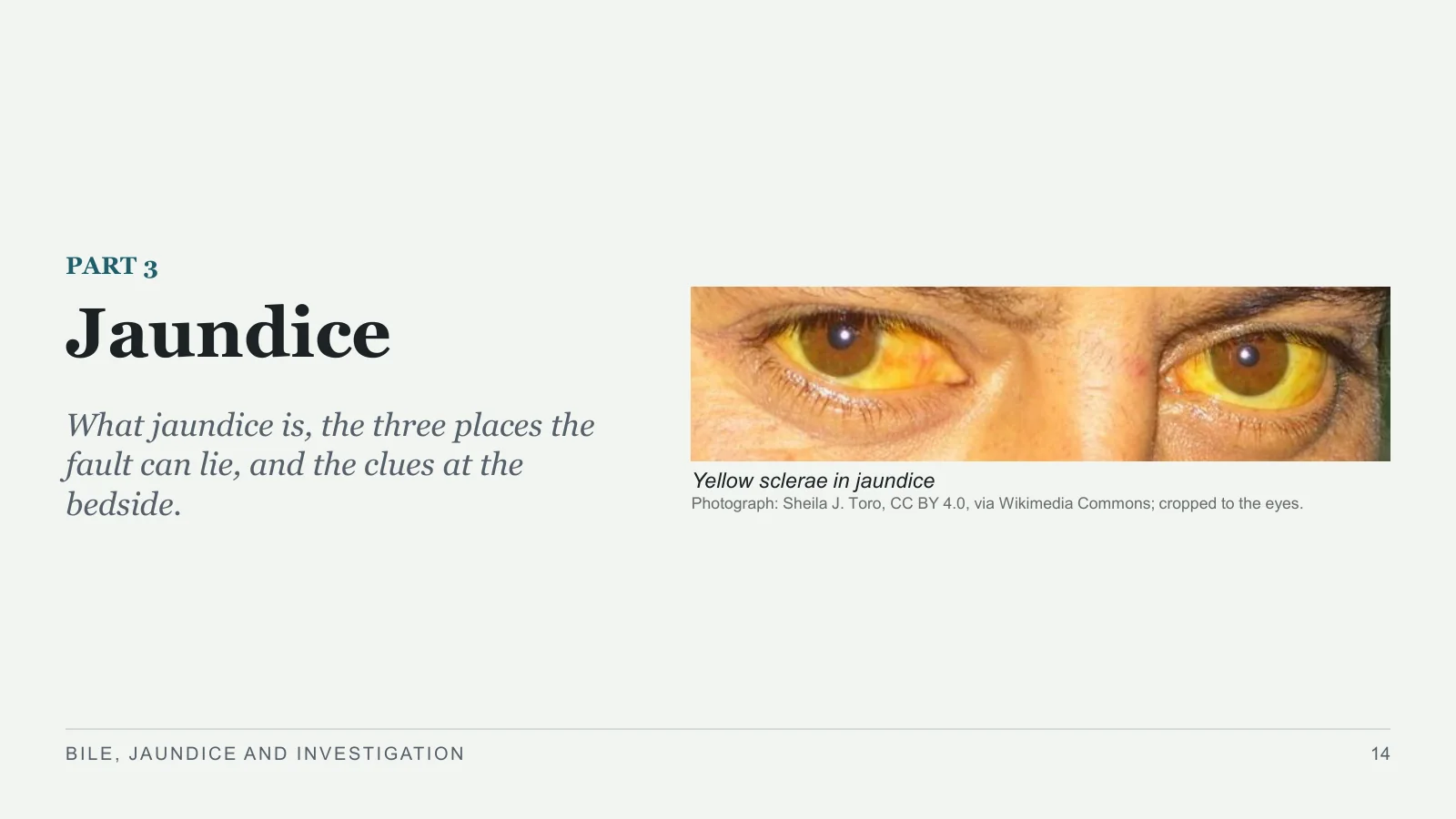
What jaundice is, the three places the fault can lie, and the clues at the bedside.
Yellow sclerae in jaundice
Photograph: Sheila J. Toro, CC BY 4.0, via Wikimedia Commons; cropped to the eyes.
BILE, JAUNDICE AND INVESTIGATION
14
Jaundice is bilirubin staining the tissues
JAUNDICE
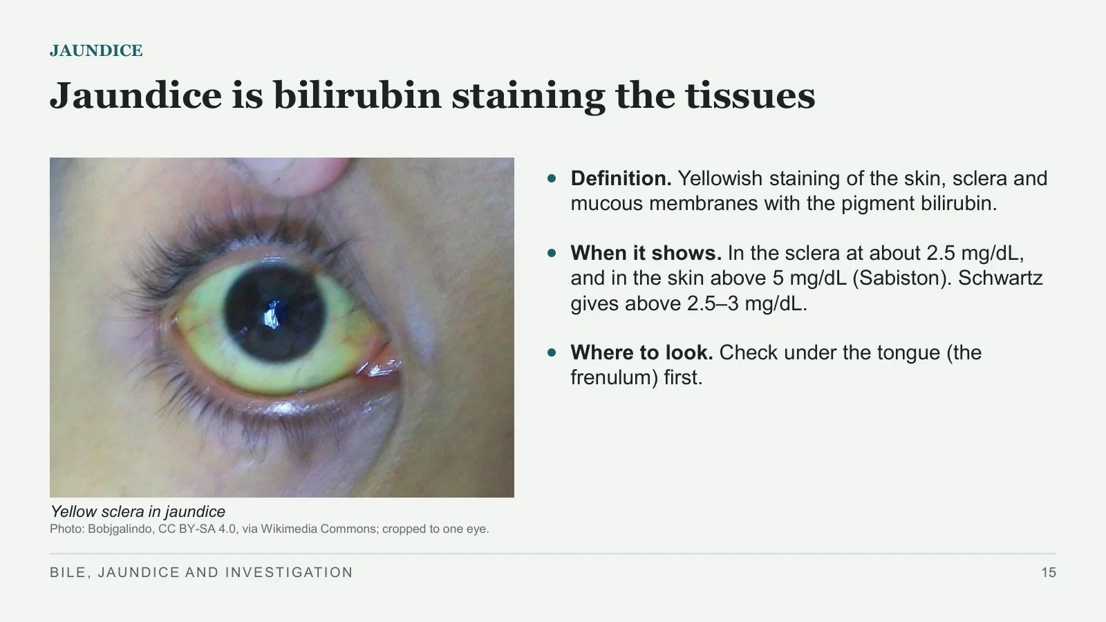
Yellow sclera in jaundice
Photo: Bobjgalindo, CC BY-SA 4.0, via Wikimedia Commons; cropped to one eye.
Definition. Yellowish staining of the skin, sclera and mucous membranes with the pigment bilirubin.
When it shows. In the sclera at about 2.5 mg/dL, and in the skin above 5 mg/dL (Sabiston). Schwartz gives above 2.5–3 mg/dL.
Where to look. Check under the tongue (the frenulum) first.
BILE, JAUNDICE AND INVESTIGATION
15
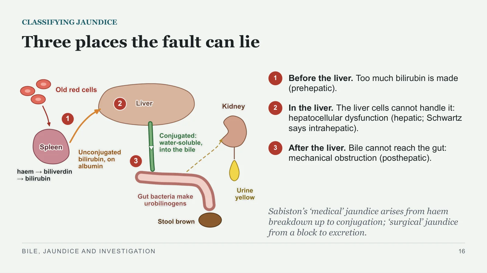
Three places the fault can lie
CLASSIFYING JAUNDICE
1
Before the liver. Too much bilirubin is made (prehepatic).
2
In the liver. The liver cells cannot handle it: hepatocellular dysfunction (hepatic; Schwartz says intrahepatic).
3
After the liver. Bile cannot reach the gut: mechanical obstruction (posthepatic).
Sabiston’s ‘medical’ jaundice arises from haem breakdown up to conjugation; ‘surgical’ jaundice from a block to excretion.
BILE, JAUNDICE AND INVESTIGATION
16
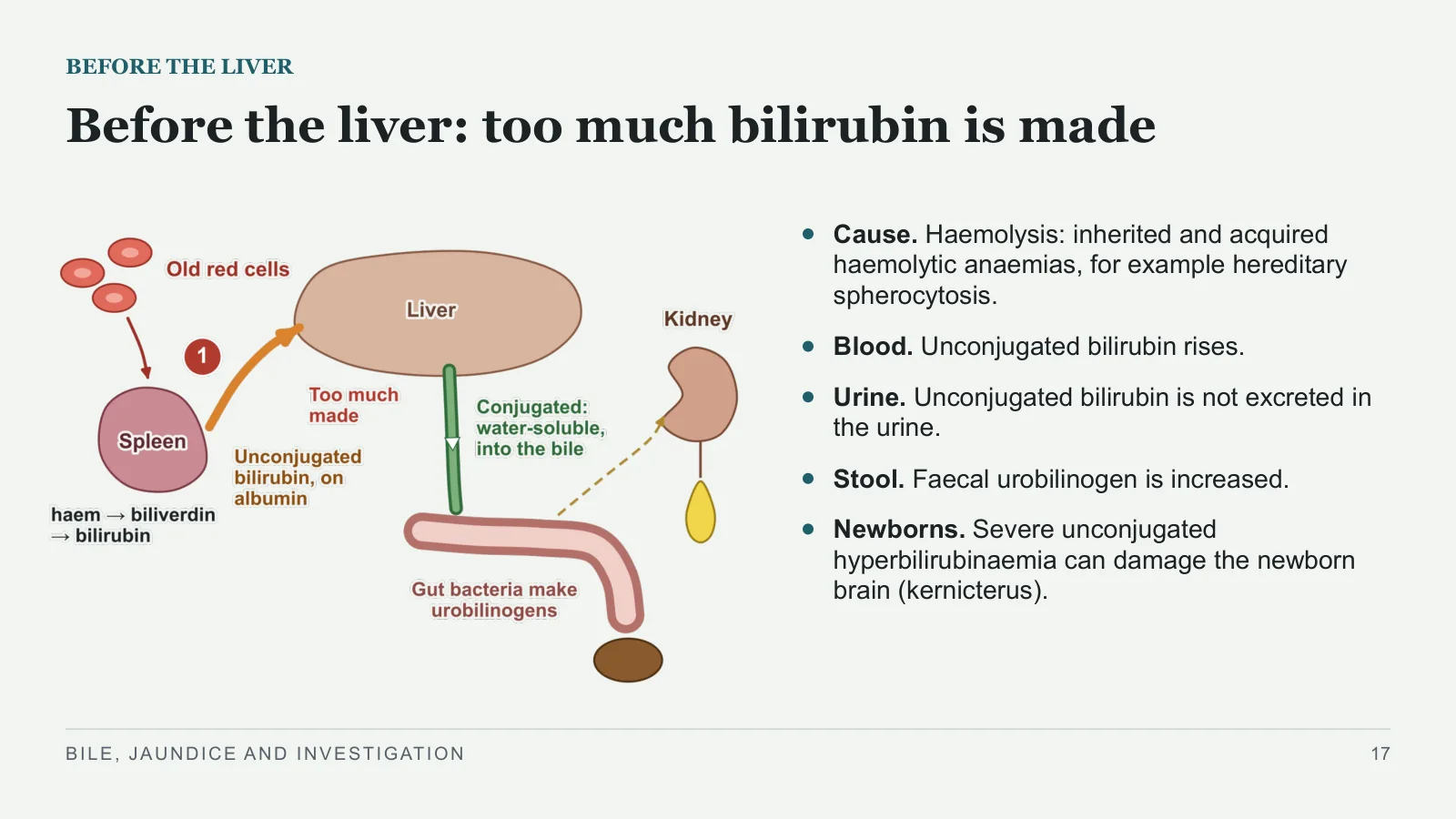
Before the liver: too much bilirubin is made
BEFORE THE LIVER
Cause. Haemolysis: inherited and acquired haemolytic anaemias, for example hereditary spherocytosis.
Blood. Unconjugated bilirubin rises.
Urine. Unconjugated bilirubin is not excreted in the urine.
Stool. Faecal urobilinogen is increased.
Newborns. Severe unconjugated hyperbilirubinaemia can damage the newborn brain (kernicterus).
BILE, JAUNDICE AND INVESTIGATION
17
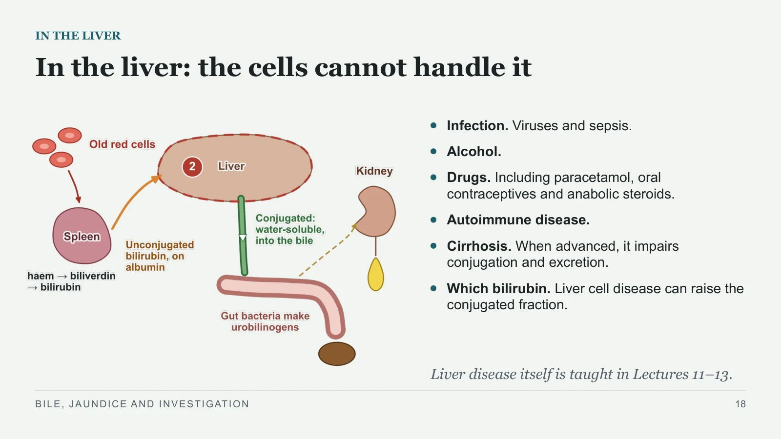
In the liver: the cells cannot handle it
IN THE LIVER
Infection. Viruses and sepsis.
Alcohol.
Drugs. Including paracetamol, oral contraceptives and anabolic steroids.
Autoimmune disease.
Cirrhosis. When advanced, it impairs conjugation and excretion.
Which bilirubin. Liver cell disease can raise the conjugated fraction.
Liver disease itself is taught in Lectures 11–13.
BILE, JAUNDICE AND INVESTIGATION
18
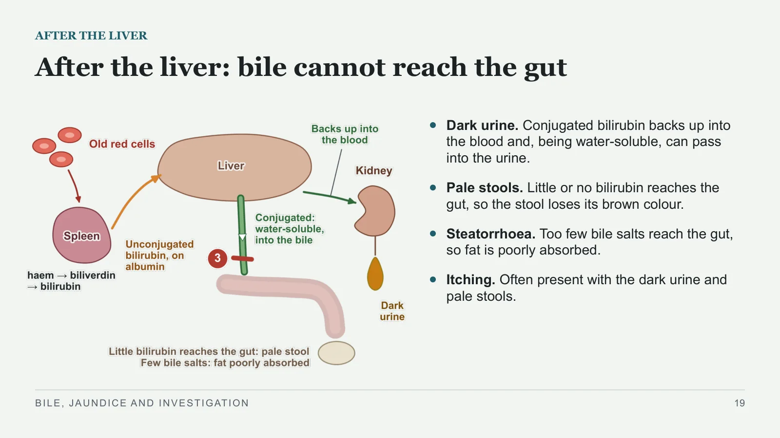
After the liver: bile cannot reach the gut
AFTER THE LIVER
Dark urine. Conjugated bilirubin backs up into the blood and, being water-soluble, can pass into the urine.
Pale stools. Little or no bilirubin reaches the gut, so the stool loses its brown colour.
Steatorrhoea. Too few bile salts reach the gut, so fat is poorly absorbed.
Itching. Often present with the dark urine and pale stools.
BILE, JAUNDICE AND INVESTIGATION
19
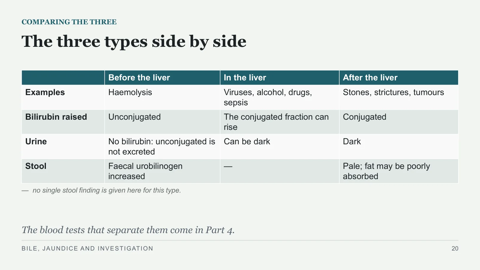
The three types side by side
COMPARING THE THREE
— no single stool finding is given here for this type.
The blood tests that separate them come in Part 4.
BILE, JAUNDICE AND INVESTIGATION
20
| Before the liver | In the liver | After the liver | |
| Examples | Haemolysis | Viruses, alcohol, drugs, sepsis | Stones, strictures, tumours |
| Bilirubin raised | Unconjugated | The conjugated fraction can rise | Conjugated |
| Urine | No bilirubin: unconjugated is not excreted | Can be dark | Dark |
| Stool | Faecal urobilinogen increased | — | Pale; fat may be poorly absorbed |
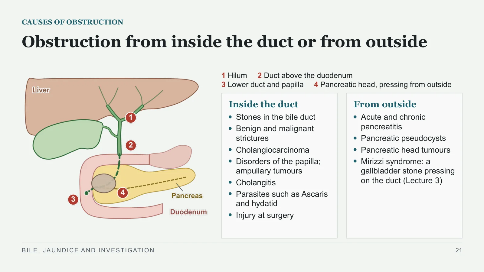
Obstruction from inside the duct or from outside
CAUSES OF OBSTRUCTION
1 Hilum 2 Duct above the duodenum
3 Lower duct and papilla 4 Pancreatic head, pressing from outside
Inside the duct
Stones in the bile duct
Benign and malignant strictures
Cholangiocarcinoma
Disorders of the papilla; ampullary tumours
Cholangitis
Parasites such as Ascaris and hydatid
Injury at surgery
From outside
Acute and chronic pancreatitis
Pancreatic pseudocysts
Pancreatic head tumours
Mirizzi syndrome: a gallbladder stone pressing on the duct (Lecture 3)
BILE, JAUNDICE AND INVESTIGATION
21
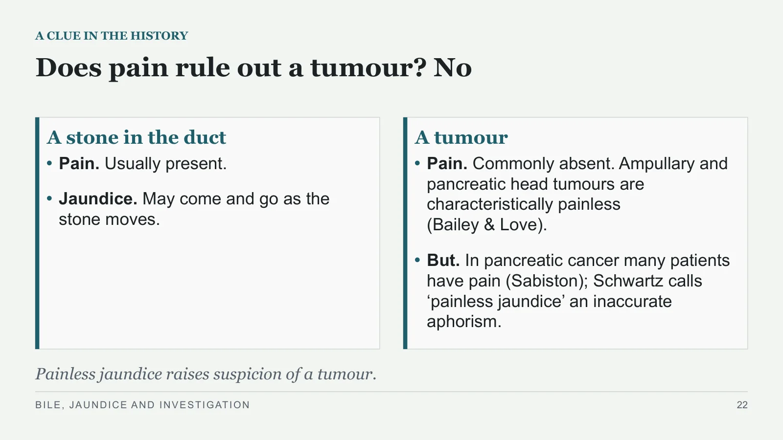
Does pain rule out a tumour? No
A CLUE IN THE HISTORY
A stone in the duct
Pain. Usually present.
Jaundice. May come and go as the stone moves.
A tumour
Pain. Commonly absent. Ampullary and pancreatic head tumours are characteristically painless (Bailey & Love).
But. In pancreatic cancer many patients have pain (Sabiston); Schwartz calls ‘painless jaundice’ an inaccurate aphorism.
Painless jaundice raises suspicion of a tumour.
BILE, JAUNDICE AND INVESTIGATION
22
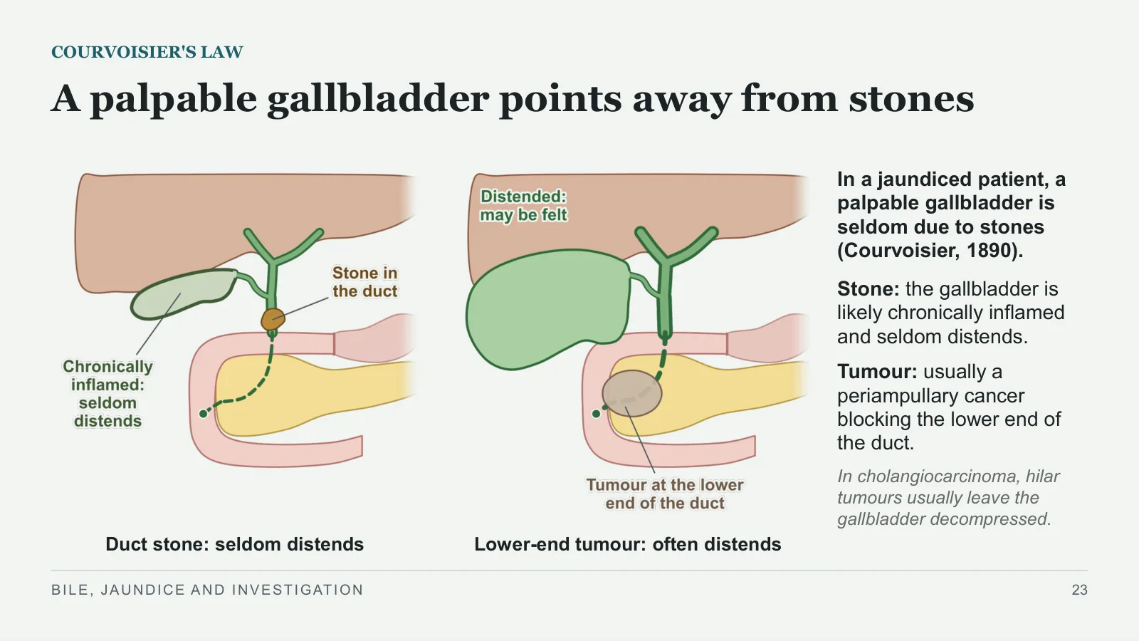
A palpable gallbladder points away from stones
COURVOISIER'S LAW
Duct stone: seldom distends
Lower-end tumour: often distends
In a jaundiced patient, a palpable gallbladder is seldom due to stones (Courvoisier, 1890).
Stone: the gallbladder is likely chronically inflamed and seldom distends.
Tumour: usually a periampullary cancer blocking the lower end of the duct.
In cholangiocarcinoma, hilar tumours usually leave the gallbladder decompressed.
BILE, JAUNDICE AND INVESTIGATION
23
+
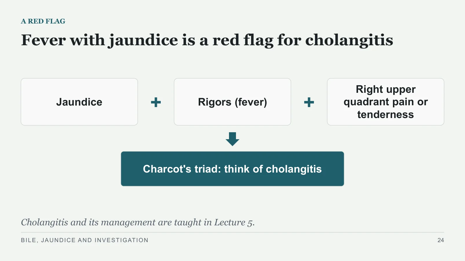
A RED FLAG
Fever with jaundice is a red flag for cholangitis
Jaundice
Rigors (fever)
+
Right upper quadrant pain or tenderness
Charcot's triad: think of cholangitis
Cholangitis and its management are taught in Lecture 5.
BILE, JAUNDICE AND INVESTIGATION
24
Liver tests
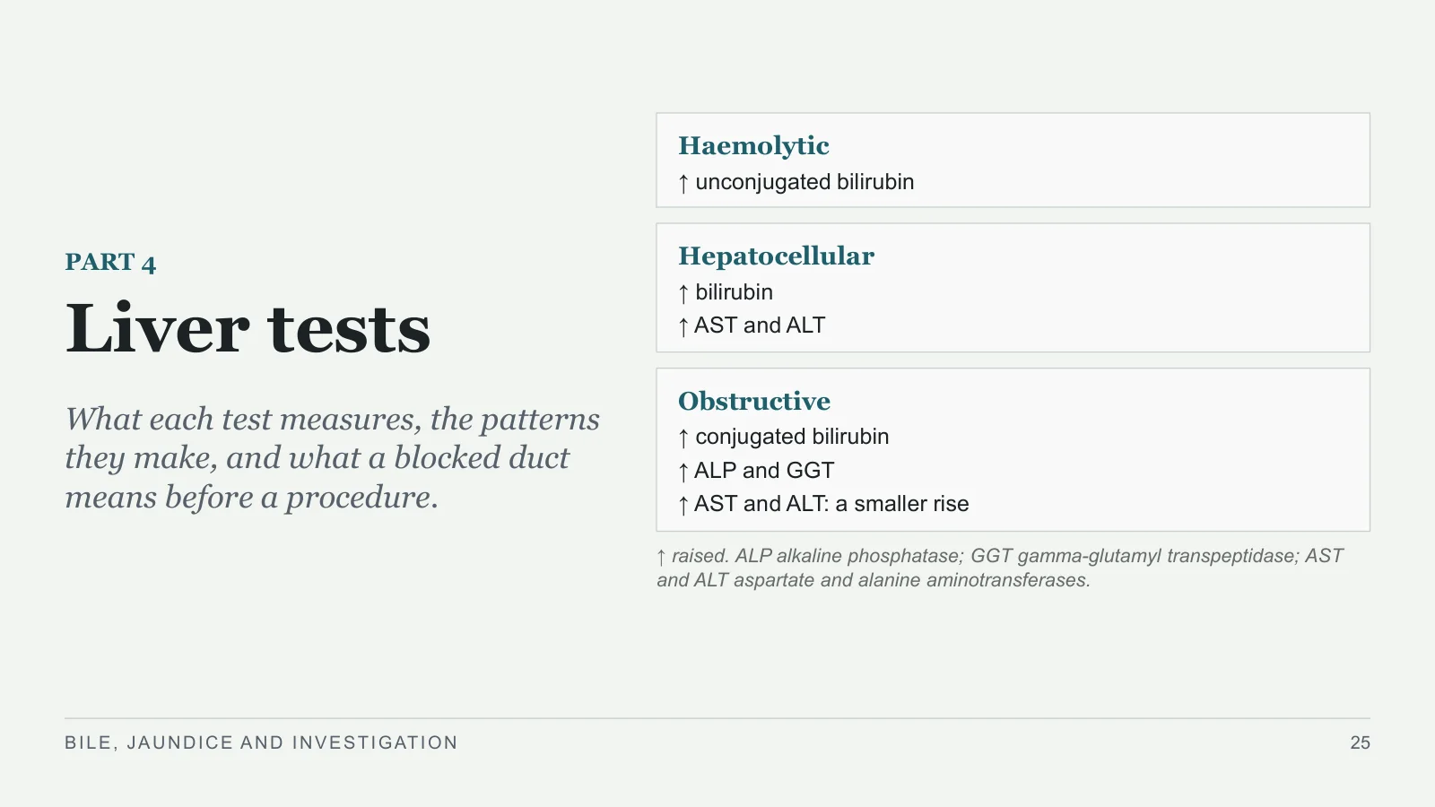
Liver tests
PART 4
What each test measures, the patterns they make, and what a blocked duct means before a procedure.
Haemolytic
↑ unconjugated bilirubin
Hepatocellular
↑ bilirubin
↑ AST and ALT
Obstructive
↑ conjugated bilirubin
↑ ALP and GGT
↑ AST and ALT: a smaller rise
↑ raised. ALP alkaline phosphatase; GGT gamma-glutamyl transpeptidase; AST and ALT aspartate and alanine aminotransferases.
BILE, JAUNDICE AND INVESTIGATION
25
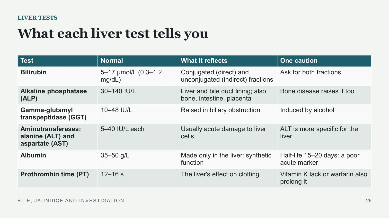
What each liver test tells you
LIVER TESTS
BILE, JAUNDICE AND INVESTIGATION
26
| Test | Normal | What it reflects | One caution |
| Bilirubin | 5–17 μmol/L (0.3–1.2 mg/dL) | Conjugated (direct) and unconjugated (indirect) fractions | Ask for both fractions |
| Alkaline phosphatase (ALP) | 30–140 IU/L | Liver and bile duct lining; also bone, intestine, placenta | Bone disease raises it too |
| Gamma-glutamyl transpeptidase (GGT) | 10–48 IU/L | Raised in biliary obstruction | Induced by alcohol |
| Aminotransferases: alanine (ALT) and aspartate (AST) | 5–40 IU/L each | Usually acute damage to liver cells | ALT is more specific for the liver |
| Albumin | 35–50 g/L | Made only in the liver: synthetic function | Half-life 15–20 days: a poor acute marker |
| Prothrombin time (PT) | 12–16 s | The liver's effect on clotting | Vitamin K lack or warfarin also prolong it |
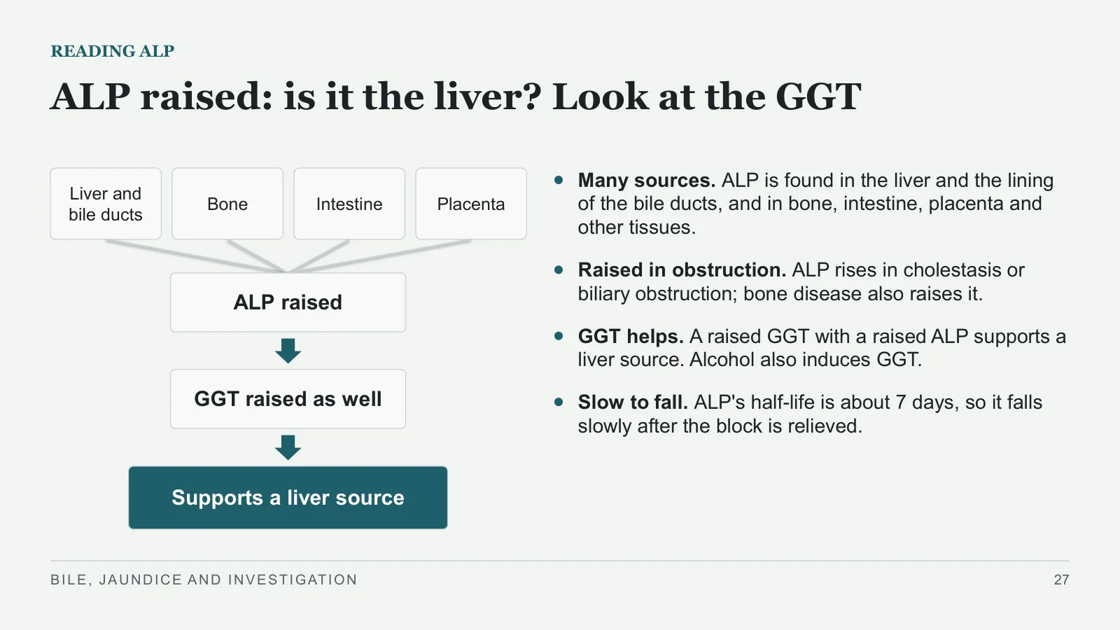
ALP raised: is it the liver? Look at the GGT
READING ALP
Liver and bile ducts
Bone
Intestine
Placenta
ALP raised
GGT raised as well
Supports a liver source
Many sources. ALP is found in the liver and the lining of the bile ducts, and in bone, intestine, placenta and other tissues.
Raised in obstruction. ALP rises in cholestasis or biliary obstruction; bone disease also raises it.
GGT helps. A raised GGT with a raised ALP supports a liver source. Alcohol also induces GGT.
Slow to fall. ALP's half-life is about 7 days, so it falls slowly after the block is relieved.
BILE, JAUNDICE AND INVESTIGATION
27
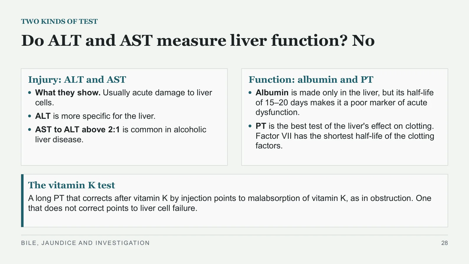
Do ALT and AST measure liver function? No
TWO KINDS OF TEST
Injury: ALT and AST
What they show. Usually acute damage to liver cells.
ALT is more specific for the liver.
AST to ALT above 2:1 is common in alcoholic liver disease.
Function: albumin and PT
Albumin is made only in the liver, but its half-life of 15–20 days makes it a poor marker of acute dysfunction.
PT is the best test of the liver's effect on clotting. Factor VII has the shortest half-life of the clotting factors.
The vitamin K test
A long PT that corrects after vitamin K by injection points to malabsorption of vitamin K, as in obstruction. One that does not correct points to liver cell failure.
BILE, JAUNDICE AND INVESTIGATION
28
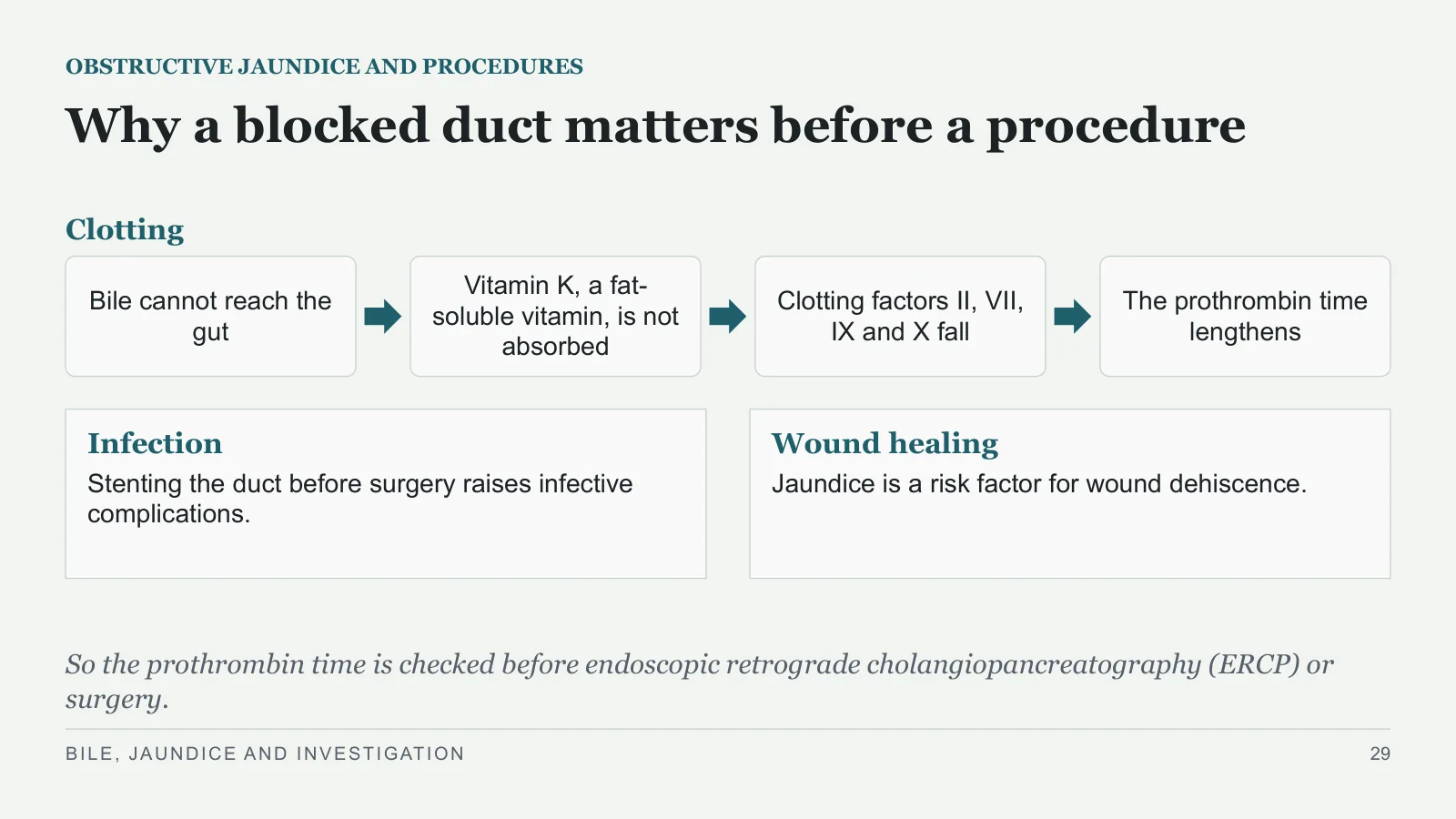
Why a blocked duct matters before a procedure
OBSTRUCTIVE JAUNDICE AND PROCEDURES
Clotting
Bile cannot reach the gut
Vitamin K, a fat-soluble vitamin, is not absorbed
Clotting factors II, VII, IX and X fall
The prothrombin time lengthens
Infection
Stenting the duct before surgery raises infective complications.
Wound healing
Jaundice is a risk factor for wound dehiscence.
So the prothrombin time is checked before endoscopic retrograde cholangiopancreatography (ERCP) or surgery.
BILE, JAUNDICE AND INVESTIGATION
29
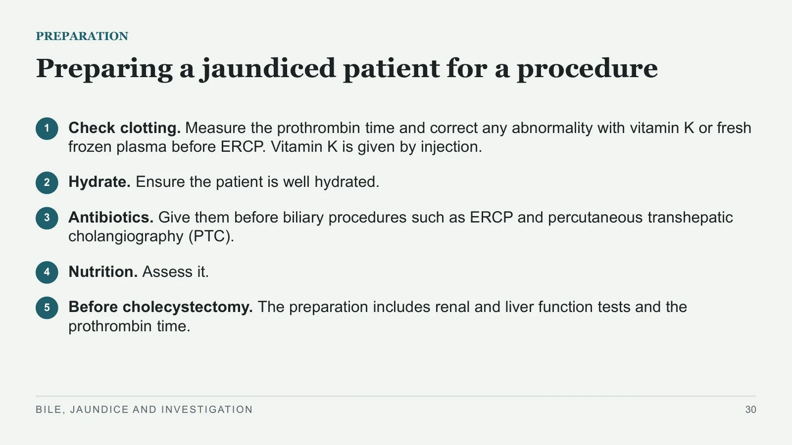
Preparing a jaundiced patient for a procedure
PREPARATION
1
Check clotting. Measure the prothrombin time and correct any abnormality with vitamin K or fresh frozen plasma before ERCP. Vitamin K is given by injection.
2
Hydrate. Ensure the patient is well hydrated.
3
Antibiotics. Give them before biliary procedures such as ERCP and percutaneous transhepatic cholangiography (PTC).
4
Nutrition. Assess it.
5
Before cholecystectomy. The preparation includes renal and liver function tests and the prothrombin time.
BILE, JAUNDICE AND INVESTIGATION
30
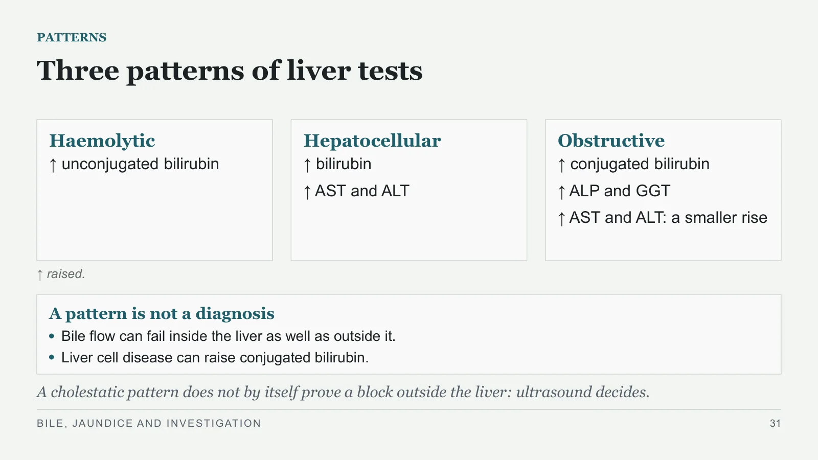
Three patterns of liver tests
PATTERNS
Haemolytic
↑ unconjugated bilirubin
Hepatocellular
↑ bilirubin
↑ AST and ALT
Obstructive
↑ conjugated bilirubin
↑ ALP and GGT
↑ AST and ALT: a smaller rise
↑ raised.
A pattern is not a diagnosis
Bile flow can fail inside the liver as well as outside it.
Liver cell disease can raise conjugated bilirubin.
A cholestatic pattern does not by itself prove a block outside the liver: ultrasound decides.
BILE, JAUNDICE AND INVESTIGATION
31
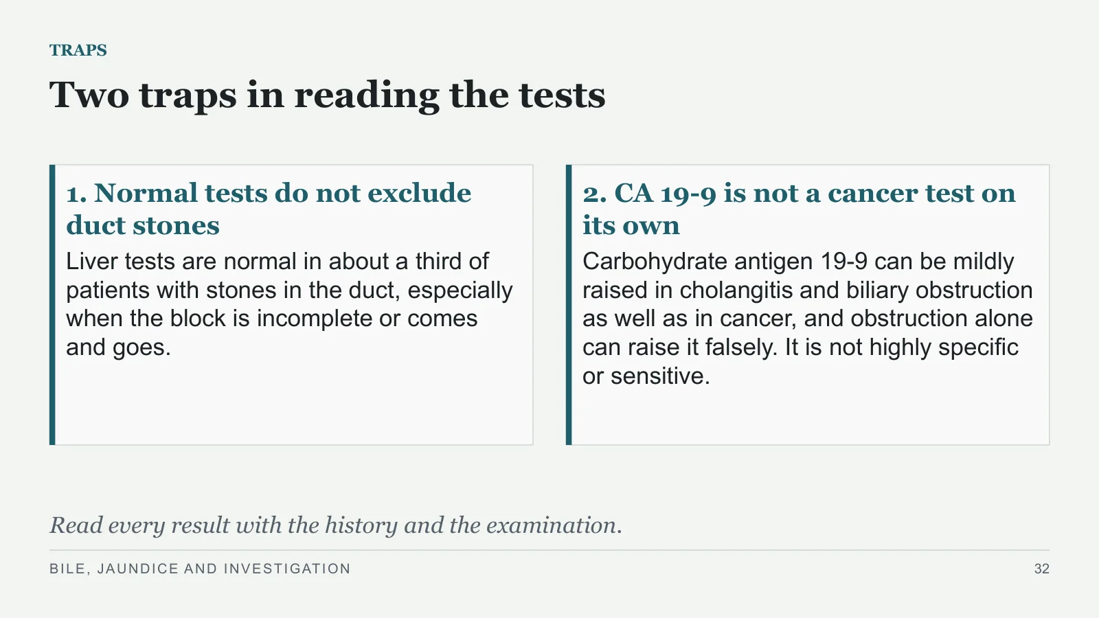
Two traps in reading the tests
TRAPS
1. Normal tests do not exclude duct stones
Liver tests are normal in about a third of patients with stones in the duct, especially when the block is incomplete or comes and goes.
2. CA 19-9 is not a cancer test on its own
Carbohydrate antigen 19-9 can be mildly raised in cholangitis and biliary obstruction as well as in cancer, and obstruction alone can raise it falsely. It is not highly specific or sensitive.
Read every result with the history and the examination.
BILE, JAUNDICE AND INVESTIGATION
32
Investigation
Imaging
PART 5
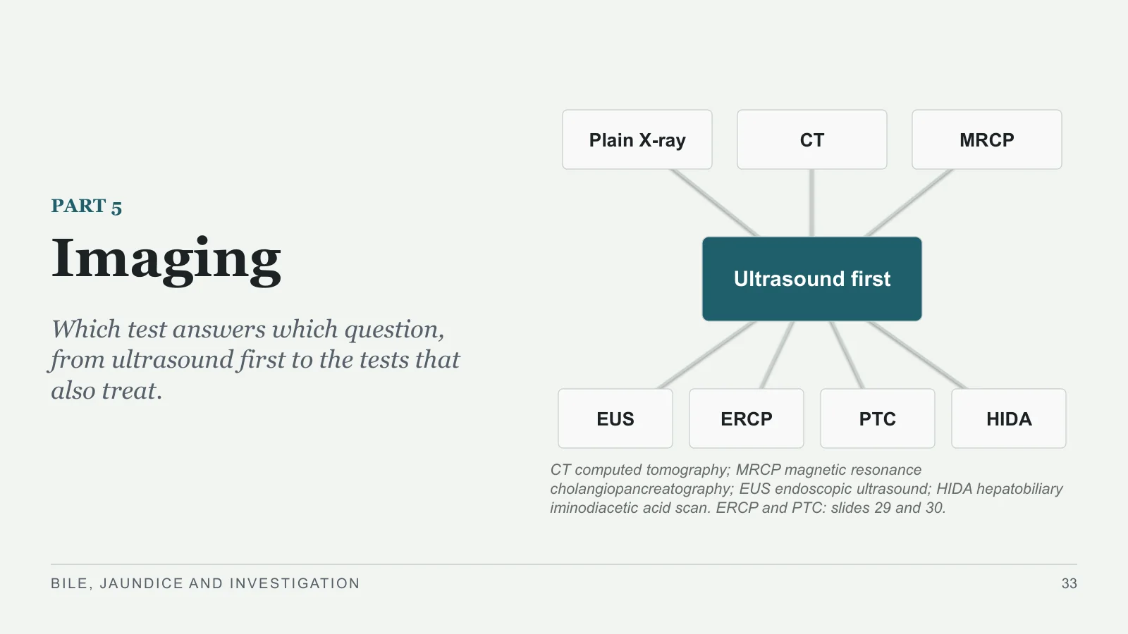
Which test answers which question, from ultrasound first to the tests that also treat.
Plain X-ray
CT
MRCP
EUS
ERCP
PTC
HIDA
Ultrasound first
CT computed tomography; MRCP magnetic resonance cholangiopancreatography; EUS endoscopic ultrasound; HIDA hepatobiliary iminodiacetic acid scan. ERCP and PTC: slides 29 and 30.
BILE, JAUNDICE AND INVESTIGATION
33
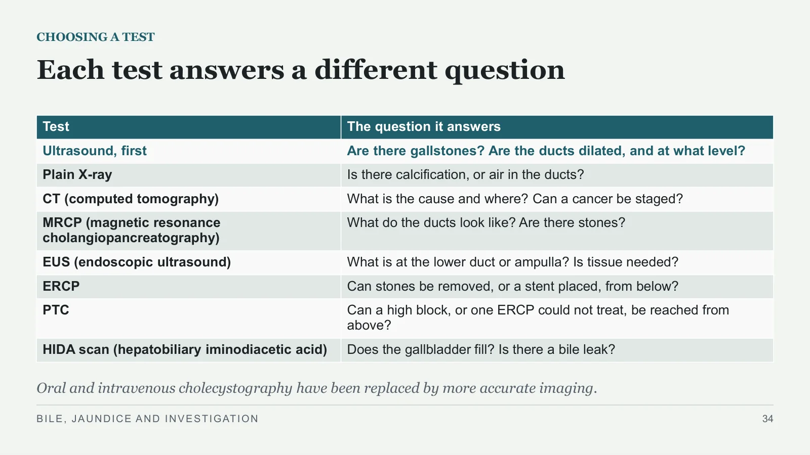
Each test answers a different question
CHOOSING A TEST
Oral and intravenous cholecystography have been replaced by more accurate imaging.
BILE, JAUNDICE AND INVESTIGATION
34
| Test | The question it answers |
| Ultrasound, first | Are there gallstones? Are the ducts dilated, and at what level? |
| Plain X-ray | Is there calcification, or air in the ducts? |
| CT (computed tomography) | What is the cause and where? Can a cancer be staged? |
| MRCP (magnetic resonance cholangiopancreatography) | What do the ducts look like? Are there stones? |
| EUS (endoscopic ultrasound) | What is at the lower duct or ampulla? Is tissue needed? |
| ERCP | Can stones be removed, or a stent placed, from below? |
| PTC | Can a high block, or one ERCP could not treat, be reached from above? |
| HIDA scan (hepatobiliary iminodiacetic acid) | Does the gallbladder fill? Is there a bile leak? |
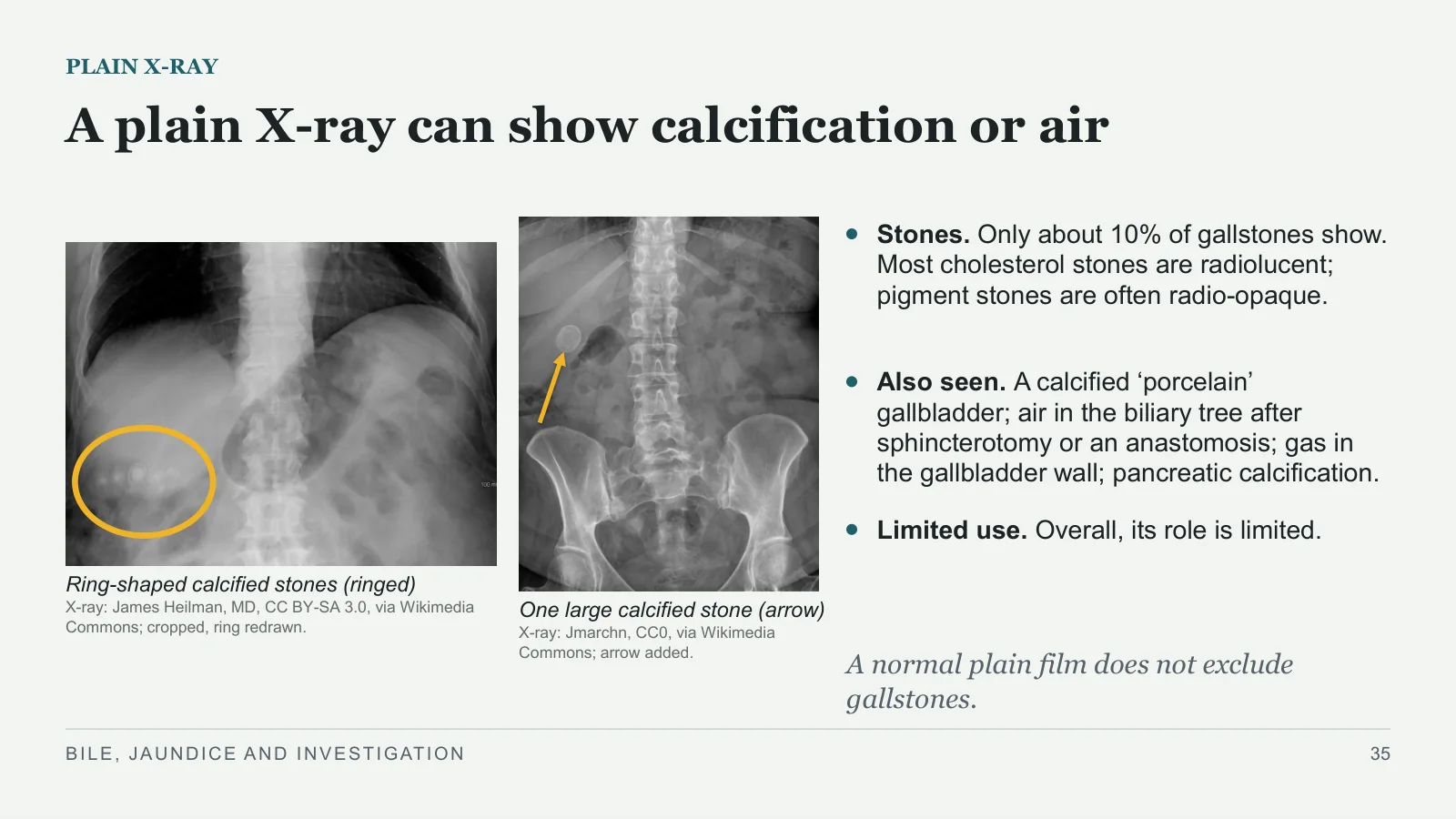
A plain X-ray can show calcification or air
PLAIN X-RAY
Ring-shaped calcified stones (ringed)
X-ray: James Heilman, MD, CC BY-SA 3.0, via Wikimedia Commons; cropped, ring redrawn.
One large calcified stone (arrow)
X-ray: Jmarchn, CC0, via Wikimedia Commons; arrow added.
Stones. Only about 10% of gallstones show. Most cholesterol stones are radiolucent; pigment stones are often radio-opaque.
Also seen. A calcified ‘porcelain’ gallbladder; air in the biliary tree after sphincterotomy or an anastomosis; gas in the gallbladder wall; pancreatic calcification.
Limited use. Overall, its role is limited.
A normal plain film does not exclude gallstones.
BILE, JAUNDICE AND INVESTIGATION
35
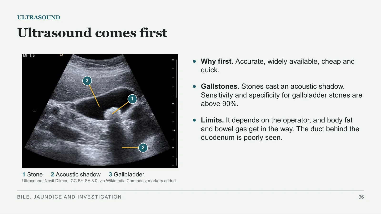
Ultrasound comes first
ULTRASOUND
1
2
3
1 Stone 2 Acoustic shadow 3 Gallbladder
Ultrasound: Nevit Dilmen, CC BY-SA 3.0, via Wikimedia Commons; markers added.
Why first. Accurate, widely available, cheap and quick.
Gallstones. Stones cast an acoustic shadow. Sensitivity and specificity for gallbladder stones are above 90%.
Limits. It depends on the operator, and body fat and bowel gas get in the way. The duct behind the duodenum is poorly seen.
BILE, JAUNDICE AND INVESTIGATION
36
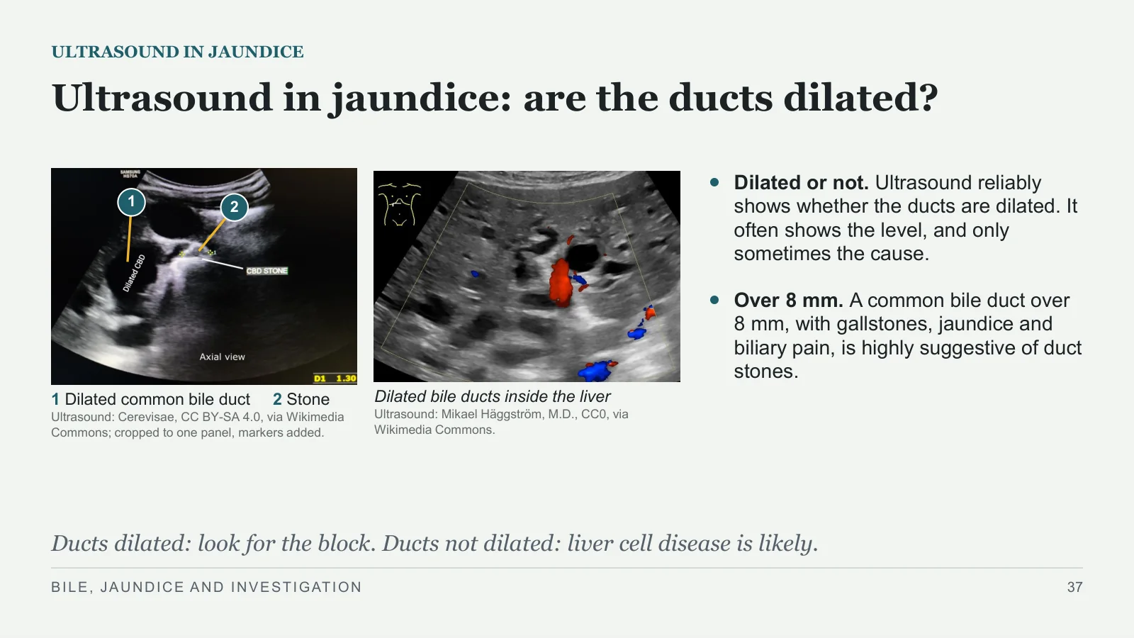
Ultrasound in jaundice: are the ducts dilated?
ULTRASOUND IN JAUNDICE
1
2
1 Dilated common bile duct 2 Stone
Ultrasound: Cerevisae, CC BY-SA 4.0, via Wikimedia Commons; cropped to one panel, markers added.
Dilated bile ducts inside the liver
Ultrasound: Mikael Häggström, M.D., CC0, via Wikimedia Commons.
Dilated or not. Ultrasound reliably shows whether the ducts are dilated. It often shows the level, and only sometimes the cause.
Over 8 mm. A common bile duct over 8 mm, with gallstones, jaundice and biliary pain, is highly suggestive of duct stones.
Ducts dilated: look for the block. Ducts not dilated: liver cell disease is likely.
BILE, JAUNDICE AND INVESTIGATION
37
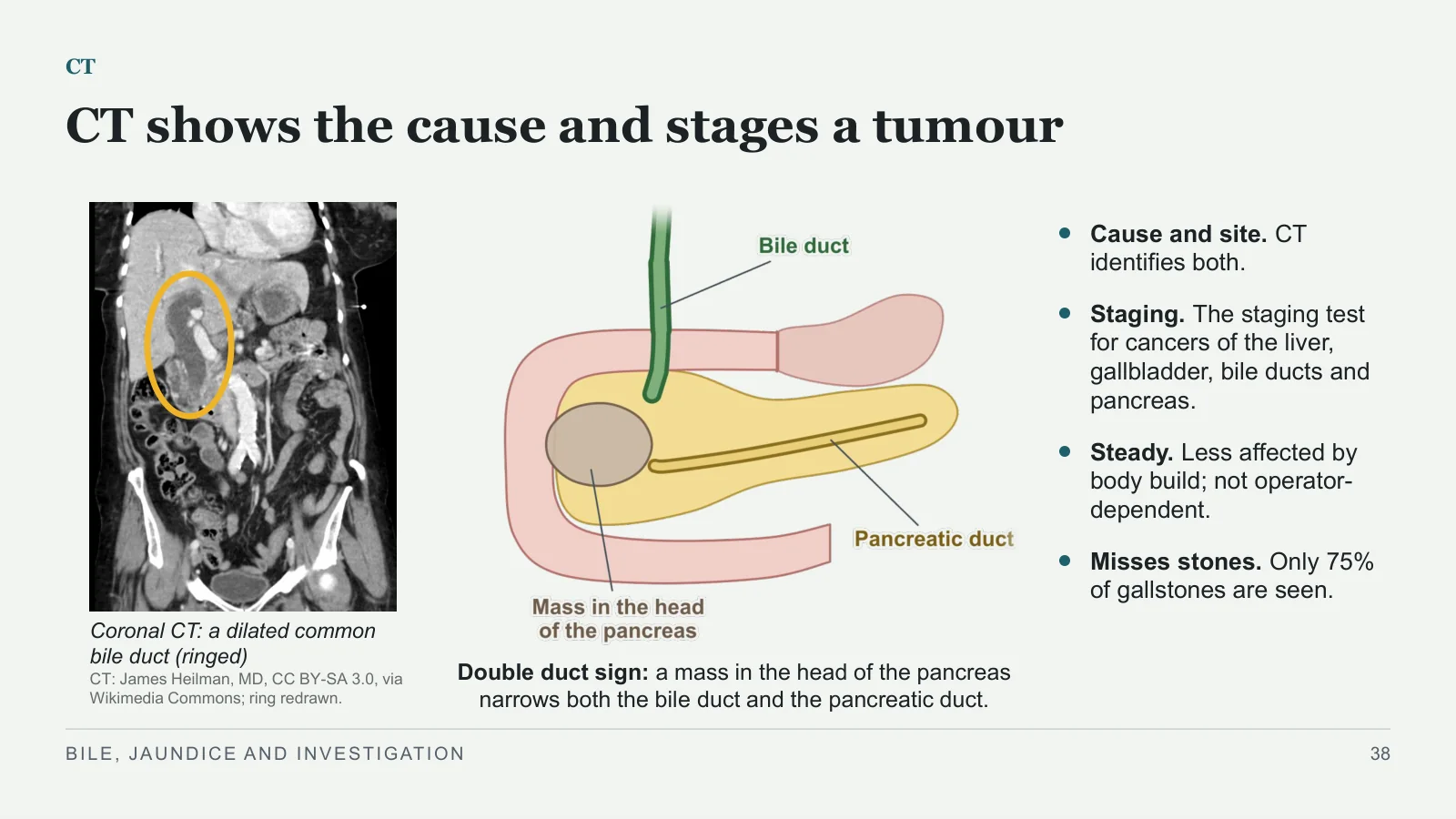
CT shows the cause and stages a tumour
CT
Coronal CT: a dilated common bile duct (ringed)
CT: James Heilman, MD, CC BY-SA 3.0, via Wikimedia Commons; ring redrawn.
Double duct sign: a mass in the head of the pancreas narrows both the bile duct and the pancreatic duct.
Cause and site. CT identifies both.
Staging. The staging test for cancers of the liver, gallbladder, bile ducts and pancreas.
Steady. Less affected by body build; not operator-dependent.
Misses stones. Only 75% of gallstones are seen.
BILE, JAUNDICE AND INVESTIGATION
38
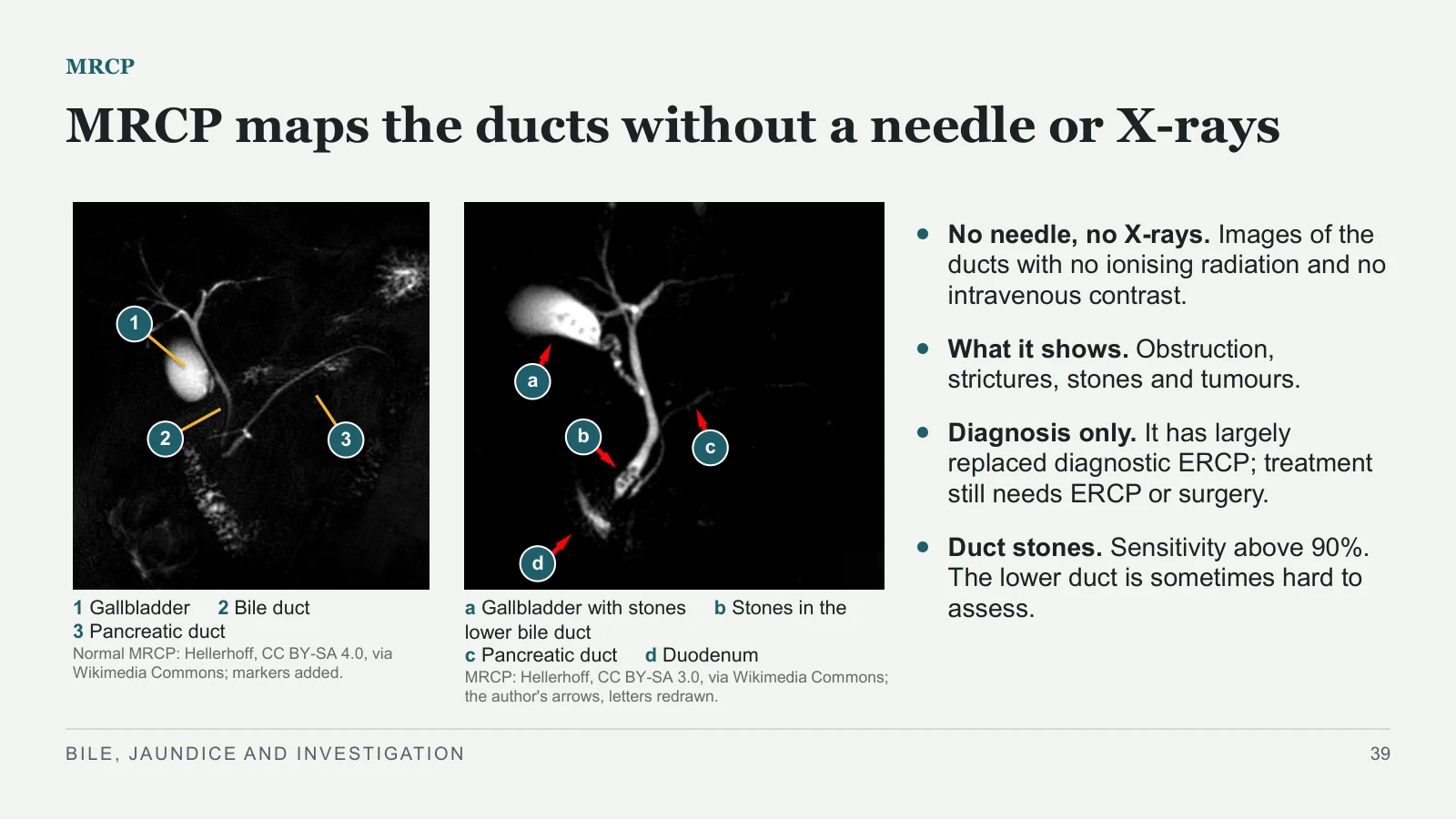
MRCP maps the ducts without a needle or X-rays
MRCP
1
2
3
1 Gallbladder 2 Bile duct
3 Pancreatic duct
Normal MRCP: Hellerhoff, CC BY-SA 4.0, via Wikimedia Commons; markers added.
a
b
c
d
a Gallbladder with stones b Stones in the lower bile duct
c Pancreatic duct d Duodenum
MRCP: Hellerhoff, CC BY-SA 3.0, via Wikimedia Commons; the author's arrows, letters redrawn.
No needle, no X-rays. Images of the ducts with no ionising radiation and no intravenous contrast.
What it shows. Obstruction, strictures, stones and tumours.
Diagnosis only. It has largely replaced diagnostic ERCP; treatment still needs ERCP or surgery.
Duct stones. Sensitivity above 90%. The lower duct is sometimes hard to assess.
BILE, JAUNDICE AND INVESTIGATION
39
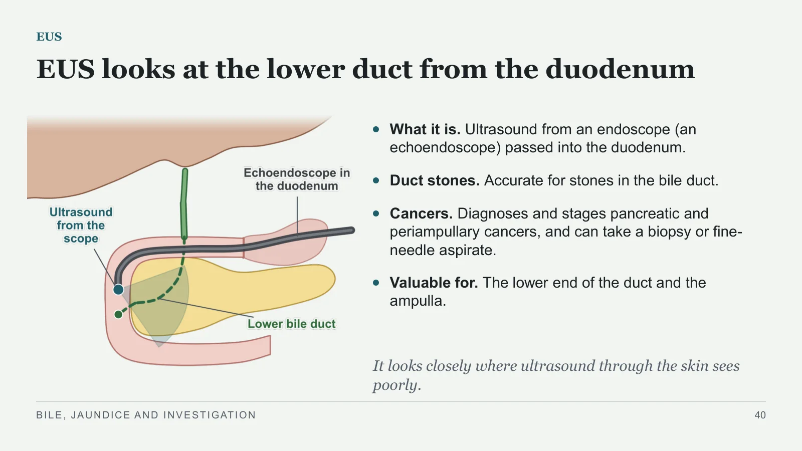
EUS looks at the lower duct from the duodenum
EUS
What it is. Ultrasound from an endoscope (an echoendoscope) passed into the duodenum.
Duct stones. Accurate for stones in the bile duct.
Cancers. Diagnoses and stages pancreatic and periampullary cancers, and can take a biopsy or fine-needle aspirate.
Valuable for. The lower end of the duct and the ampulla.
It looks closely where ultrasound through the skin sees poorly.
BILE, JAUNDICE AND INVESTIGATION
40
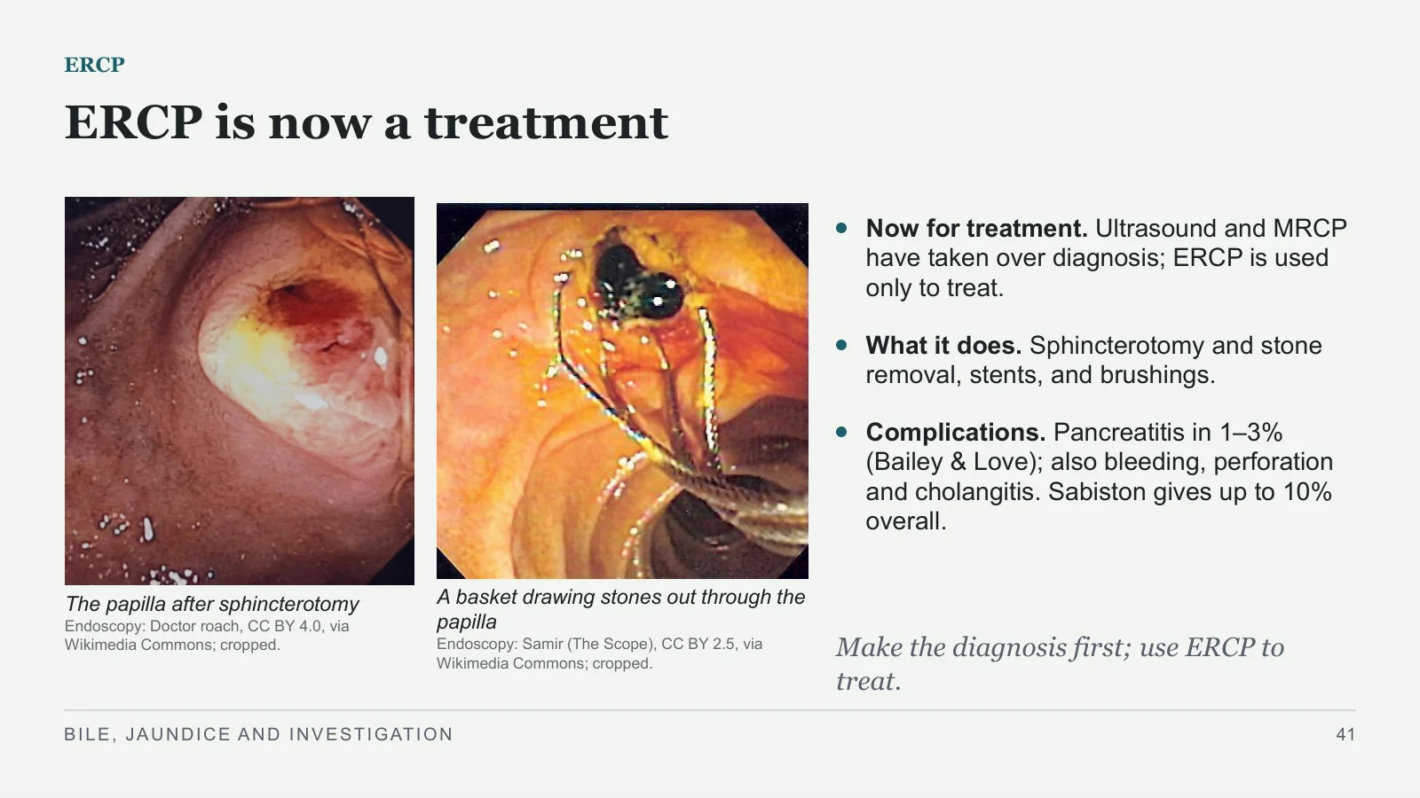
ERCP is now a treatment
ERCP
The papilla after sphincterotomy
Endoscopy: Doctor roach, CC BY 4.0, via Wikimedia Commons; cropped.
A basket drawing stones out through the papilla
Endoscopy: Samir (The Scope), CC BY 2.5, via Wikimedia Commons; cropped.
Now for treatment. Ultrasound and MRCP have taken over diagnosis; ERCP is used only to treat.
What it does. Sphincterotomy and stone removal, stents, and brushings.
Complications. Pancreatitis in 1–3% (Bailey & Love); also bleeding, perforation and cholangitis. Sabiston gives up to 10% overall.
Make the diagnosis first; use ERCP to treat.
BILE, JAUNDICE AND INVESTIGATION
41
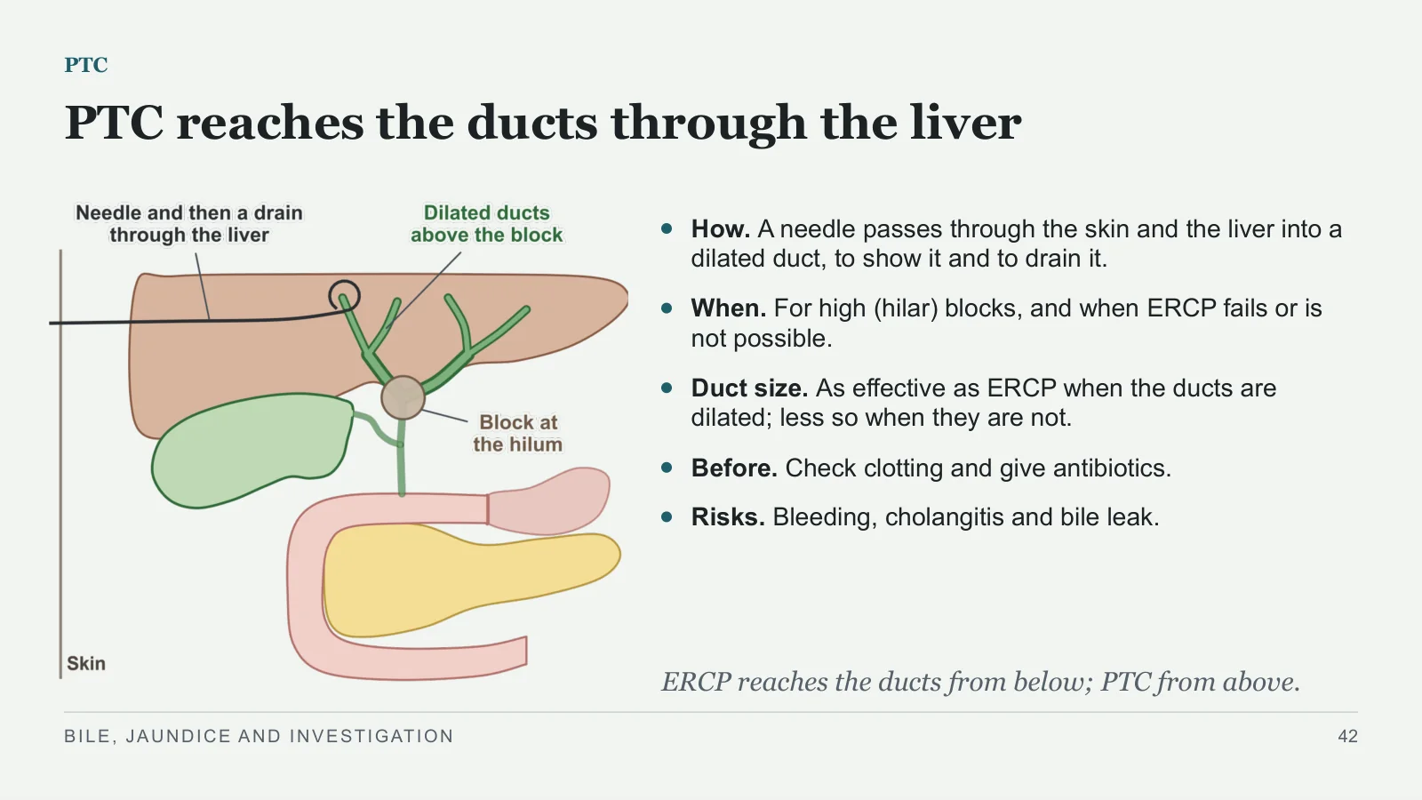
PTC reaches the ducts through the liver
PTC
How. A needle passes through the skin and the liver into a dilated duct, to show it and to drain it.
When. For high (hilar) blocks, and when ERCP fails or is not possible.
Duct size. As effective as ERCP when the ducts are dilated; less so when they are not.
Before. Check clotting and give antibiotics.
Risks. Bleeding, cholangitis and bile leak.
ERCP reaches the ducts from below; PTC from above.
BILE, JAUNDICE AND INVESTIGATION
42
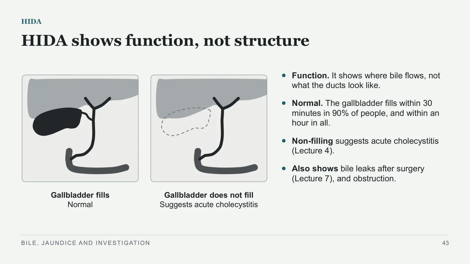
HIDA shows function, not structure
HIDA
Gallbladder fills
Normal
Gallbladder does not fill
Suggests acute cholecystitis
Function. It shows where bile flows, not what the ducts look like.
Normal. The gallbladder fills within 30 minutes in 90% of people, and within an hour in all.
Non-filling suggests acute cholecystitis (Lecture 4).
Also shows bile leaks after surgery (Lecture 7), and obstruction.
BILE, JAUNDICE AND INVESTIGATION
43
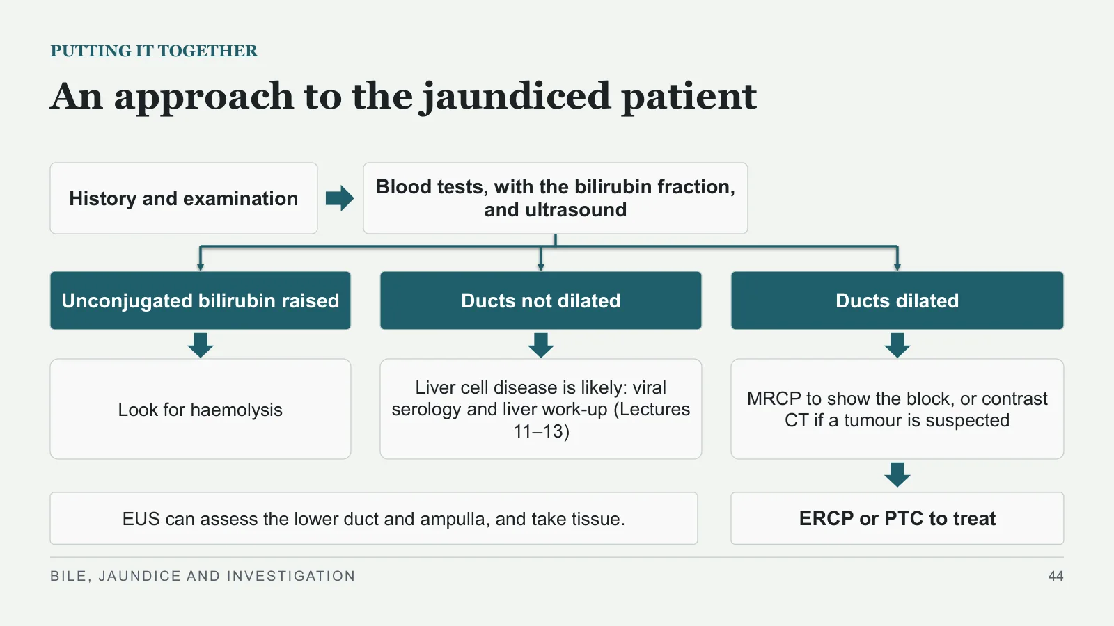
An approach to the jaundiced patient
PUTTING IT TOGETHER
History and examination
Blood tests, with the bilirubin fraction, and ultrasound
Unconjugated bilirubin raised
Ducts not dilated
Ducts dilated
Look for haemolysis
Liver cell disease is likely: viral serology and liver work-up (Lectures 11–13)
MRCP to show the block, or contrast CT if a tumour is suspected
ERCP or PTC to treat
EUS can assess the lower duct and ampulla, and take tissue.
BILE, JAUNDICE AND INVESTIGATION
44
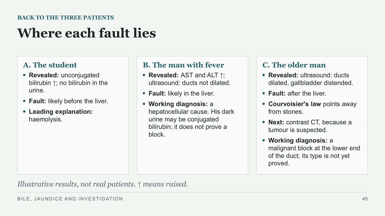
Where each fault lies
BACK TO THE THREE PATIENTS
A. The student
Revealed: unconjugated bilirubin ↑; no bilirubin in the urine.
Fault: likely before the liver.
Leading explanation: haemolysis.
B. The man with fever
Revealed: AST and ALT ↑; ultrasound: ducts not dilated.
Fault: likely in the liver.
Working diagnosis: a hepatocellular cause. His dark urine may be conjugated bilirubin; it does not prove a block.
C. The older man
Revealed: ultrasound: ducts dilated, gallbladder distended.
Fault: after the liver.
Courvoisier's law points away from stones.
Next: contrast CT, because a tumour is suspected.
Working diagnosis: a malignant block at the lower end of the duct; its type is not yet proved.
Illustrative results, not real patients. ↑ means raised.
BILE, JAUNDICE AND INVESTIGATION
45
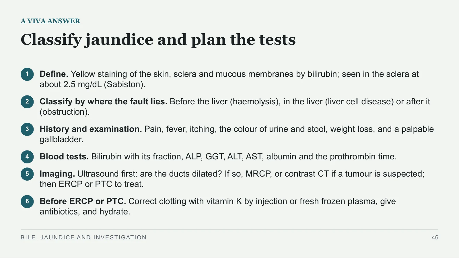
Classify jaundice and plan the tests
A VIVA ANSWER
1
Define. Yellow staining of the skin, sclera and mucous membranes by bilirubin; seen in the sclera at about 2.5 mg/dL (Sabiston).
2
Classify by where the fault lies. Before the liver (haemolysis), in the liver (liver cell disease) or after it (obstruction).
3
History and examination. Pain, fever, itching, the colour of urine and stool, weight loss, and a palpable gallbladder.
4
Blood tests. Bilirubin with its fraction, ALP, GGT, ALT, AST, albumin and the prothrombin time.
5
Imaging. Ultrasound first: are the ducts dilated? If so, MRCP, or contrast CT if a tumour is suspected; then ERCP or PTC to treat.
6
Before ERCP or PTC. Correct clotting with vitamin K by injection or fresh frozen plasma, give antibiotics, and hydrate.
BILE, JAUNDICE AND INVESTIGATION
46
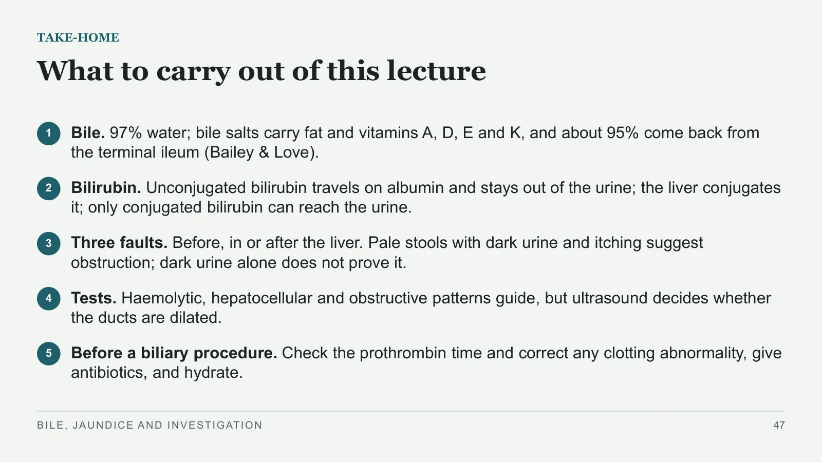
What to carry out of this lecture
TAKE-HOME
1
Bile. 97% water; bile salts carry fat and vitamins A, D, E and K, and about 95% come back from the terminal ileum (Bailey & Love).
2
Bilirubin. Unconjugated bilirubin travels on albumin and stays out of the urine; the liver conjugates it; only conjugated bilirubin can reach the urine.
3
Three faults. Before, in or after the liver. Pale stools with dark urine and itching suggest obstruction; dark urine alone does not prove it.
4
Tests. Haemolytic, hepatocellular and obstructive patterns guide, but ultrasound decides whether the ducts are dilated.
5
Before a biliary procedure. Check the prothrombin time and correct any clotting abnormality, give antibiotics, and hydrate.
BILE, JAUNDICE AND INVESTIGATION
47
References
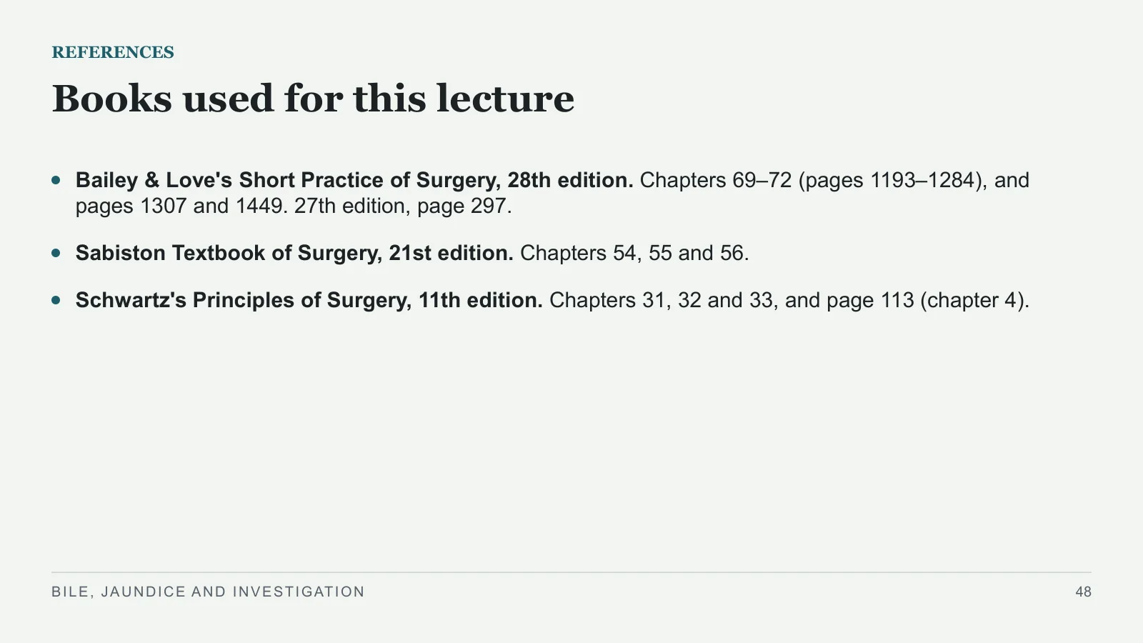
Books used for this lecture
REFERENCES
Bailey & Love's Short Practice of Surgery, 28th edition. Chapters 69–72 (pages 1193–1284), and pages 1307 and 1449. 27th edition, page 297.
Sabiston Textbook of Surgery, 21st edition. Chapters 54, 55 and 56.
Schwartz's Principles of Surgery, 11th edition. Chapters 31, 32 and 33, and page 113 (chapter 4).
BILE, JAUNDICE AND INVESTIGATION
48
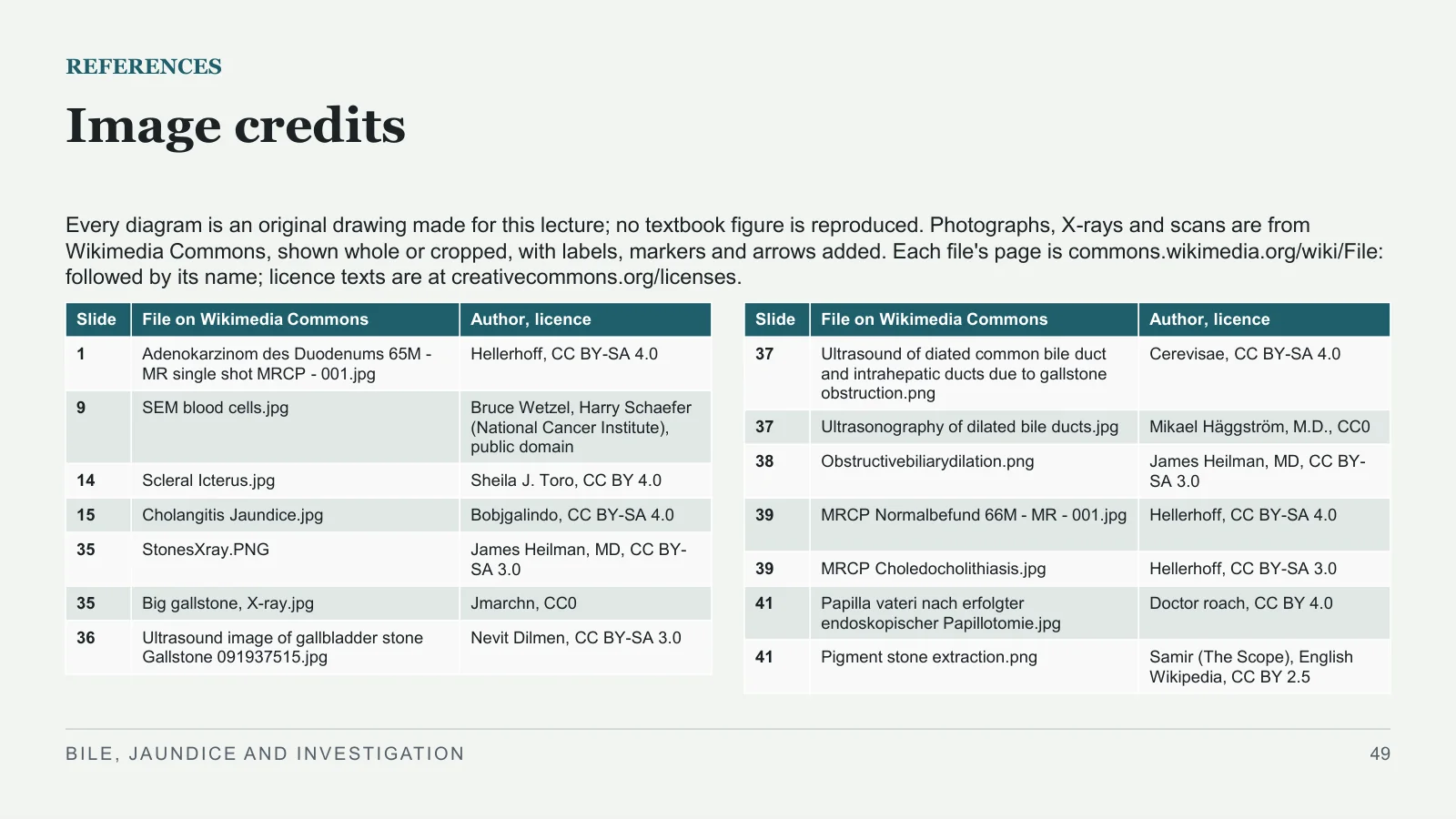
Image credits
REFERENCES
Every diagram is an original drawing made for this lecture; no textbook figure is reproduced. Photographs, X-rays and scans are from Wikimedia Commons, shown whole or cropped, with labels, markers and arrows added. Each file's page is commons.wikimedia.org/wiki/File: followed by its name; licence texts are at creativecommons.org/licenses.
BILE, JAUNDICE AND INVESTIGATION
49
| Slide | File on Wikimedia Commons | Author, licence |
| 1 | Adenokarzinom des Duodenums 65M - MR single shot MRCP - 001.jpg | Hellerhoff, CC BY-SA 4.0 |
| 9 | SEM blood cells.jpg | Bruce Wetzel, Harry Schaefer (National Cancer Institute), public domain |
| 14 | Scleral Icterus.jpg | Sheila J. Toro, CC BY 4.0 |
| 15 | Cholangitis Jaundice.jpg | Bobjgalindo, CC BY-SA 4.0 |
| 35 | StonesXray.PNG | James Heilman, MD, CC BY-SA 3.0 |
| 35 | Big gallstone, X-ray.jpg | Jmarchn, CC0 |
| 36 | Ultrasound image of gallbladder stone Gallstone 091937515.jpg | Nevit Dilmen, CC BY-SA 3.0 |
| Slide | File on Wikimedia Commons | Author, licence |
| 37 | Ultrasound of diated common bile duct and intrahepatic ducts due to gallstone obstruction.png | Cerevisae, CC BY-SA 4.0 |
| 37 | Ultrasonography of dilated bile ducts.jpg | Mikael Häggström, M.D., CC0 |
| 38 | Obstructivebiliarydilation.png | James Heilman, MD, CC BY-SA 3.0 |
| 39 | MRCP Normalbefund 66M - MR - 001.jpg | Hellerhoff, CC BY-SA 4.0 |
| 39 | MRCP Choledocholithiasis.jpg | Hellerhoff, CC BY-SA 3.0 |
| 41 | Papilla vateri nach erfolgter endoskopischer Papillotomie.jpg | Doctor roach, CC BY 4.0 |
| 41 | Pigment stone extraction.png | Samir (The Scope), English Wikipedia, CC BY 2.5 |
Sources and image credits
Original lecture diagrams and source/image credits are preserved from the PowerPoint. Each photograph or scan below links to its source and applicable licence. Changes are described in the slide captions; adapted ShareAlike images retain that licence.
Images
- Slide 1: Adenokarzinom des Duodenums 65M - MR single shot MRCP - 001.jpg · Hellerhoff, CC BY-SA 4.0. Slide caption: MRCP: Hellerhoff, CC BY-SA 4.0, via Wikimedia Commons. Adaptations of this image retain the same Creative Commons ShareAlike licence.
- Licence — Adenokarzinom des Duodenums 65M - MR single shot MRCP - 001.jpg · CC BY-SA 4.0
- Slide 9: SEM blood cells.jpg · Bruce Wetzel, Harry Schaefer (National Cancer Institute), public domain. Slide caption: Micrograph: Bruce Wetzel, Harry Schaefer (National Cancer Institute), public domain, via Wikimedia Commons.
- Slide 14: Scleral Icterus.jpg · Sheila J. Toro, CC BY 4.0. Slide caption: Photograph: Sheila J. Toro, CC BY 4.0, via Wikimedia Commons; cropped to the eyes.
- Licence — Scleral Icterus.jpg · CC BY 4.0
- Slide 15: Cholangitis Jaundice.jpg · Bobjgalindo, CC BY-SA 4.0. Slide caption: Photo: Bobjgalindo, CC BY-SA 4.0, via Wikimedia Commons; cropped to one eye. Adaptations of this image retain the same Creative Commons ShareAlike licence.
- Licence — Cholangitis Jaundice.jpg · CC BY-SA 4.0
- Slide 35: StonesXray.PNG · James Heilman, MD, CC BY-SA 3.0. Slide caption: X-ray: James Heilman, MD, CC BY-SA 3.0, via Wikimedia Commons; cropped, ring redrawn. X-ray: Jmarchn, CC0, via Wikimedia Commons; arrow added. Adaptations of this image retain the same Creative Commons ShareAlike licence.
- Licence — StonesXray.PNG · CC BY-SA 3.0
- Slide 35: Big gallstone, X-ray.jpg · Jmarchn, CC0. Slide caption: X-ray: James Heilman, MD, CC BY-SA 3.0, via Wikimedia Commons; cropped, ring redrawn. X-ray: Jmarchn, CC0, via Wikimedia Commons; arrow added.
- Licence — Big gallstone, X-ray.jpg · CC0
- Slide 36: Ultrasound image of gallbladder stone Gallstone 091937515.jpg · Nevit Dilmen, CC BY-SA 3.0. Source attribution: © Nevit Dilmen. Slide caption: Ultrasound: Nevit Dilmen, CC BY-SA 3.0, via Wikimedia Commons; markers added. Adaptations of this image retain the same Creative Commons ShareAlike licence.
- Licence — Ultrasound image of gallbladder stone Gallstone 091937515.jpg · CC BY-SA 3.0
- Slide 37: Ultrasound of diated common bile duct and intrahepatic ducts due to gallstone obstruction.png · Cerevisae, CC BY-SA 4.0. Slide caption: Ultrasound: Cerevisae, CC BY-SA 4.0, via Wikimedia Commons; cropped to one panel, markers added. Ultrasound: Mikael Häggström, M.D., CC0, via Wikimedia Commons. Adaptations of this image retain the same Creative Commons ShareAlike licence.
- Licence — Ultrasound of diated common bile duct and intrahepatic ducts due to gallstone obstruction.png · CC BY-SA 4.0
- Slide 37: Ultrasonography of dilated bile ducts.jpg · Mikael Häggström, M.D., CC0. Slide caption: Ultrasound: Cerevisae, CC BY-SA 4.0, via Wikimedia Commons; cropped to one panel, markers added. Ultrasound: Mikael Häggström, M.D., CC0, via Wikimedia Commons.
- Licence — Ultrasonography of dilated bile ducts.jpg · CC0
- Slide 38: Obstructivebiliarydilation.png · James Heilman, MD, CC BY-SA 3.0. Slide caption: CT: James Heilman, MD, CC BY-SA 3.0, via Wikimedia Commons; ring redrawn. Adaptations of this image retain the same Creative Commons ShareAlike licence.
- Licence — Obstructivebiliarydilation.png · CC BY-SA 3.0
- Slide 39: MRCP Normalbefund 66M - MR - 001.jpg · Hellerhoff, CC BY-SA 4.0. Slide caption: Normal MRCP: Hellerhoff, CC BY-SA 4.0, via Wikimedia Commons; markers added. MRCP: Hellerhoff, CC BY-SA 3.0, via Wikimedia Commons; the author's arrows, letters redrawn. Adaptations of this image retain the same Creative Commons ShareAlike licence.
- Licence — MRCP Normalbefund 66M - MR - 001.jpg · CC BY-SA 4.0
- Slide 39: MRCP Choledocholithiasis.jpg · Hellerhoff, CC BY-SA 3.0. Slide caption: Normal MRCP: Hellerhoff, CC BY-SA 4.0, via Wikimedia Commons; markers added. MRCP: Hellerhoff, CC BY-SA 3.0, via Wikimedia Commons; the author's arrows, letters redrawn. Adaptations of this image retain the same Creative Commons ShareAlike licence.
- Licence — MRCP Choledocholithiasis.jpg · CC BY-SA 3.0
- Slide 41: Papilla vateri nach erfolgter endoskopischer Papillotomie.jpg · Doctor roach, CC BY 4.0. Slide caption: Endoscopy: Doctor roach, CC BY 4.0, via Wikimedia Commons; cropped. Endoscopy: Samir (The Scope), CC BY 2.5, via Wikimedia Commons; cropped.
- Licence — Papilla vateri nach erfolgter endoskopischer Papillotomie.jpg · CC BY 4.0
- Slide 41: Pigment stone extraction.png · Samir (The Scope), English Wikipedia, CC BY 2.5. Source attribution: Samir (The Scope), English Wikipedia. Slide caption: Endoscopy: Doctor roach, CC BY 4.0, via Wikimedia Commons; cropped. Endoscopy: Samir (The Scope), CC BY 2.5, via Wikimedia Commons; cropped.
- Licence — Pigment stone extraction.png · CC BY 2.5


Sources
- [1] REFERENCES
- [2] Books used for this lecture
- [3] Bailey & Love's Short Practice of Surgery, 28th edition. Chapters 69–72 (pages 1193–1284), and pages 1307 and 1449. 27th edition, page 297.
- [4] Sabiston Textbook of Surgery, 21st edition. Chapters 54, 55 and 56.
- [5] Schwartz's Principles of Surgery, 11th edition. Chapters 31, 32 and 33, and page 113 (chapter 4).