Hepatobiliary surgery · Lecture 3
Gallstones · Chronic gallbladder disease · 47 slides
Gallstones and chronic gallbladder disease.
How stones form, why they hurt, and which ones need an operation: from biliary pain and chronic inflammation to Mirizzi syndrome and gallstone ileus.
About the downloads
PowerPoint without speaker notes. SHA-256 checksums verify the downloaded files.
Contents · 8 parts
Introduction
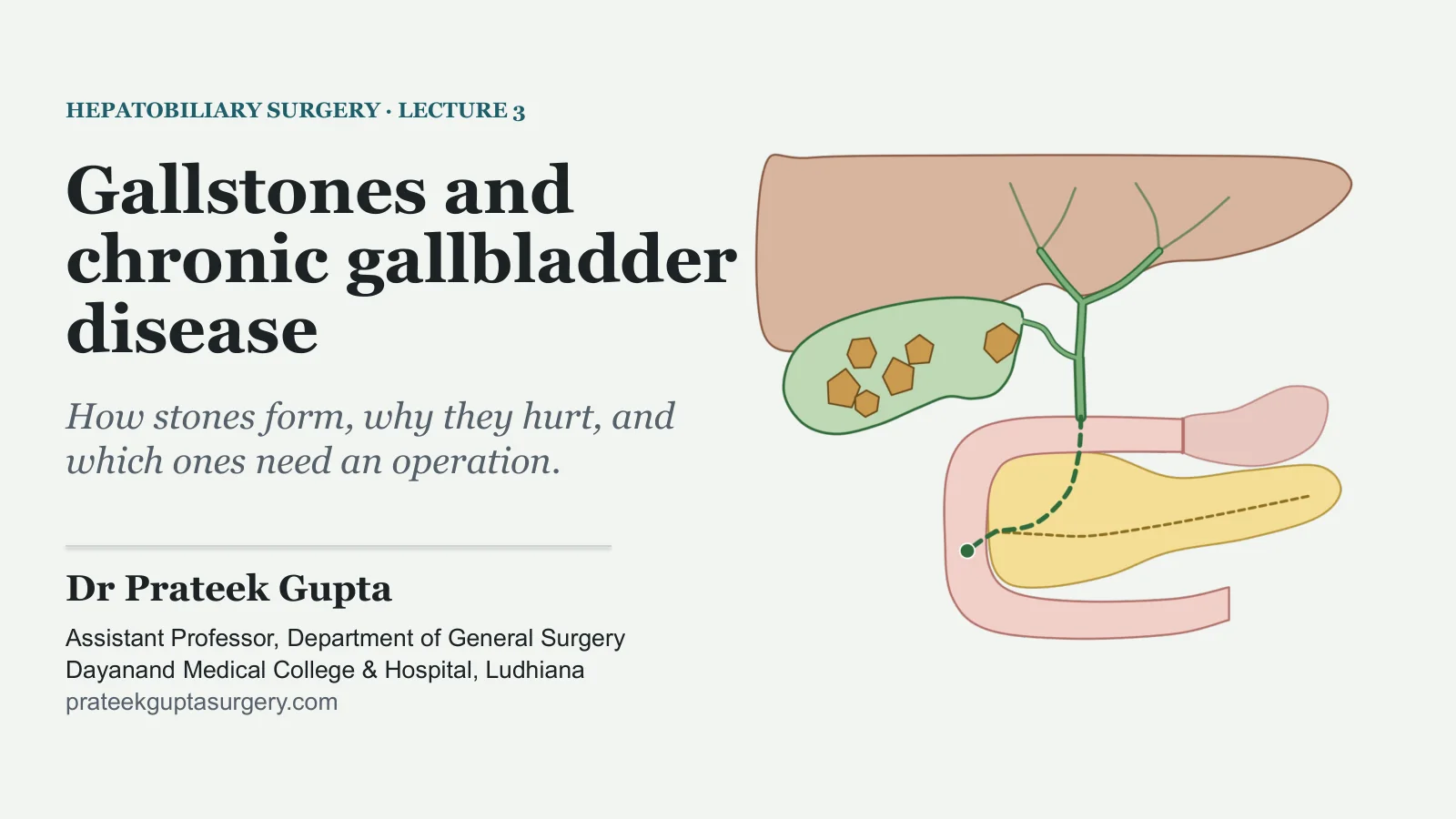
Gallstones and chronic gallbladder disease
HEPATOBILIARY SURGERY · LECTURE 3
Gallstones and chronic gallbladder disease
How stones form, why they hurt, and which ones need an operation.
Dr Prateek Gupta
Assistant Professor, Department of General Surgery Dayanand Medical College & Hospital, Ludhiana prateekguptasurgery.com
Two patients with gallstones
TWO PATIENTS IN THE SURGICAL OUTPATIENT CLINIC
A. A 42-year-old woman
Attacks of severe, steady pain in the upper abdomen, going through to the back. Each lasts a few hours, often wakes her at night, and comes with vomiting. She is well between attacks. Ultrasound shows gallstones.
B. A 78-year-old woman
Three days of colicky abdominal pain, vomiting and a distended abdomen.
Questions to keep in mind
1
Is A's pain biliary pain?
2
Does A need an operation? Her sister's stones were found by chance and have never hurt: does she?
3
How can a gallstone block B's bowel?
4
What will B's imaging show?
Illustrative cases, not real patients. We return to them at the end.
GALLSTONES AND CHRONIC GALLBLADDER DISEASE
2
Gallstone formation
Stones
PART 1
How common they are, the three kinds, how they form, and who gets them.
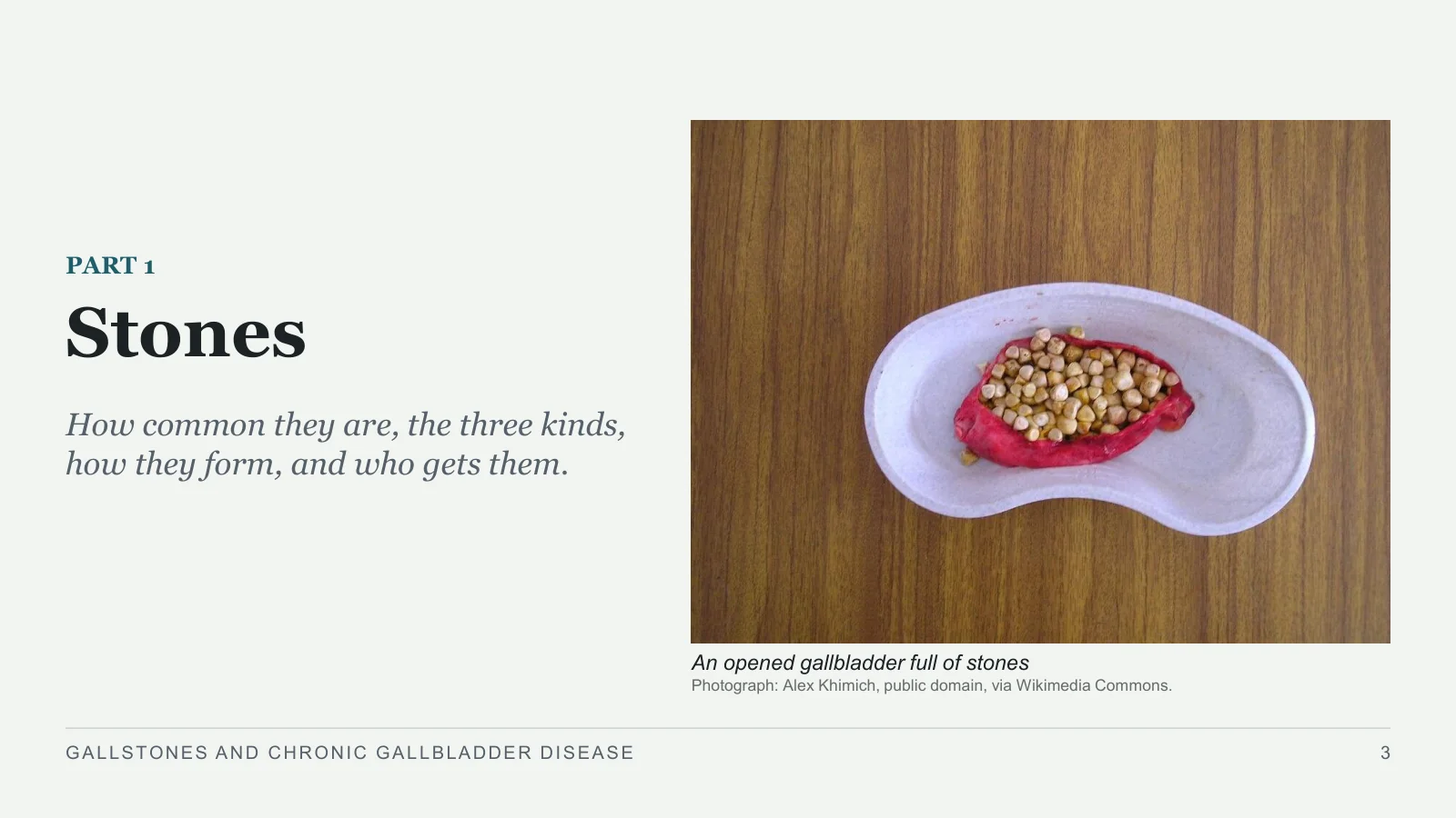
An opened gallbladder full of stones
Photograph: Alex Khimich, public domain, via Wikimedia Commons.
GALLSTONES AND CHRONIC GALLBLADDER DISEASE
3
10–15%
HOW COMMON
Gallstones are common
of the population in western societies have gallstones.
17%
of people in the UK have gallstones at the time of death.
More common in Native American and Latin American people.
India? None of the three books gives a figure.
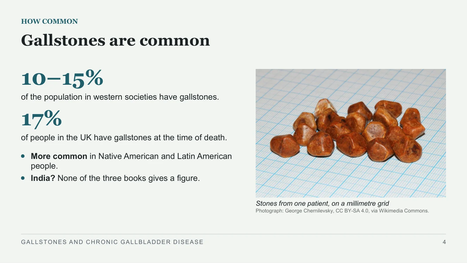
Stones from one patient, on a millimetre grid
Photograph: George Chernilevsky, CC BY-SA 4.0, via Wikimedia Commons.
GALLSTONES AND CHRONIC GALLBLADDER DISEASE
4
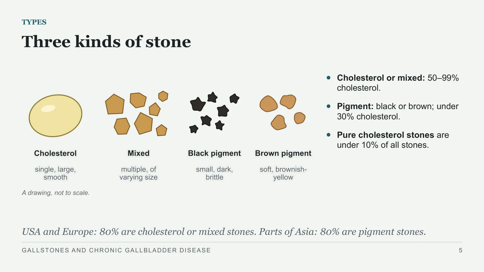
Three kinds of stone
TYPES
A drawing, not to scale.
Cholesterol or mixed: 50–99% cholesterol.
Pigment: black or brown; under 30% cholesterol.
Pure cholesterol stones are under 10% of all stones.
USA and Europe: 80% are cholesterol or mixed stones. Parts of Asia: 80% are pigment stones.
GALLSTONES AND CHRONIC GALLBLADDER DISEASE
5
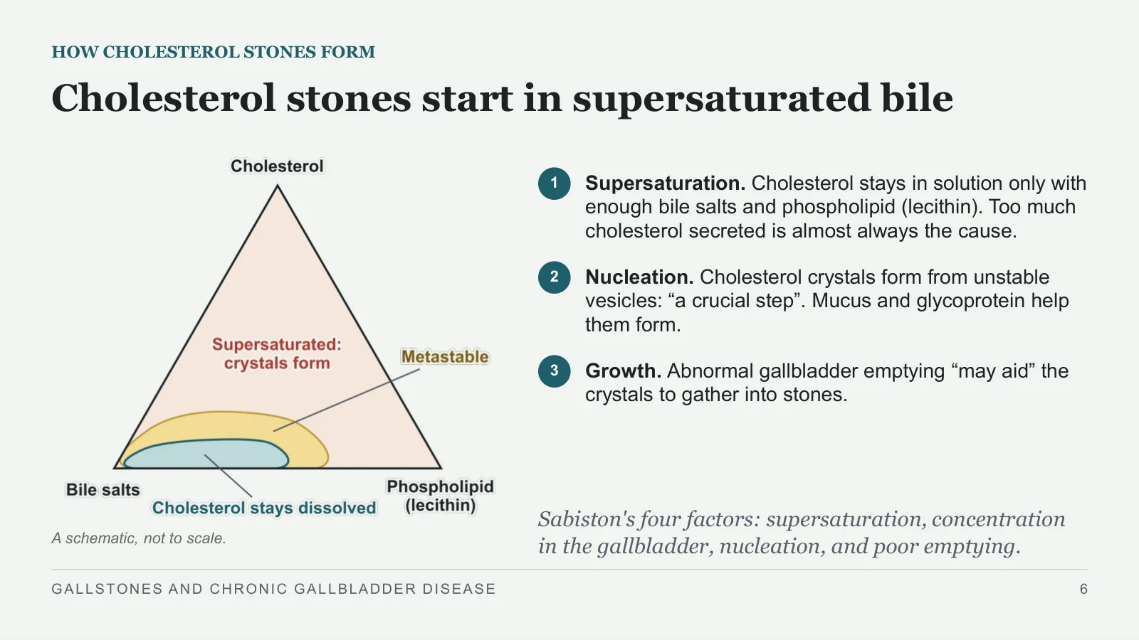
Cholesterol stones start in supersaturated bile
HOW CHOLESTEROL STONES FORM
A schematic, not to scale.
1
Supersaturation. Cholesterol stays in solution only with enough bile salts and phospholipid (lecithin). Too much cholesterol secreted is almost always the cause.
2
Nucleation. Cholesterol crystals form from unstable vesicles: “a crucial step”. Mucus and glycoprotein help them form.
3
Growth. Abnormal gallbladder emptying “may aid” the crystals to gather into stones.
Sabiston's four factors: supersaturation, concentration in the gallbladder, nucleation, and poor emptying.
GALLSTONES AND CHRONIC GALLBLADDER DISEASE
6
Black and brown pigment stones differ
PIGMENT STONES
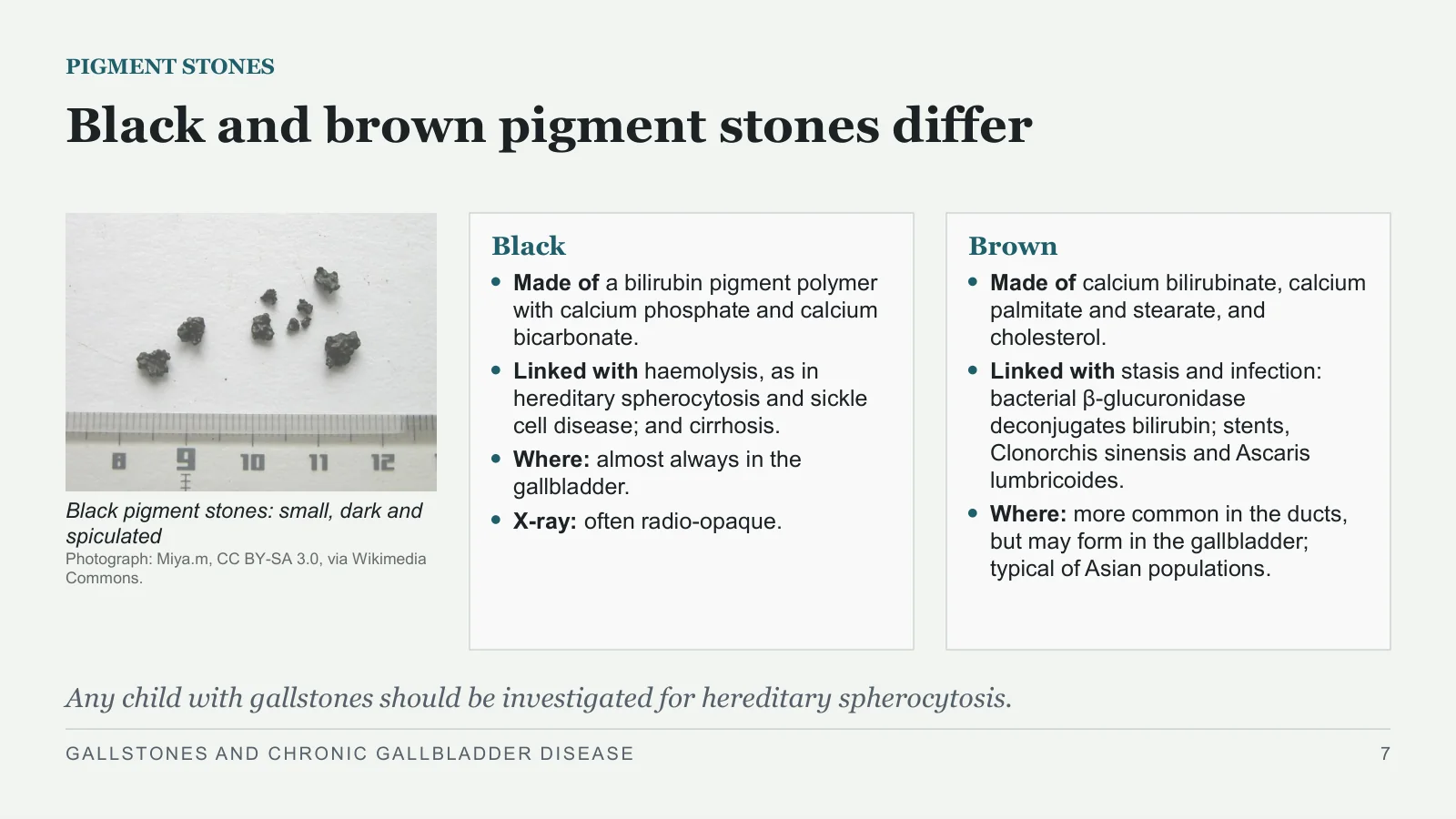
Black pigment stones: small, dark and spiculated
Photograph: Miya.m, CC BY-SA 3.0, via Wikimedia Commons.
Black
Made of a bilirubin pigment polymer with calcium phosphate and calcium bicarbonate.
Linked with haemolysis, as in hereditary spherocytosis and sickle cell disease; and cirrhosis.
Where: almost always in the gallbladder.
X-ray: often radio-opaque.
Brown
Made of calcium bilirubinate, calcium palmitate and stearate, and cholesterol.
Linked with stasis and infection: bacterial β-glucuronidase deconjugates bilirubin; stents, Clonorchis sinensis and Ascaris lumbricoides.
Where: more common in the ducts, but may form in the gallbladder; typical of Asian populations.
Any child with gallstones should be investigated for hereditary spherocytosis.
GALLSTONES AND CHRONIC GALLBLADDER DISEASE
7
Risk factors for gallstones
WHO GETS THEM
Who
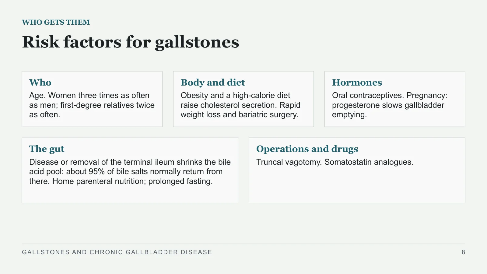
Age. Women three times as often as men; first-degree relatives twice as often.
Body and diet
Obesity and a high-calorie diet raise cholesterol secretion. Rapid weight loss and bariatric surgery.
Hormones
Oral contraceptives. Pregnancy: progesterone slows gallbladder emptying.
The gut
Disease or removal of the terminal ileum shrinks the bile acid pool: about 95% of bile salts normally return from there. Home parenteral nutrition; prolonged fasting.
Operations and drugs
Truncal vagotomy. Somatostatin analogues.
GALLSTONES AND CHRONIC GALLBLADDER DISEASE
8
Biliary pain and investigation
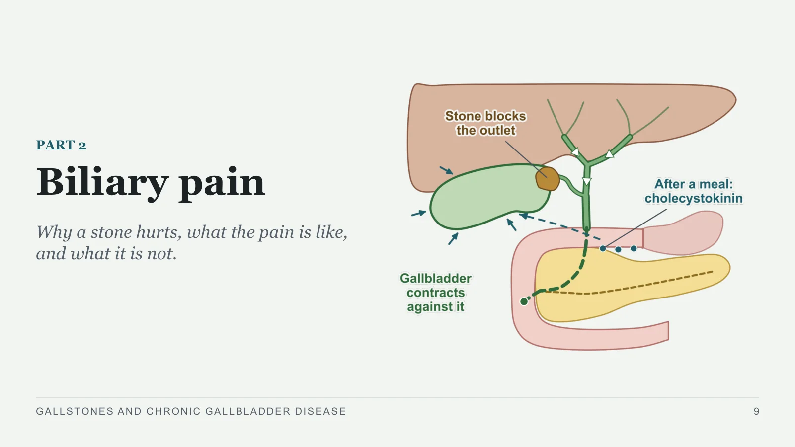
Biliary pain
PART 2
Why a stone hurts, what the pain is like, and what it is not.
GALLSTONES AND CHRONIC GALLBLADDER DISEASE
9
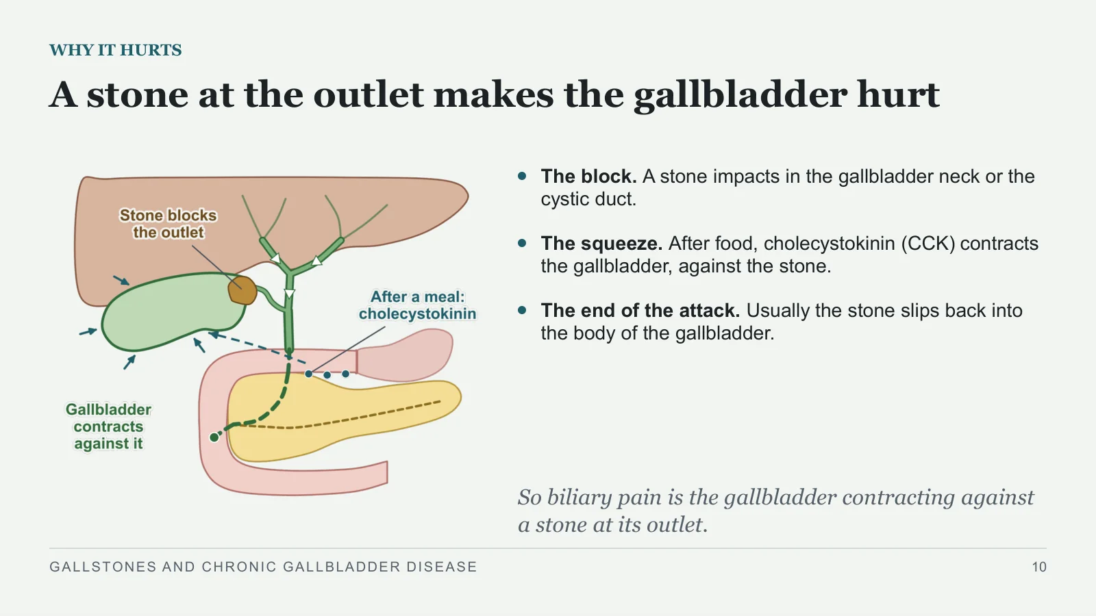
A stone at the outlet makes the gallbladder hurt
WHY IT HURTS
The block. A stone impacts in the gallbladder neck or the cystic duct.
The squeeze. After food, cholecystokinin (CCK) contracts the gallbladder, against the stone.
The end of the attack. Usually the stone slips back into the body of the gallbladder.
So biliary pain is the gallbladder contracting against a stone at its outlet.
GALLSTONES AND CHRONIC GALLBLADDER DISEASE
10
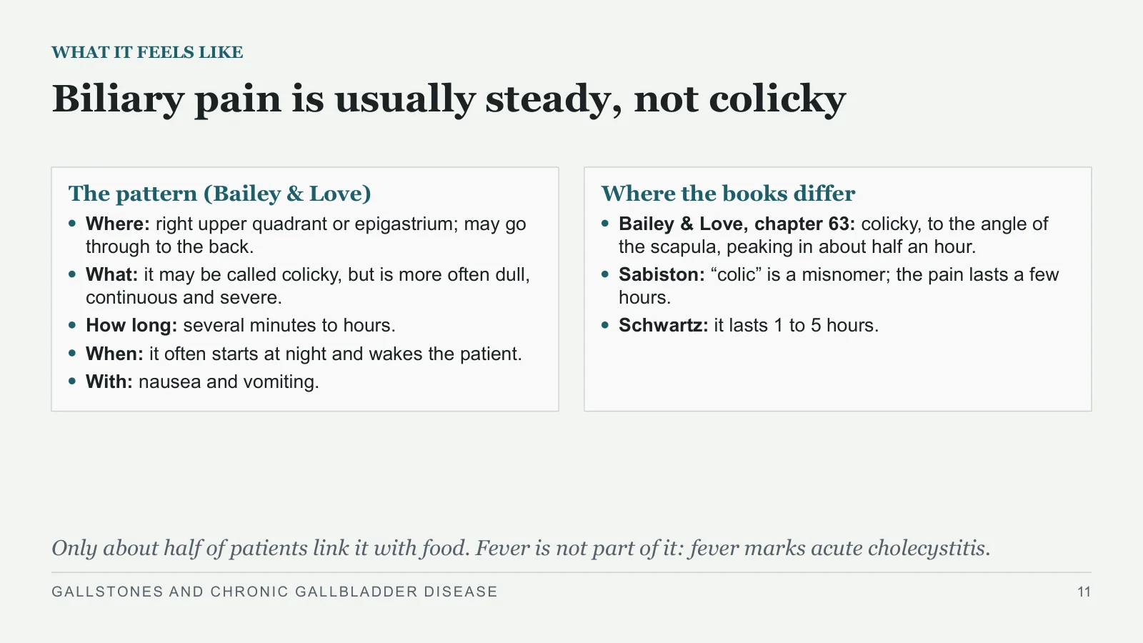
Biliary pain is usually steady, not colicky
WHAT IT FEELS LIKE
The pattern (Bailey & Love)
Where: right upper quadrant or epigastrium; may go through to the back.
What: it may be called colicky, but is more often dull, continuous and severe.
How long: several minutes to hours.
When: it often starts at night and wakes the patient.
With: nausea and vomiting.
Where the books differ
Bailey & Love, chapter 63: colicky, to the angle of the scapula, peaking in about half an hour.
Sabiston: “colic” is a misnomer; the pain lasts a few hours.
Schwartz: it lasts 1 to 5 hours.
Only about half of patients link it with food. Fever is not part of it: fever marks acute cholecystitis.
GALLSTONES AND CHRONIC GALLBLADDER DISEASE
11
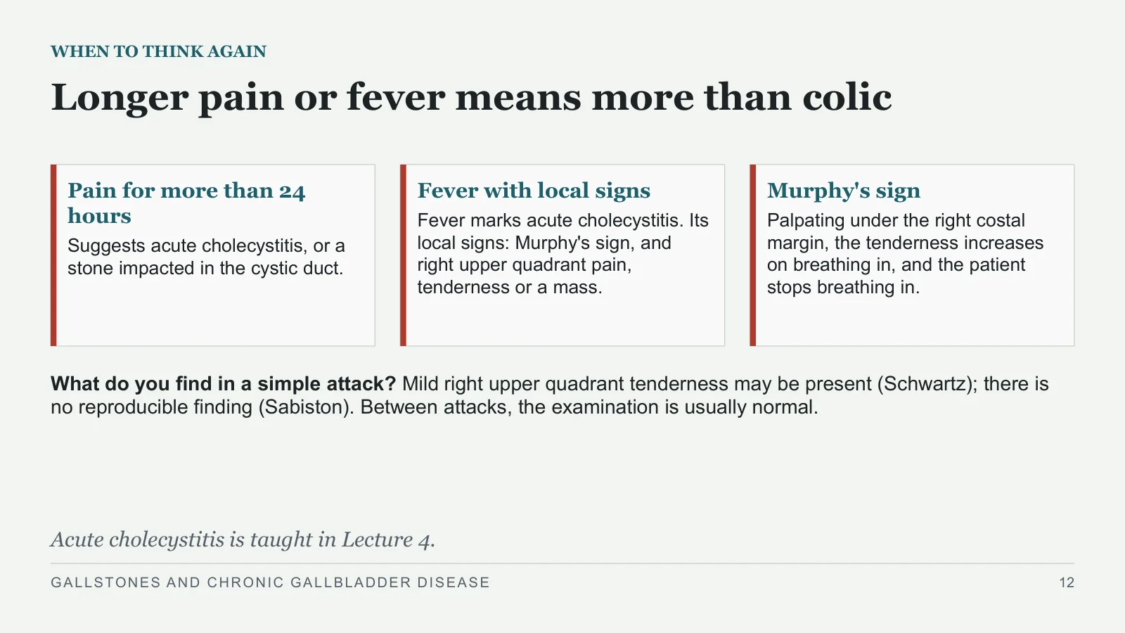
Longer pain or fever means more than colic
WHEN TO THINK AGAIN
Pain for more than 24 hours
Suggests acute cholecystitis, or a stone impacted in the cystic duct.
Fever with local signs
Fever marks acute cholecystitis. Its local signs: Murphy's sign, and right upper quadrant pain, tenderness or a mass.
Murphy's sign
Palpating under the right costal margin, the tenderness increases on breathing in, and the patient stops breathing in.
What do you find in a simple attack? Mild right upper quadrant tenderness may be present (Schwartz); there is no reproducible finding (Sabiston). Between attacks, the examination is usually normal.
Acute cholecystitis is taught in Lecture 4.
GALLSTONES AND CHRONIC GALLBLADDER DISEASE
12
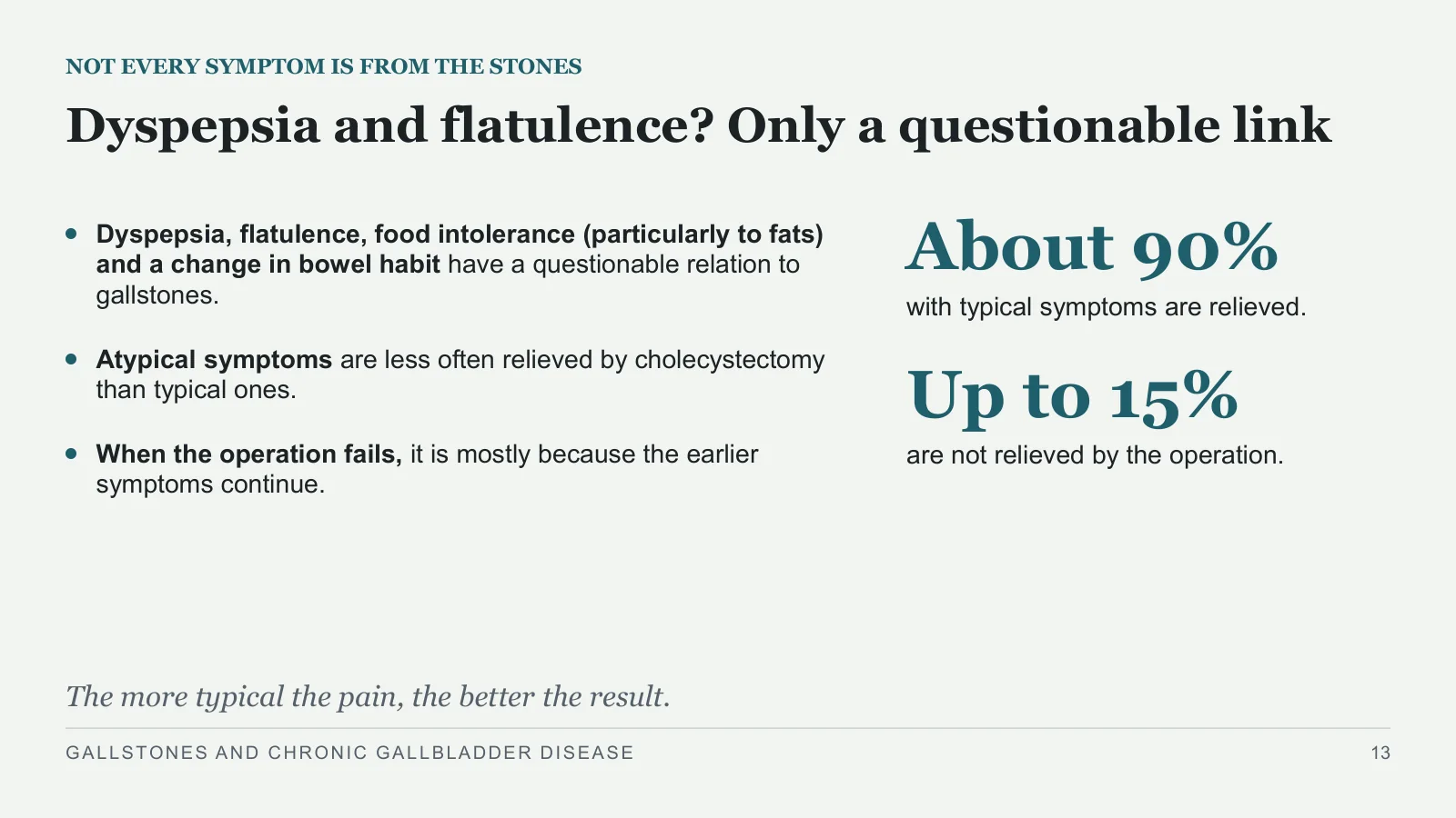
About 90%
NOT EVERY SYMPTOM IS FROM THE STONES
Dyspepsia and flatulence? Only a questionable link
Dyspepsia, flatulence, food intolerance (particularly to fats) and a change in bowel habit have a questionable relation to gallstones.
Atypical symptoms are less often relieved by cholecystectomy than typical ones.
When the operation fails, it is mostly because the earlier symptoms continue.
with typical symptoms are relieved.
Up to 15%
are not relieved by the operation.
The more typical the pain, the better the result.
GALLSTONES AND CHRONIC GALLBLADDER DISEASE
13
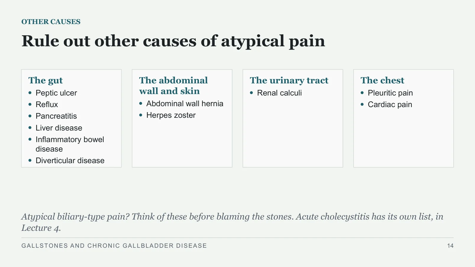
Rule out other causes of atypical pain
OTHER CAUSES
The gut
Peptic ulcer
Reflux
Pancreatitis
Liver disease
Inflammatory bowel disease
Diverticular disease
The abdominal wall and skin
Abdominal wall hernia
Herpes zoster
The urinary tract
Renal calculi
The chest
Pleuritic pain
Cardiac pain
Atypical biliary-type pain? Think of these before blaming the stones. Acute cholecystitis has its own list, in Lecture 4.
GALLSTONES AND CHRONIC GALLBLADDER DISEASE
14
Ultrasound confirms the stones
INVESTIGATION
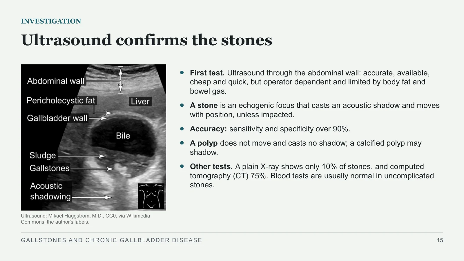
Ultrasound: Mikael Häggström, M.D., CC0, via Wikimedia Commons; the author's labels.
First test. Ultrasound through the abdominal wall: accurate, available, cheap and quick, but operator dependent and limited by body fat and bowel gas.
A stone is an echogenic focus that casts an acoustic shadow and moves with position, unless impacted.
Accuracy: sensitivity and specificity over 90%.
A polyp does not move and casts no shadow; a calcified polyp may shadow.
Other tests. A plain X-ray shows only 10% of stones, and computed tomography (CT) 75%. Blood tests are usually normal in uncomplicated stones.
GALLSTONES AND CHRONIC GALLBLADDER DISEASE
15
Sludge with typical attacks fits biliary colic
SLUDGE
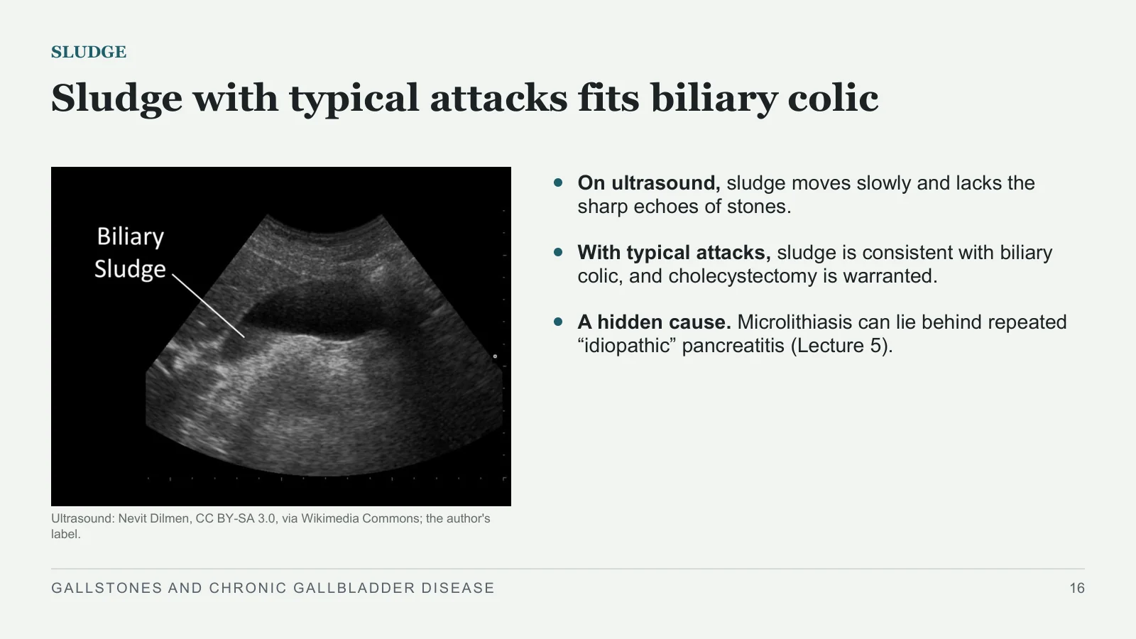
Ultrasound: Nevit Dilmen, CC BY-SA 3.0, via Wikimedia Commons; the author's label.
On ultrasound, sludge moves slowly and lacks the sharp echoes of stones.
With typical attacks, sludge is consistent with biliary colic, and cholecystectomy is warranted.
A hidden cause. Microlithiasis can lie behind repeated “idiopathic” pancreatitis (Lecture 5).
GALLSTONES AND CHRONIC GALLBLADDER DISEASE
16
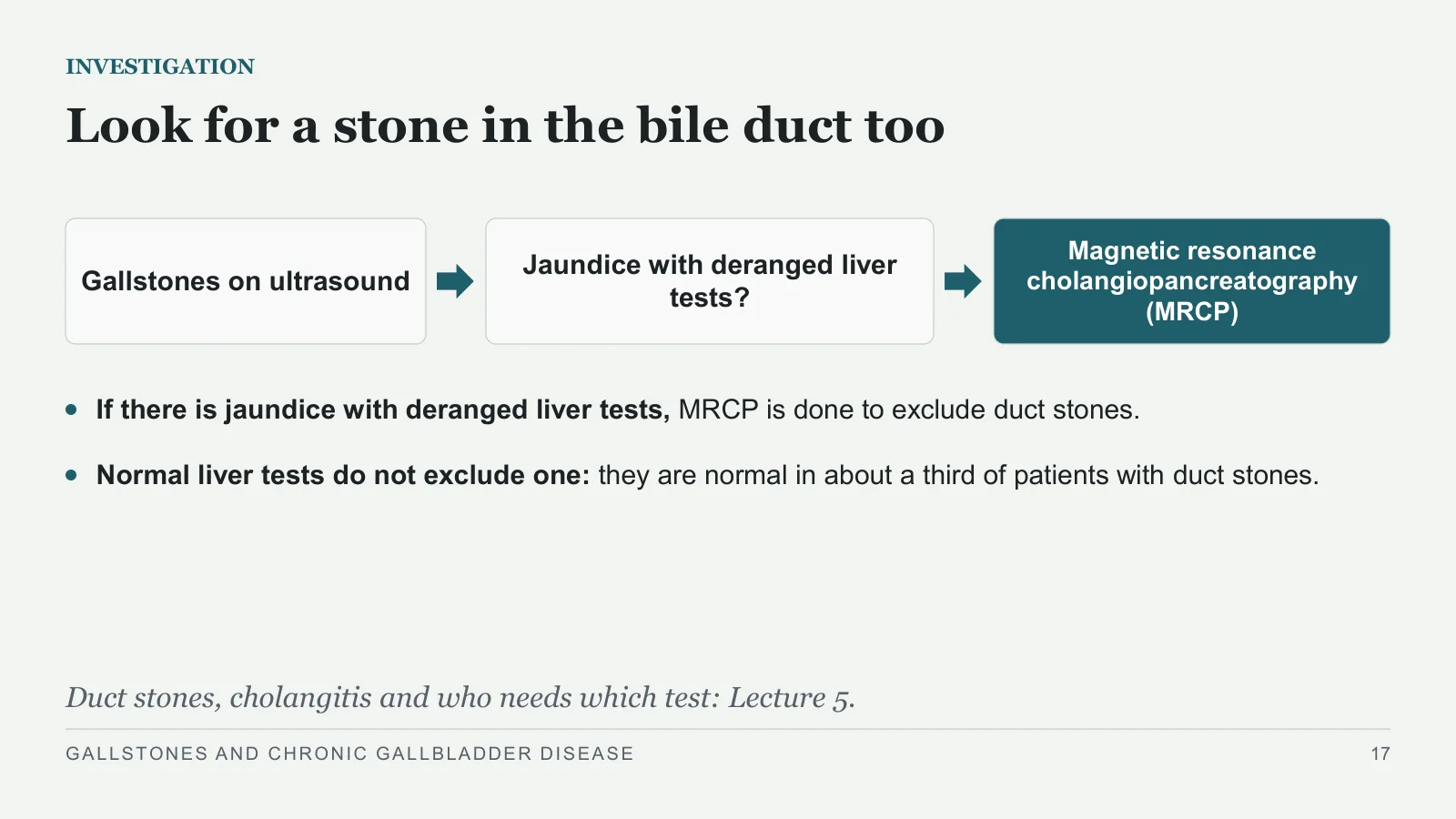
Look for a stone in the bile duct too
INVESTIGATION
Gallstones on ultrasound
Jaundice with deranged liver tests?
Magnetic resonance cholangiopancreatography (MRCP)
If there is jaundice with deranged liver tests, MRCP is done to exclude duct stones.
Normal liver tests do not exclude one: they are normal in about a third of patients with duct stones.
Duct stones, cholangitis and who needs which test: Lecture 5.
GALLSTONES AND CHRONIC GALLBLADDER DISEASE
17
Natural history and treatment
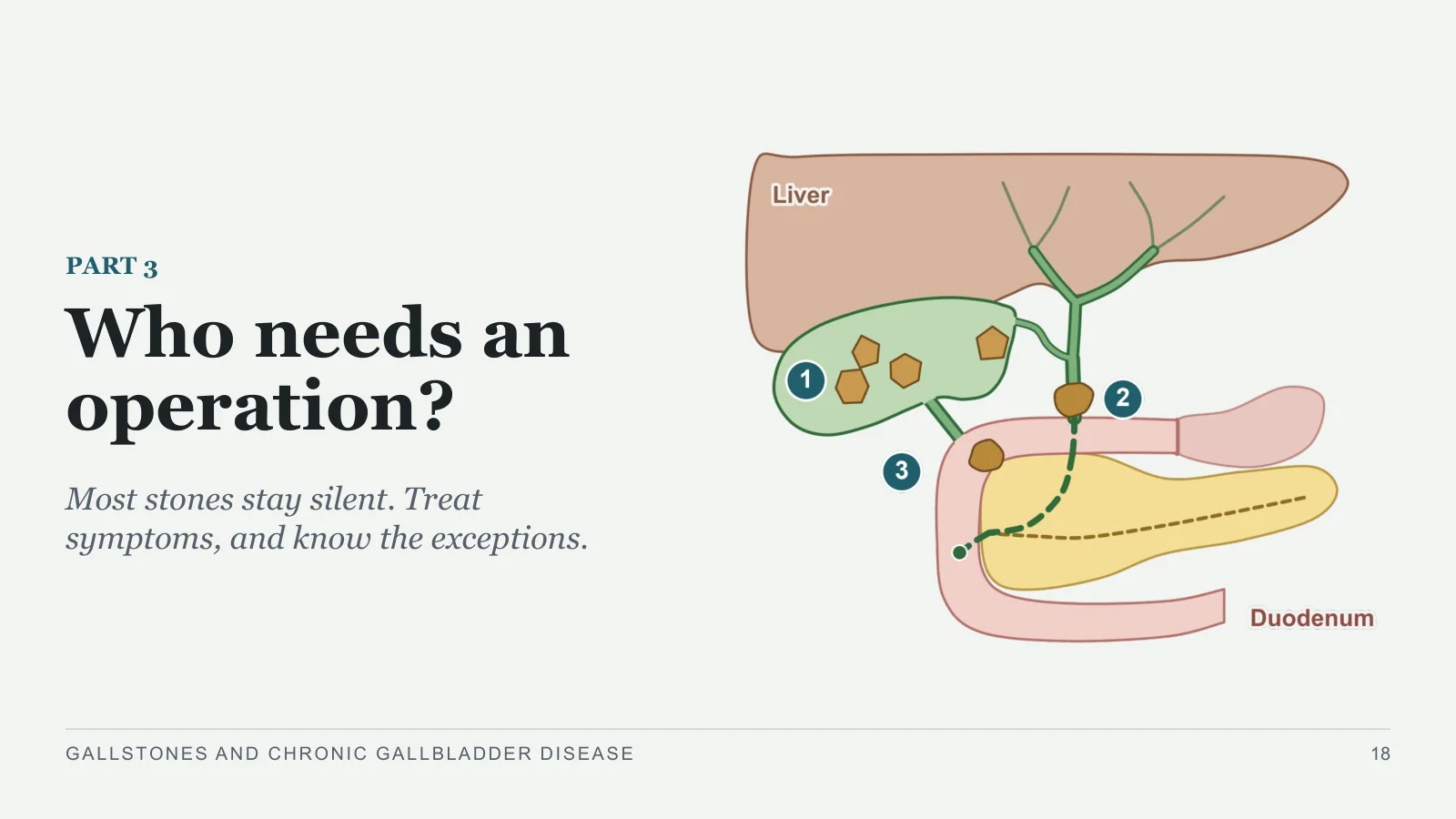
Who needs an operation?
PART 3
Most stones stay silent. Treat symptoms, and know the exceptions.
GALLSTONES AND CHRONIC GALLBLADDER DISEASE
18
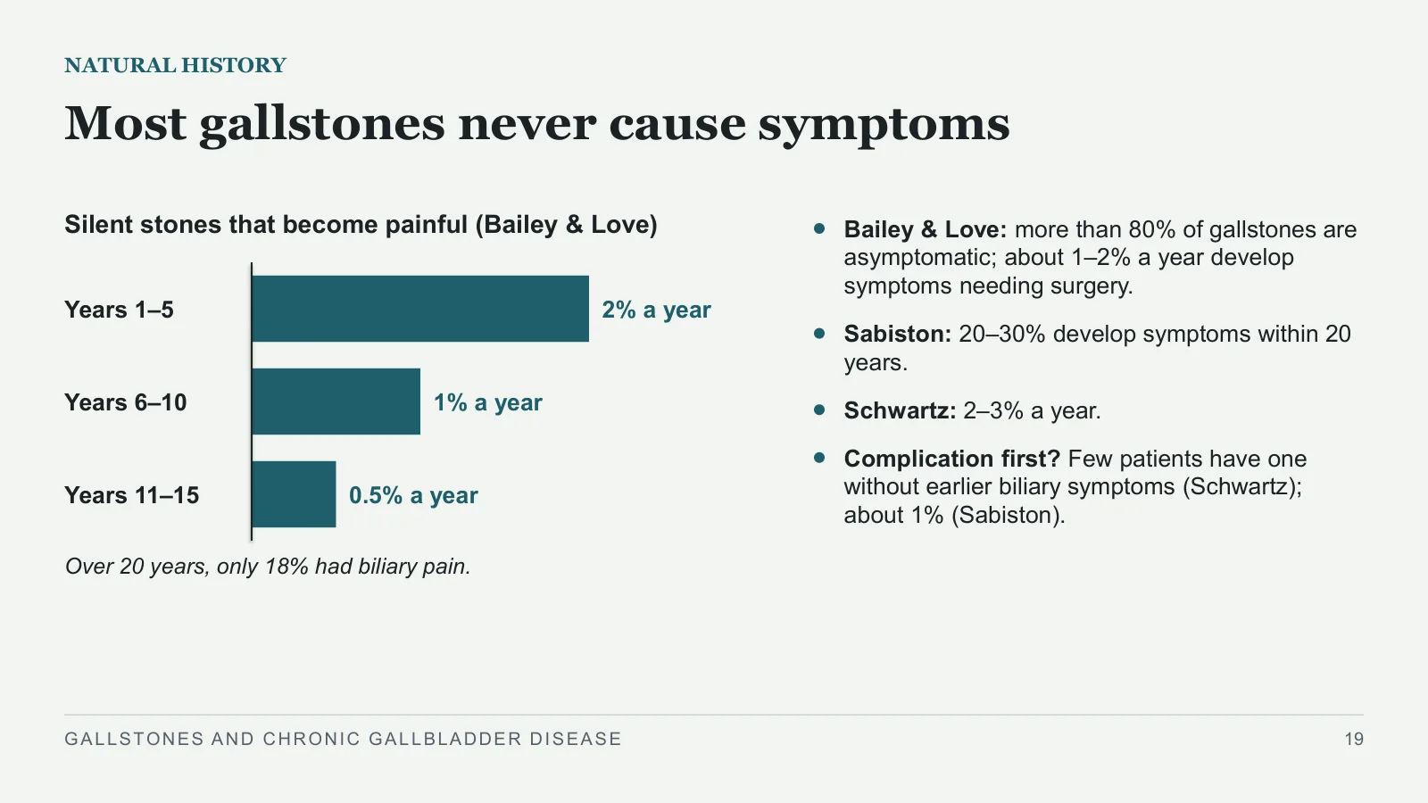
Most gallstones never cause symptoms
NATURAL HISTORY
Silent stones that become painful (Bailey & Love)
Years 1–5
2% a year
Years 6–10
1% a year
Years 11–15
0.5% a year
Over 20 years, only 18% had biliary pain.
Bailey & Love: more than 80% of gallstones are asymptomatic; about 1–2% a year develop symptoms needing surgery.
Sabiston: 20–30% develop symptoms within 20 years.
Schwartz: 2–3% a year.
Complication first? Few patients have one without earlier biliary symptoms (Schwartz); about 1% (Sabiston).
GALLSTONES AND CHRONIC GALLBLADDER DISEASE
19
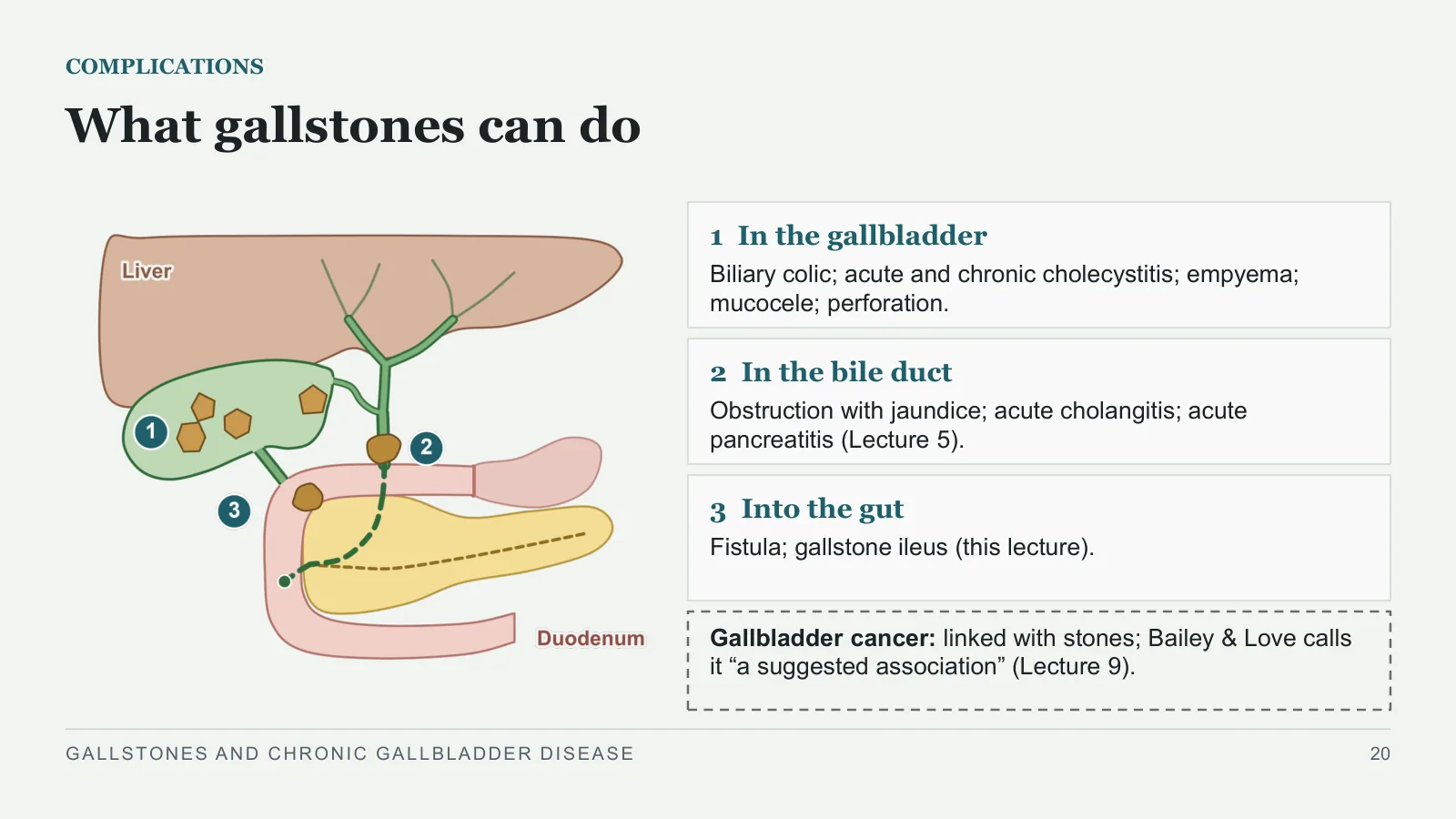
What gallstones can do
COMPLICATIONS
1 In the gallbladder
Biliary colic; acute and chronic cholecystitis; empyema; mucocele; perforation.
2 In the bile duct
Obstruction with jaundice; acute cholangitis; acute pancreatitis (Lecture 5).
3 Into the gut
Fistula; gallstone ileus (this lecture).
Gallbladder cancer: linked with stones; Bailey & Love calls it “a suggested association” (Lecture 9).
GALLSTONES AND CHRONIC GALLBLADDER DISEASE
20
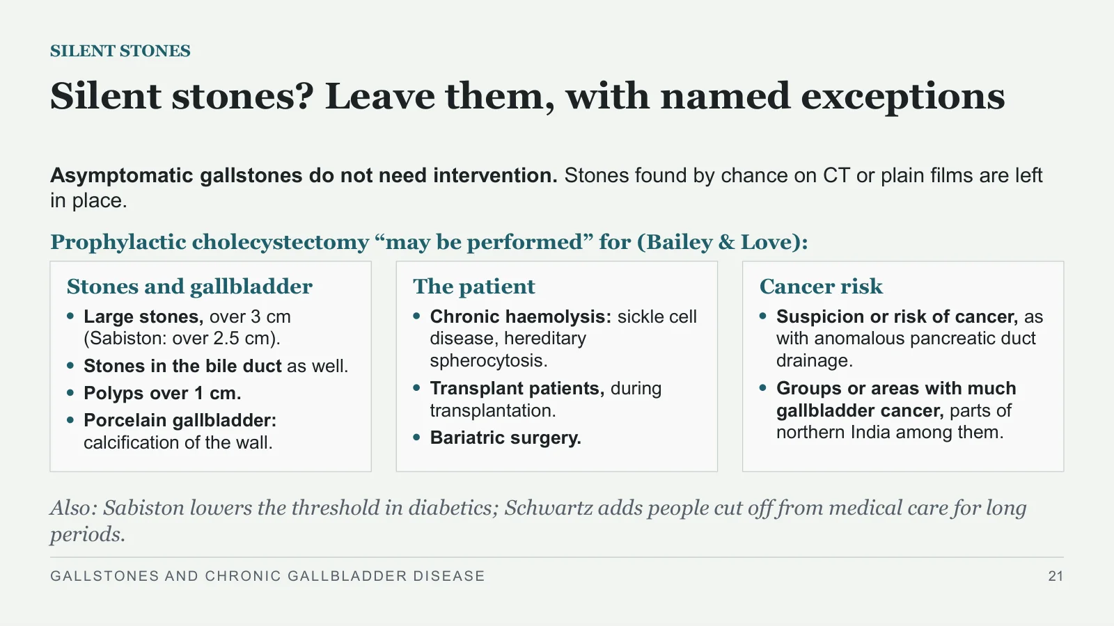
Silent stones? Leave them, with named exceptions
SILENT STONES
Asymptomatic gallstones do not need intervention. Stones found by chance on CT or plain films are left in place.
Prophylactic cholecystectomy “may be performed” for (Bailey & Love):
Stones and gallbladder
Large stones, over 3 cm (Sabiston: over 2.5 cm).
Stones in the bile duct as well.
Polyps over 1 cm.
Porcelain gallbladder: calcification of the wall.
The patient
Chronic haemolysis: sickle cell disease, hereditary spherocytosis.
Transplant patients, during transplantation.
Bariatric surgery.
Cancer risk
Suspicion or risk of cancer, as with anomalous pancreatic duct drainage.
Groups or areas with much gallbladder cancer, parts of northern India among them.
Also: Sabiston lowers the threshold in diabetics; Schwartz adds people cut off from medical care for long periods.
GALLSTONES AND CHRONIC GALLBLADDER DISEASE
21
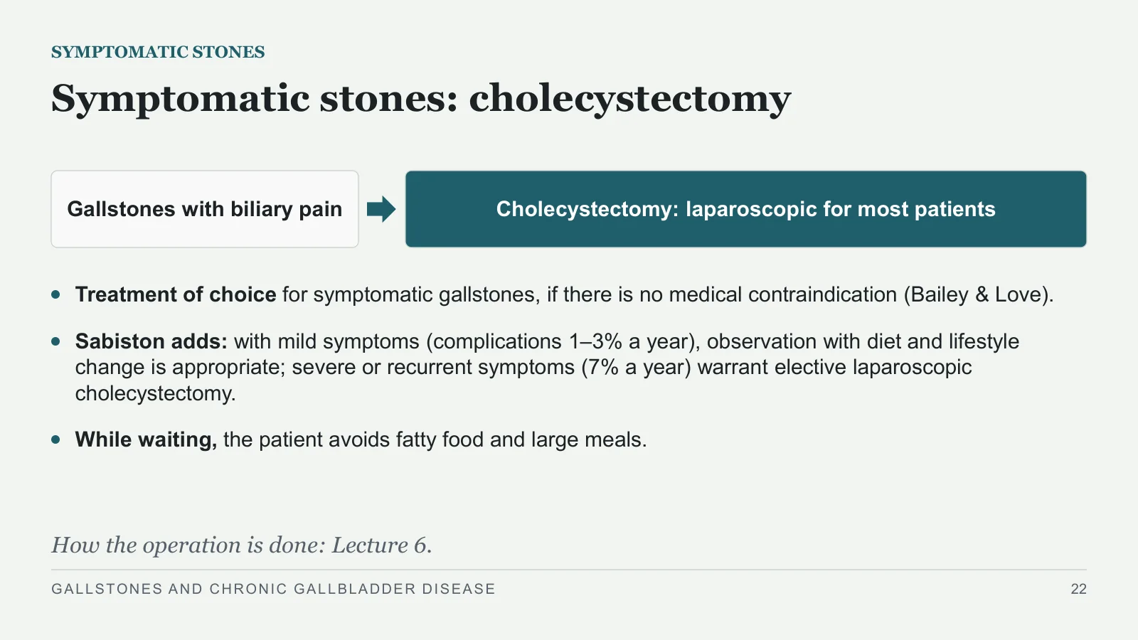
Symptomatic stones: cholecystectomy
SYMPTOMATIC STONES
Gallstones with biliary pain
Cholecystectomy: laparoscopic for most patients
Treatment of choice for symptomatic gallstones, if there is no medical contraindication (Bailey & Love).
Sabiston adds: with mild symptoms (complications 1–3% a year), observation with diet and lifestyle change is appropriate; severe or recurrent symptoms (7% a year) warrant elective laparoscopic cholecystectomy.
While waiting, the patient avoids fatty food and large meals.
How the operation is done: Lecture 6.
GALLSTONES AND CHRONIC GALLBLADDER DISEASE
22
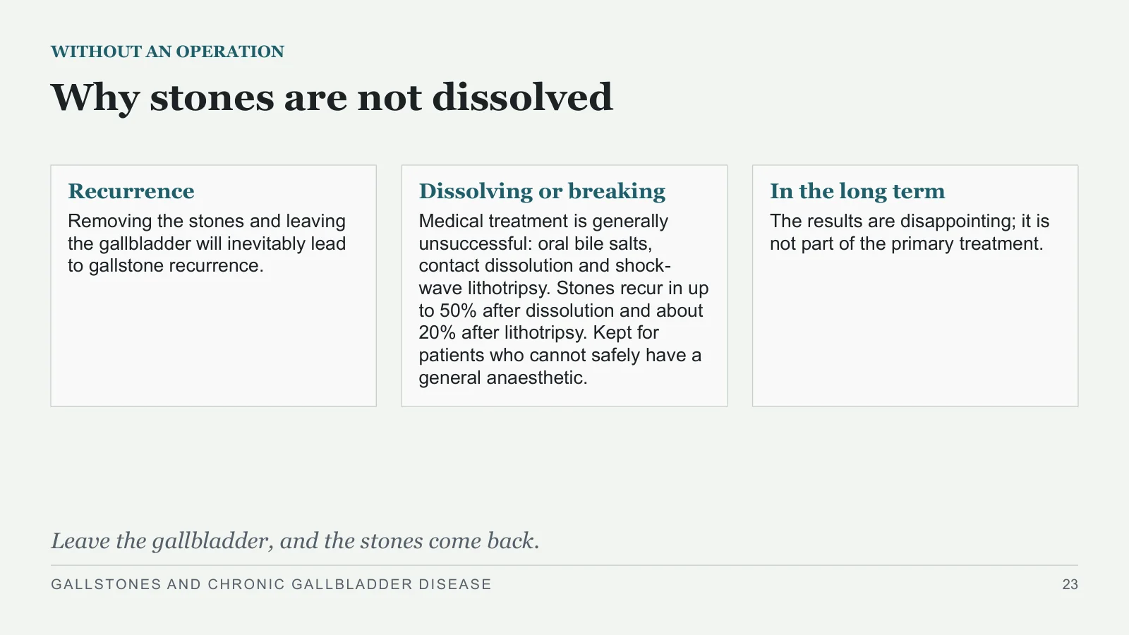
Why stones are not dissolved
WITHOUT AN OPERATION
Recurrence
Removing the stones and leaving the gallbladder will inevitably lead to gallstone recurrence.
Dissolving or breaking
Medical treatment is generally unsuccessful: oral bile salts, contact dissolution and shock-wave lithotripsy. Stones recur in up to 50% after dissolution and about 20% after lithotripsy. Kept for patients who cannot safely have a general anaesthetic.
In the long term
The results are disappointing; it is not part of the primary treatment.
Leave the gallbladder, and the stones come back.
GALLSTONES AND CHRONIC GALLBLADDER DISEASE
23
Chronic cholecystitis and mucocele
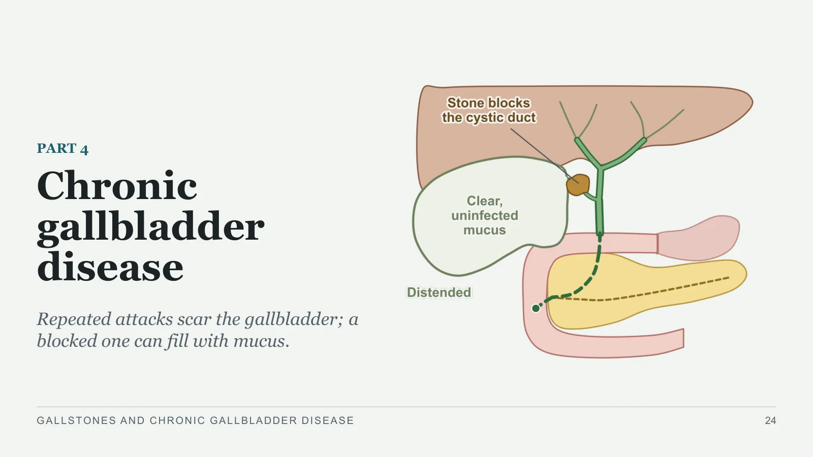
Chronic gallbladder disease
PART 4
Repeated attacks scar the gallbladder; a blocked one can fill with mucus.
GALLSTONES AND CHRONIC GALLBLADDER DISEASE
24
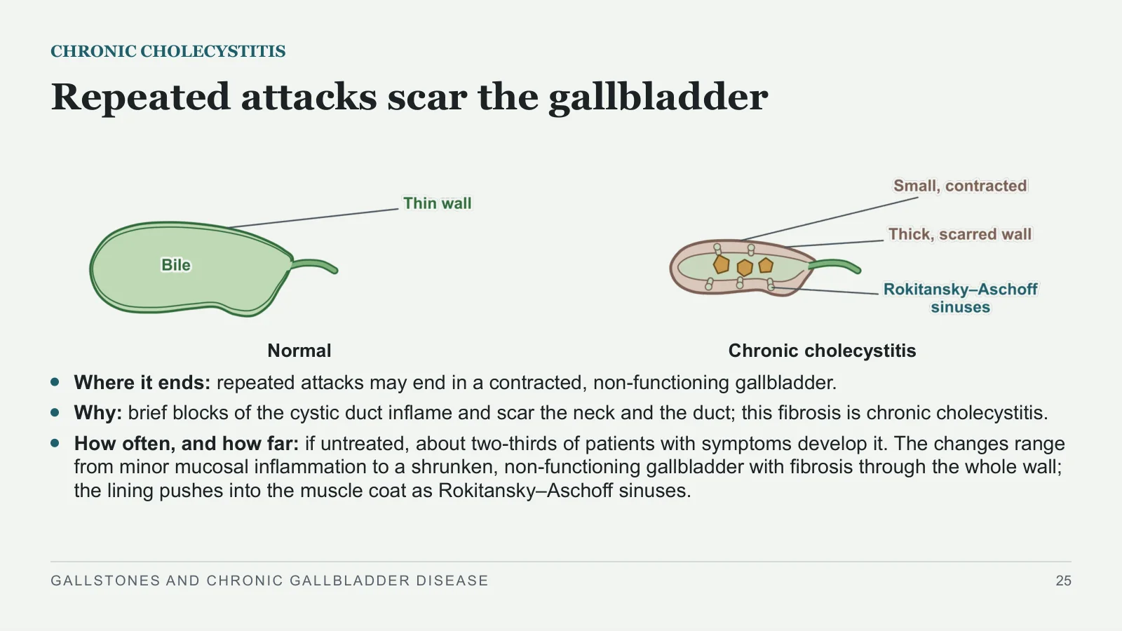
Repeated attacks scar the gallbladder
CHRONIC CHOLECYSTITIS
Normal
Chronic cholecystitis
Where it ends: repeated attacks may end in a contracted, non-functioning gallbladder.
Why: brief blocks of the cystic duct inflame and scar the neck and the duct; this fibrosis is chronic cholecystitis.
How often, and how far: if untreated, about two-thirds of patients with symptoms develop it. The changes range from minor mucosal inflammation to a shrunken, non-functioning gallbladder with fibrosis through the whole wall; the lining pushes into the muscle coat as Rokitansky–Aschoff sinuses.
GALLSTONES AND CHRONIC GALLBLADDER DISEASE
25
Chronic cholecystitis looks like biliary colic
CHRONIC CHOLECYSTITIS
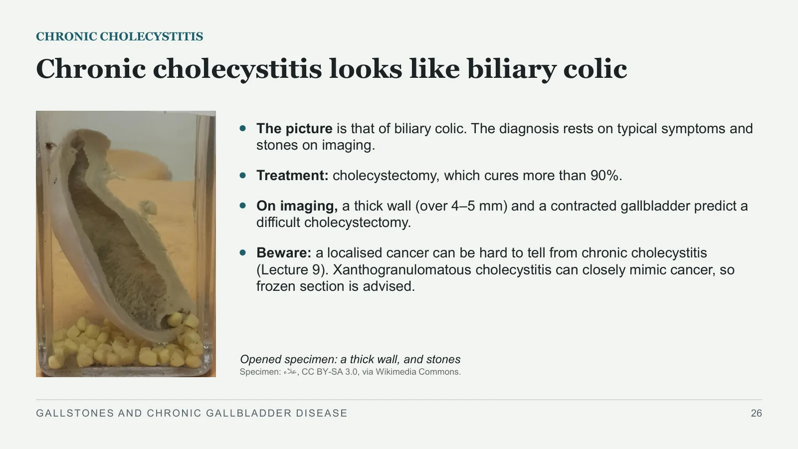
Opened specimen: a thick wall, and stones
Specimen: علاء, CC BY-SA 3.0, via Wikimedia Commons.
The picture is that of biliary colic. The diagnosis rests on typical symptoms and stones on imaging.
Treatment: cholecystectomy, which cures more than 90%.
On imaging, a thick wall (over 4–5 mm) and a contracted gallbladder predict a difficult cholecystectomy.
Beware: a localised cancer can be hard to tell from chronic cholecystitis (Lecture 9). Xanthogranulomatous cholecystitis can closely mimic cancer, so frozen section is advised.
GALLSTONES AND CHRONIC GALLBLADDER DISEASE
26
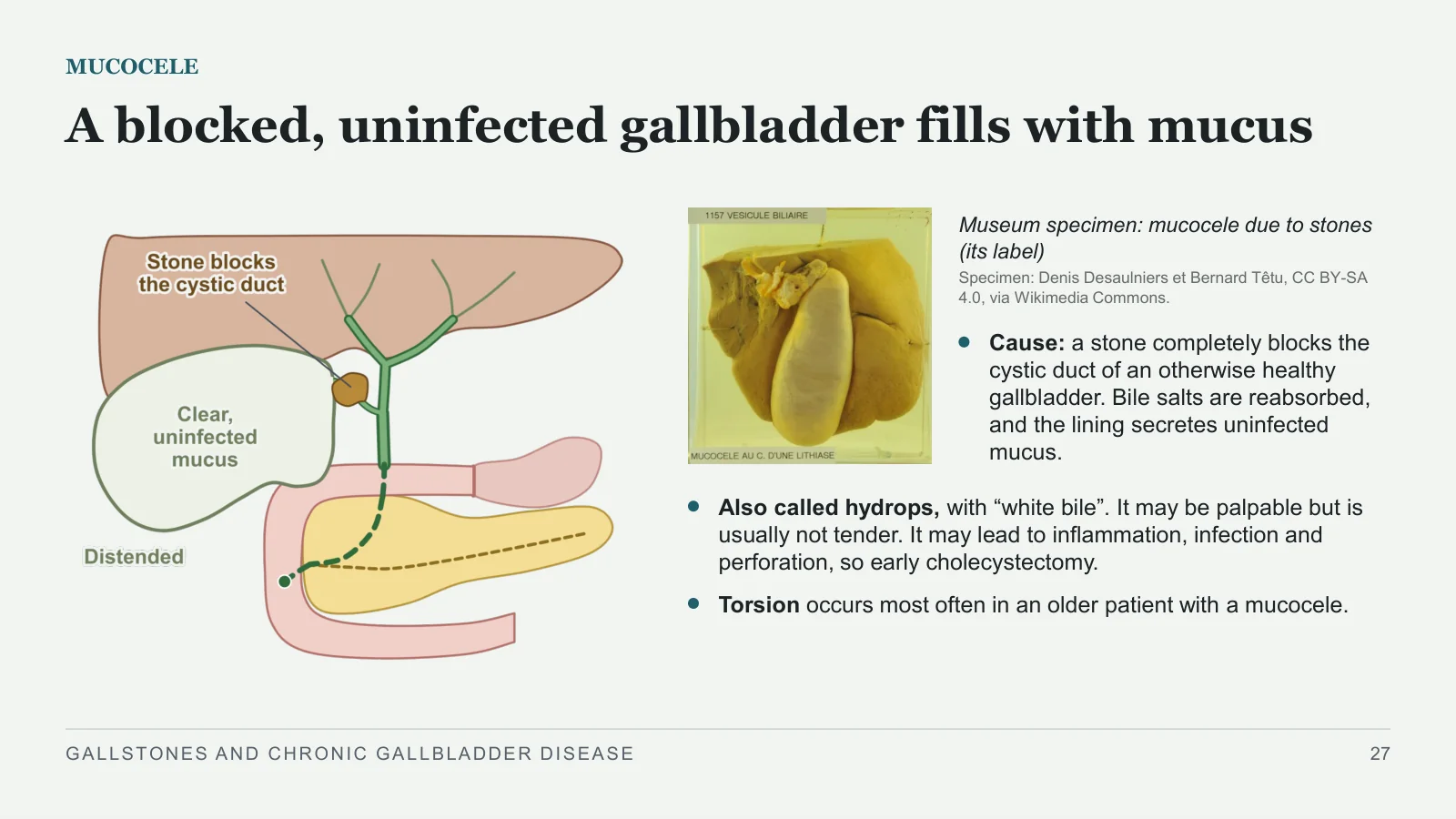
A blocked, uninfected gallbladder fills with mucus
MUCOCELE
Museum specimen: mucocele due to stones (its label)
Specimen: Denis Desaulniers et Bernard Têtu, CC BY-SA 4.0, via Wikimedia Commons.
Cause: a stone completely blocks the cystic duct of an otherwise healthy gallbladder. Bile salts are reabsorbed, and the lining secretes uninfected mucus.
Also called hydrops, with “white bile”. It may be palpable but is usually not tender. It may lead to inflammation, infection and perforation, so early cholecystectomy.
Torsion occurs most often in an older patient with a mucocele.
GALLSTONES AND CHRONIC GALLBLADDER DISEASE
27
Mirizzi fistula and ileus
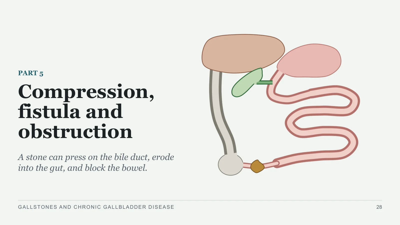
Compression, fistula and obstruction
PART 5
A stone can press on the bile duct, erode into the gut, and block the bowel.
GALLSTONES AND CHRONIC GALLBLADDER DISEASE
28
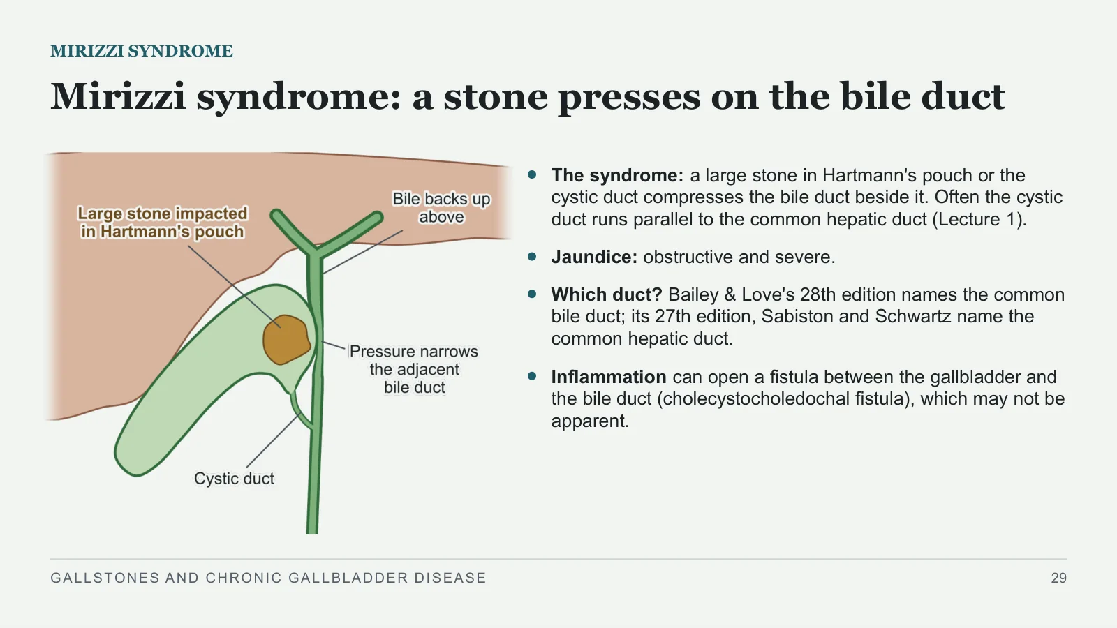
Mirizzi syndrome: a stone presses on the bile duct
MIRIZZI SYNDROME
The syndrome: a large stone in Hartmann's pouch or the cystic duct compresses the bile duct beside it. Often the cystic duct runs parallel to the common hepatic duct (Lecture 1).
Jaundice: obstructive and severe.
Which duct? Bailey & Love's 28th edition names the common bile duct; its 27th edition, Sabiston and Schwartz name the common hepatic duct.
Inflammation can open a fistula between the gallbladder and the bile duct (cholecystocholedochal fistula), which may not be apparent.
GALLSTONES AND CHRONIC GALLBLADDER DISEASE
29
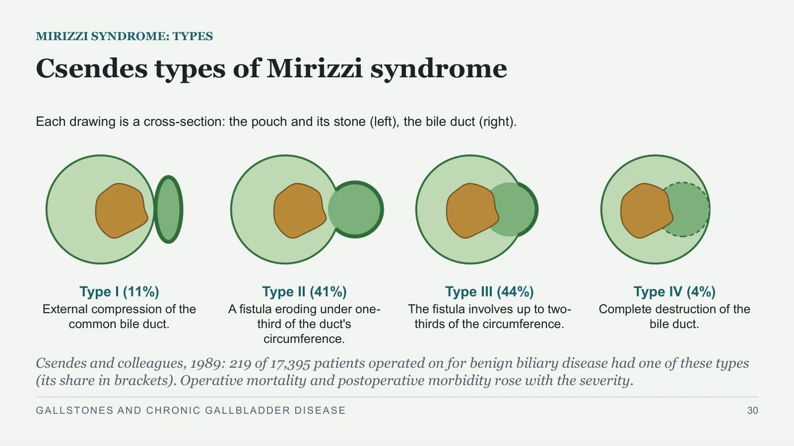
Csendes types of Mirizzi syndrome
MIRIZZI SYNDROME: TYPES
Each drawing is a cross-section: the pouch and its stone (left), the bile duct (right).
Type I (11%)
External compression of the common bile duct.
Type II (41%)
A fistula eroding under one-third of the duct's circumference.
Type III (44%)
The fistula involves up to two-thirds of the circumference.
Type IV (4%)
Complete destruction of the bile duct.
Csendes and colleagues, 1989: 219 of 17,395 patients operated on for benign biliary disease had one of these types (its share in brackets). Operative mortality and postoperative morbidity rose with the severity.
GALLSTONES AND CHRONIC GALLBLADDER DISEASE
30
Mirizzi syndrome is a trap at operation
MIRIZZI SYNDROME AT OPERATION
Why it is a trap
A red flag for a difficult cholecystectomy, and an imaging predictor of one.
The main danger: damage to the bile duct.
Dissecting out the cystic duct completely only injures the common hepatic or common bile duct.
What the books do
Bailey & Love, 27th edition: open the infundibulum, remove the stone, and oversew it.
Sabiston: cholecystectomy, which may need repair of the common duct; a large fistula may need a choledochojejunostomy.
In any difficult cholecystectomy, Bailey & Love's bail-out is a subtotal cholecystectomy.
How these operations are done: Lecture 6.
GALLSTONES AND CHRONIC GALLBLADDER DISEASE
31
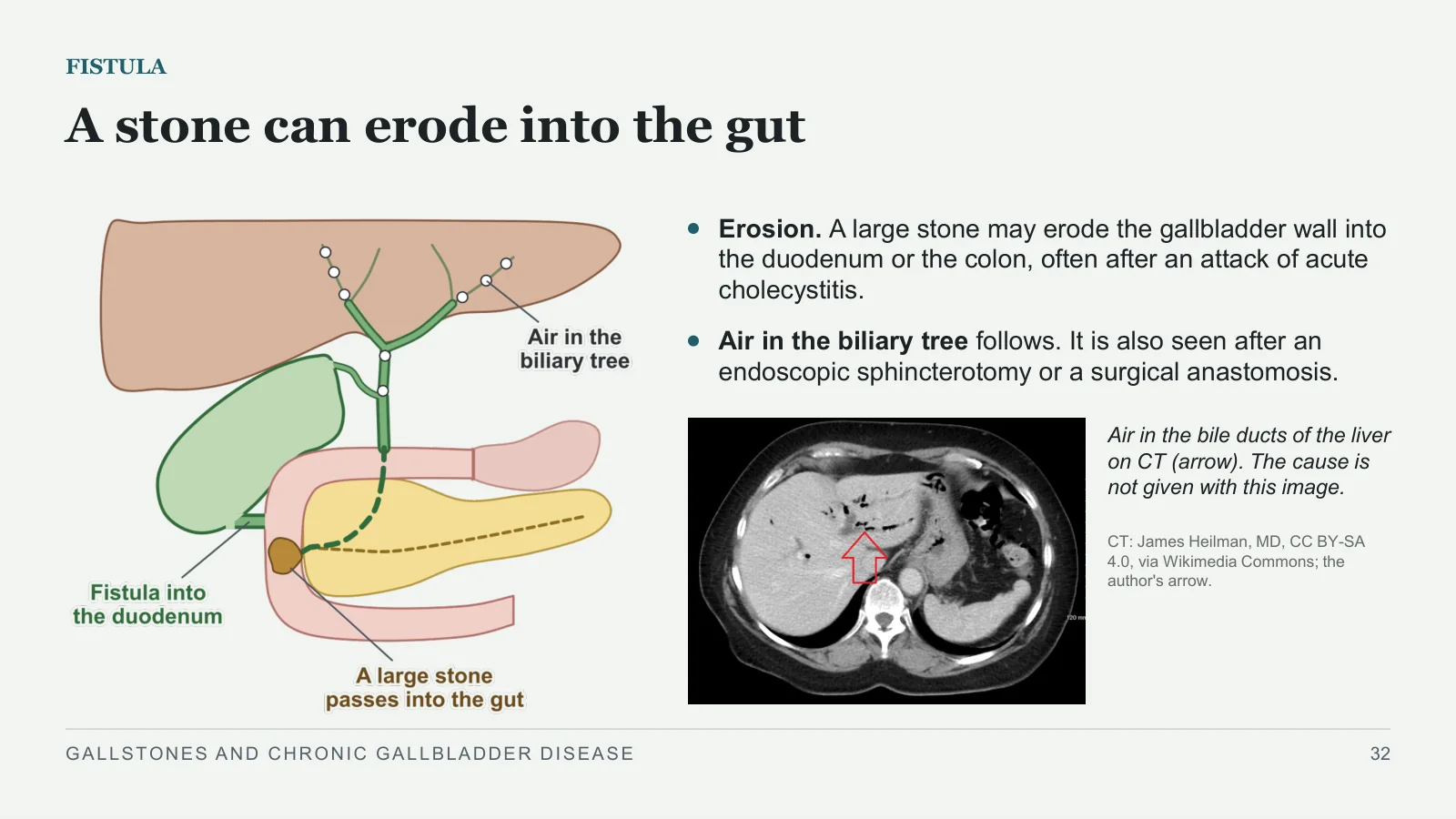
A stone can erode into the gut
FISTULA
Erosion. A large stone may erode the gallbladder wall into the duodenum or the colon, often after an attack of acute cholecystitis.
Air in the biliary tree follows. It is also seen after an endoscopic sphincterotomy or a surgical anastomosis.
Air in the bile ducts of the liver on CT (arrow). The cause is not given with this image.
CT: James Heilman, MD, CC BY-SA 4.0, via Wikimedia Commons; the author's arrow.
GALLSTONES AND CHRONIC GALLBLADDER DISEASE
32
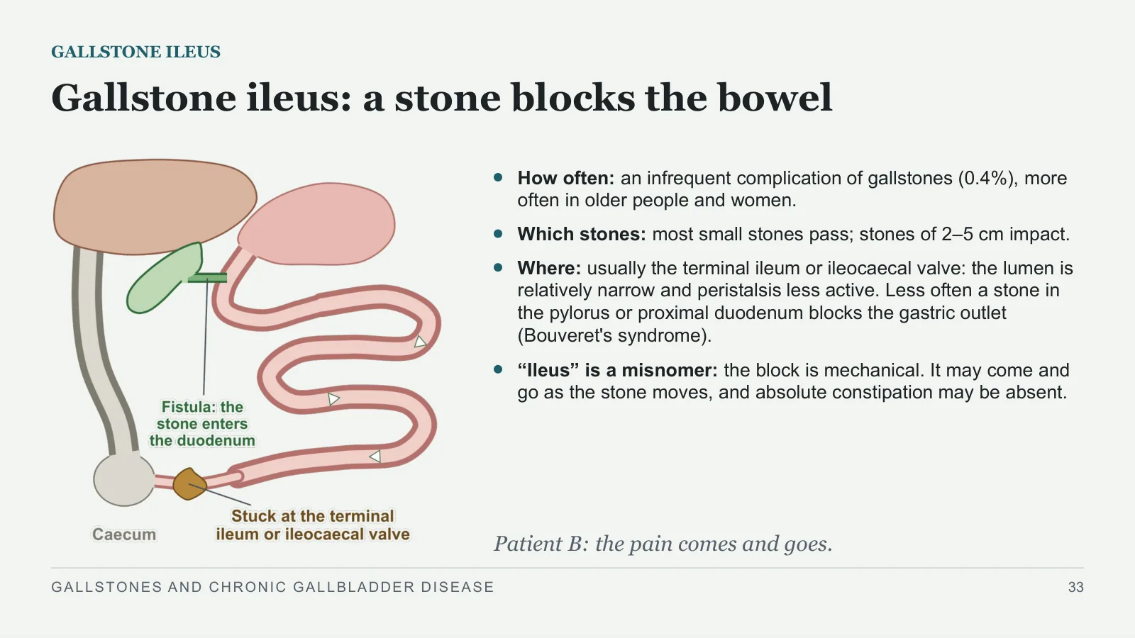
Gallstone ileus: a stone blocks the bowel
GALLSTONE ILEUS
How often: an infrequent complication of gallstones (0.4%), more often in older people and women.
Which stones: most small stones pass; stones of 2–5 cm impact.
Where: usually the terminal ileum or ileocaecal valve: the lumen is relatively narrow and peristalsis less active. Less often a stone in the pylorus or proximal duodenum blocks the gastric outlet (Bouveret's syndrome).
“Ileus” is a misnomer: the block is mechanical. It may come and go as the stone moves, and absolute constipation may be absent.
Patient B: the pain comes and goes.
GALLSTONES AND CHRONIC GALLBLADDER DISEASE
33
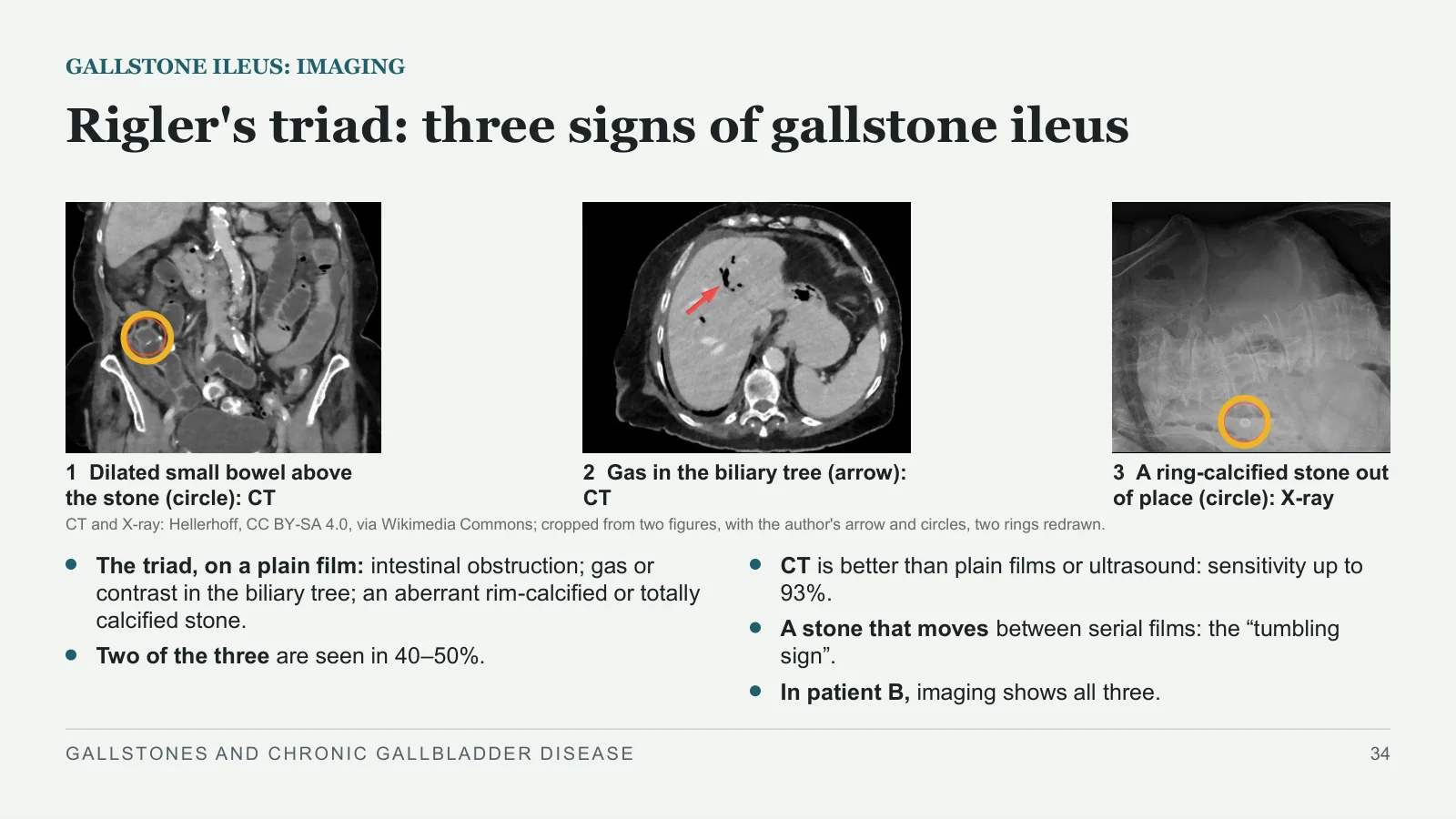
Rigler's triad: three signs of gallstone ileus
GALLSTONE ILEUS: IMAGING
1 Dilated small bowel above the stone (circle): CT
2 Gas in the biliary tree (arrow): CT
3 A ring-calcified stone out of place (circle): X-ray
CT and X-ray: Hellerhoff, CC BY-SA 4.0, via Wikimedia Commons; cropped from two figures, with the author's arrow and circles, two rings redrawn.
The triad, on a plain film: intestinal obstruction; gas or contrast in the biliary tree; an aberrant rim-calcified or totally calcified stone.
Two of the three are seen in 40–50%.
CT is better than plain films or ultrasound: sensitivity up to 93%.
A stone that moves between serial films: the “tumbling sign”.
In patient B, imaging shows all three.
GALLSTONES AND CHRONIC GALLBLADDER DISEASE
34
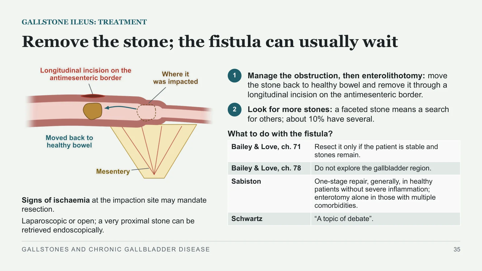
Remove the stone; the fistula can usually wait
GALLSTONE ILEUS: TREATMENT
Signs of ischaemia at the impaction site may mandate resection.
Laparoscopic or open; a very proximal stone can be retrieved endoscopically.
1
Manage the obstruction, then enterolithotomy: move the stone back to healthy bowel and remove it through a longitudinal incision on the antimesenteric border.
2
Look for more stones: a faceted stone means a search for others; about 10% have several.
What to do with the fistula?
GALLSTONES AND CHRONIC GALLBLADDER DISEASE
35
| Bailey & Love, ch. 71 | Resect it only if the patient is stable and stones remain. |
| Bailey & Love, ch. 78 | Do not explore the gallbladder region. |
| Sabiston | One-stage repair, generally, in healthy patients without severe inflammation; enterotomy alone in those with multiple comorbidities. |
| Schwartz | “A topic of debate”. |
Other gallbladder conditions
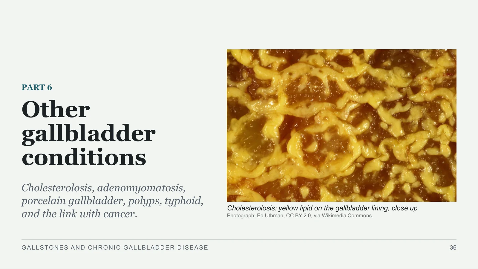
Other gallbladder conditions
PART 6
Cholesterolosis, adenomyomatosis, porcelain gallbladder, polyps, typhoid, and the link with cancer.
Cholesterolosis: yellow lipid on the gallbladder lining, close up
Photograph: Ed Uthman, CC BY 2.0, via Wikimedia Commons.
GALLSTONES AND CHRONIC GALLBLADDER DISEASE
36
Cholesterolosis and adenomyomatosis
THE CHOLECYSTOSES
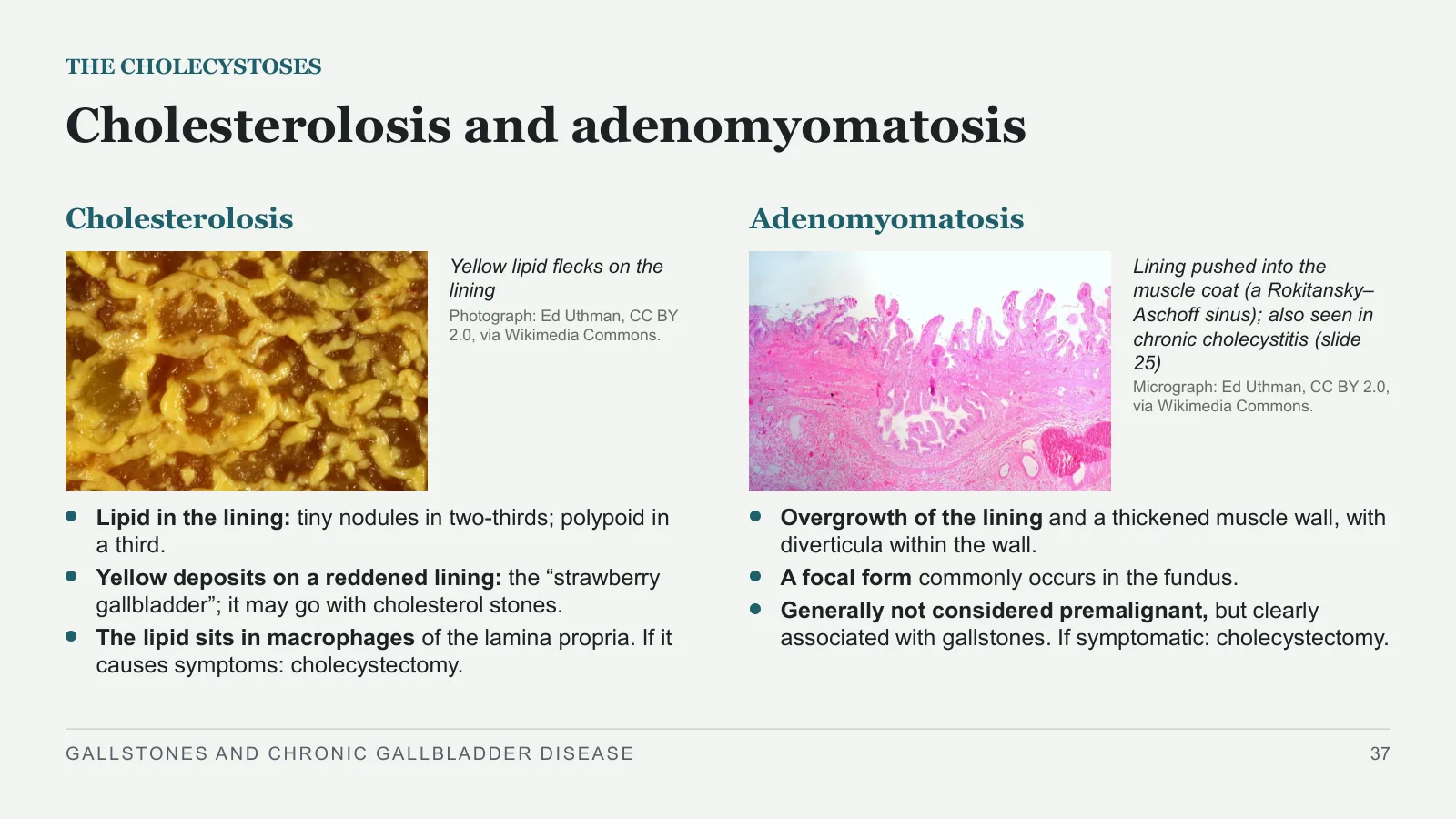
Cholesterolosis
Yellow lipid flecks on the lining
Photograph: Ed Uthman, CC BY 2.0, via Wikimedia Commons.
Lipid in the lining: tiny nodules in two-thirds; polypoid in a third.
Yellow deposits on a reddened lining: the “strawberry gallbladder”; it may go with cholesterol stones.
The lipid sits in macrophages of the lamina propria. If it causes symptoms: cholecystectomy.
Adenomyomatosis
Lining pushed into the muscle coat (a Rokitansky–Aschoff sinus); also seen in chronic cholecystitis (slide 25)
Micrograph: Ed Uthman, CC BY 2.0, via Wikimedia Commons.
Overgrowth of the lining and a thickened muscle wall, with diverticula within the wall.
A focal form commonly occurs in the fundus.
Generally not considered premalignant, but clearly associated with gallstones. If symptomatic: cholecystectomy.
GALLSTONES AND CHRONIC GALLBLADDER DISEASE
37
Porcelain gallbladder: a calcified wall
PORCELAIN GALLBLADDER
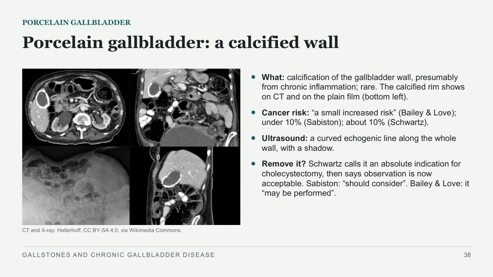
CT and X-ray: Hellerhoff, CC BY-SA 4.0, via Wikimedia Commons.
What: calcification of the gallbladder wall, presumably from chronic inflammation; rare. The calcified rim shows on CT and on the plain film (bottom left).
Cancer risk: “a small increased risk” (Bailey & Love); under 10% (Sabiston); about 10% (Schwartz).
Ultrasound: a curved echogenic line along the whole wall, with a shadow.
Remove it? Schwartz calls it an absolute indication for cholecystectomy, then says observation is now acceptable. Sabiston: “should consider”. Bailey & Love: it “may be performed”.
GALLSTONES AND CHRONIC GALLBLADDER DISEASE
38
Gallbladder polyps: size and risk features decide
GALLBLADDER POLYPS
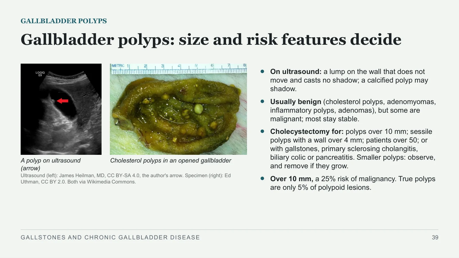
A polyp on ultrasound (arrow)
Cholesterol polyps in an opened gallbladder
Ultrasound (left): James Heilman, MD, CC BY-SA 4.0, the author's arrow. Specimen (right): Ed Uthman, CC BY 2.0. Both via Wikimedia Commons.
On ultrasound: a lump on the wall that does not move and casts no shadow; a calcified polyp may shadow.
Usually benign (cholesterol polyps, adenomyomas, inflammatory polyps, adenomas), but some are malignant; most stay stable.
Cholecystectomy for: polyps over 10 mm; sessile polyps with a wall over 4 mm; patients over 50; or with gallstones, primary sclerosing cholangitis, biliary colic or pancreatitis. Smaller polyps: observe, and remove if they grow.
Over 10 mm, a 25% risk of malignancy. True polyps are only 5% of polypoid lesions.
GALLSTONES AND CHRONIC GALLBLADDER DISEASE
39
Typhoid can make the gallbladder a carrier
TYPHOID
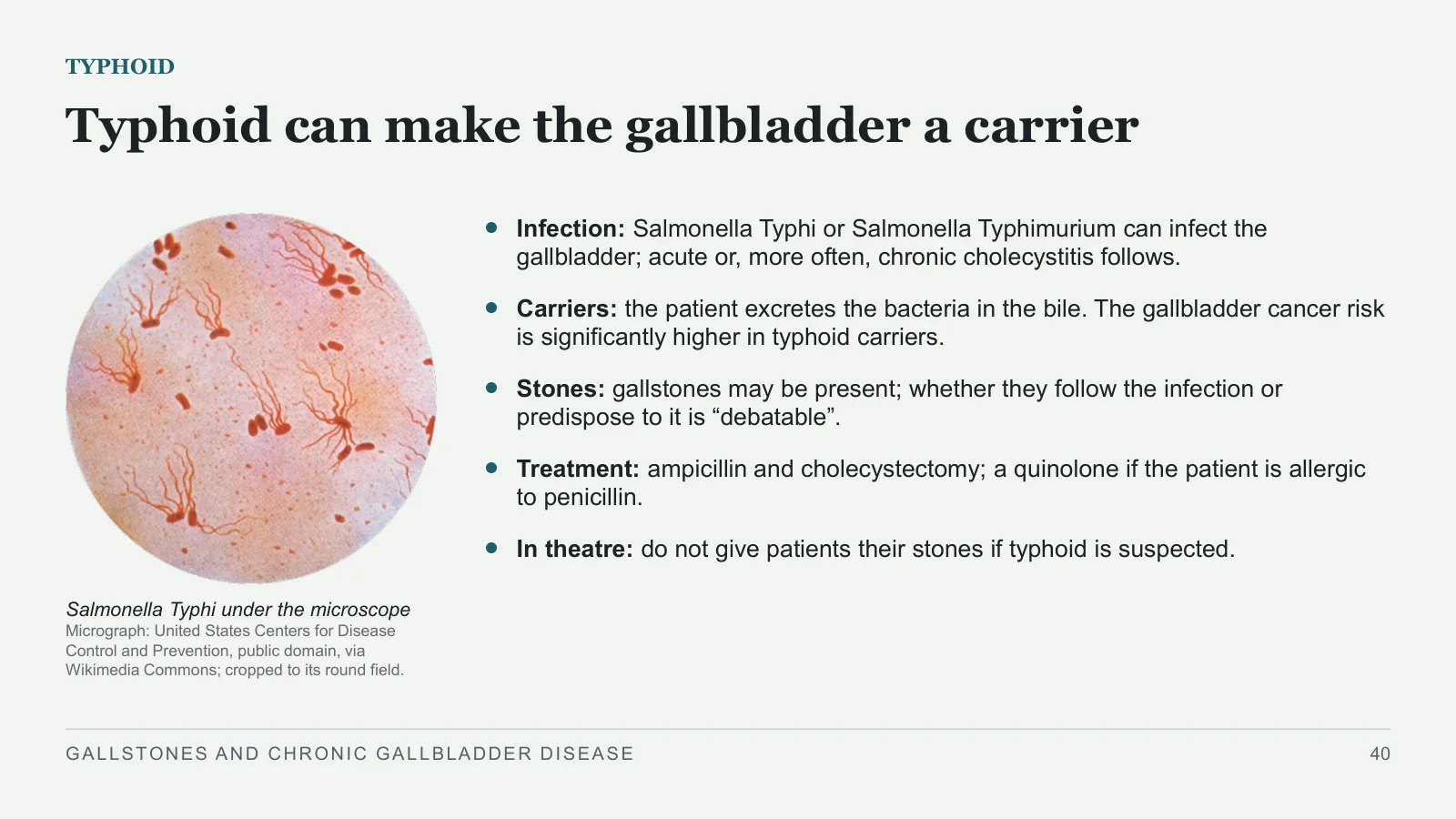
Salmonella Typhi under the microscope
Micrograph: United States Centers for Disease Control and Prevention, public domain, via Wikimedia Commons; cropped to its round field.
Infection: Salmonella Typhi or Salmonella Typhimurium can infect the gallbladder; acute or, more often, chronic cholecystitis follows.
Carriers: the patient excretes the bacteria in the bile. The gallbladder cancer risk is significantly higher in typhoid carriers.
Stones: gallstones may be present; whether they follow the infection or predispose to it is “debatable”.
Treatment: ampicillin and cholecystectomy; a quinolone if the patient is allergic to penicillin.
In theatre: do not give patients their stones if typhoid is suspected.
GALLSTONES AND CHRONIC GALLBLADDER DISEASE
40
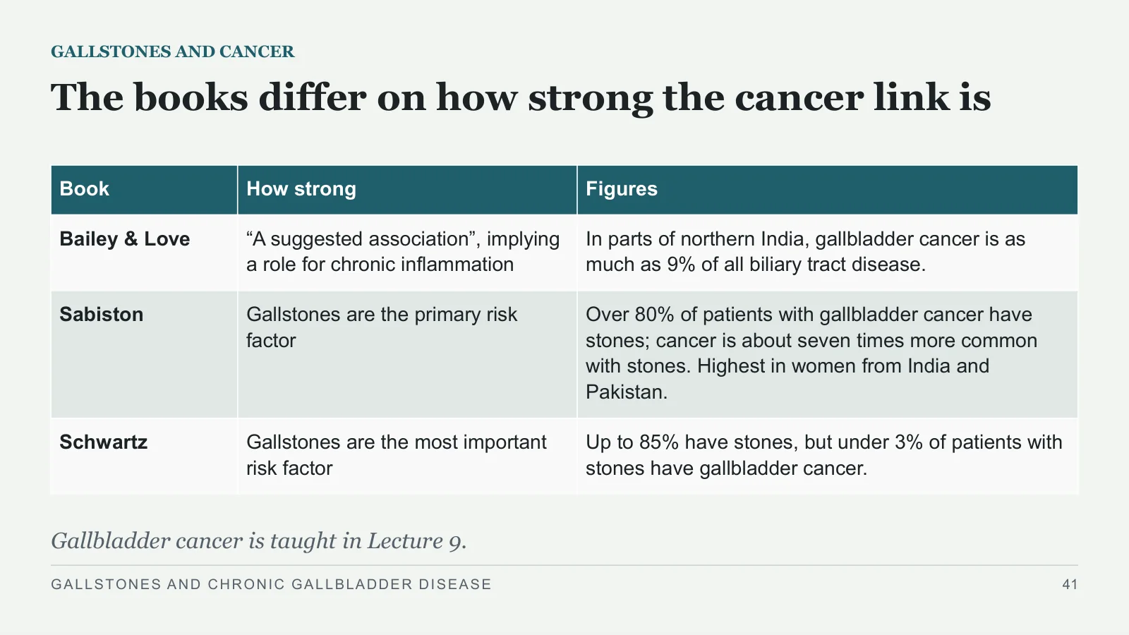
The books differ on how strong the cancer link is
GALLSTONES AND CANCER
Gallbladder cancer is taught in Lecture 9.
GALLSTONES AND CHRONIC GALLBLADDER DISEASE
41
| Book | How strong | Figures |
| Bailey & Love | “A suggested association”, implying a role for chronic inflammation | In parts of northern India, gallbladder cancer is as much as 9% of all biliary tract disease. |
| Sabiston | Gallstones are the primary risk factor | Over 80% of patients with gallbladder cancer have stones; cancer is about seven times more common with stones. Highest in women from India and Pakistan. |
| Schwartz | Gallstones are the most important risk factor | Up to 85% have stones, but under 3% of patients with stones have gallbladder cancer. |
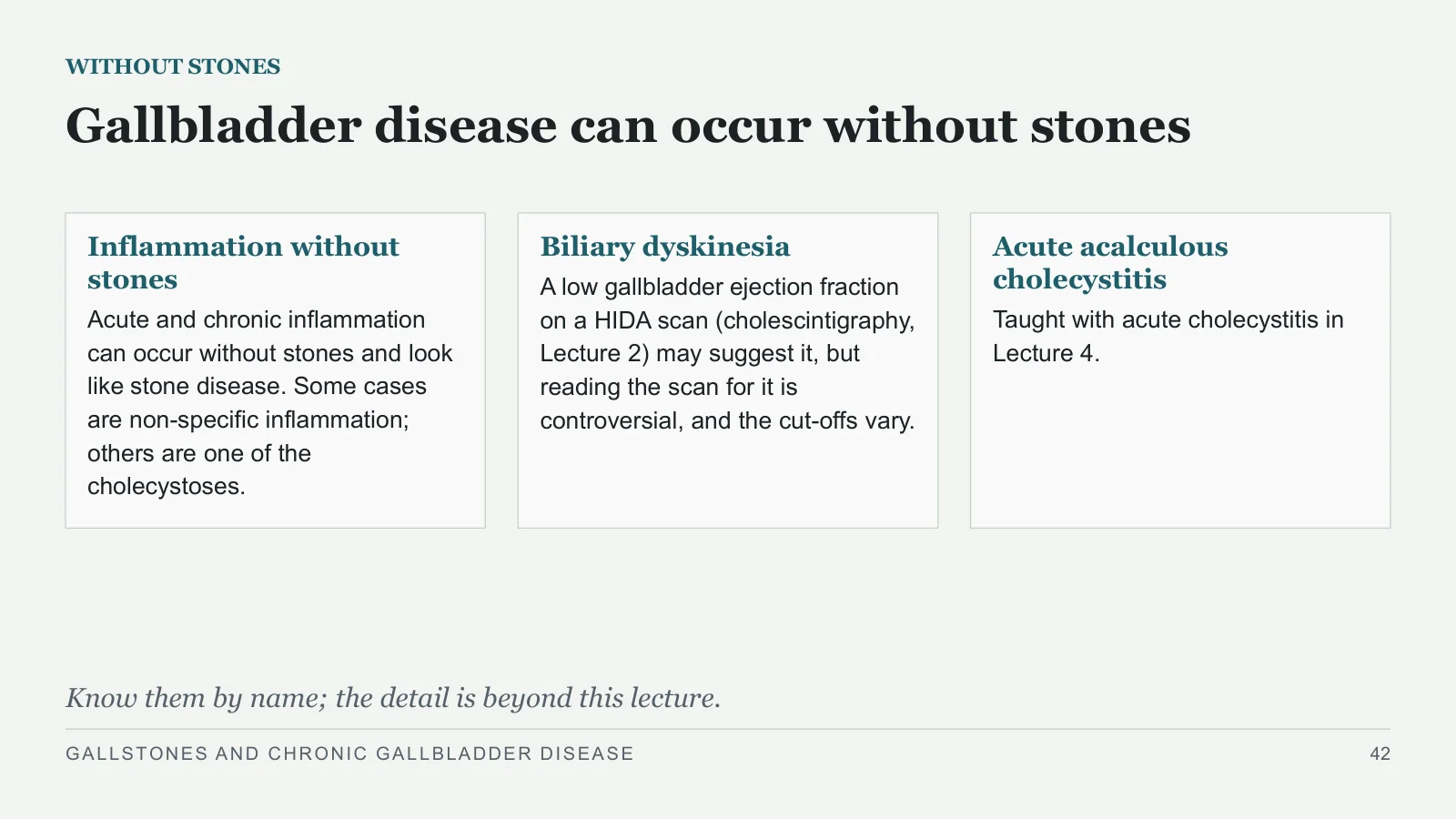
Gallbladder disease can occur without stones
WITHOUT STONES
Inflammation without stones
Acute and chronic inflammation can occur without stones and look like stone disease. Some cases are non-specific inflammation; others are one of the cholecystoses.
Biliary dyskinesia
A low gallbladder ejection fraction on a HIDA scan (cholescintigraphy, Lecture 2) may suggest it, but reading the scan for it is controversial, and the cut-offs vary.
Acute acalculous cholecystitis
Taught with acute cholecystitis in Lecture 4.
Know them by name; the detail is beyond this lecture.
GALLSTONES AND CHRONIC GALLBLADDER DISEASE
42
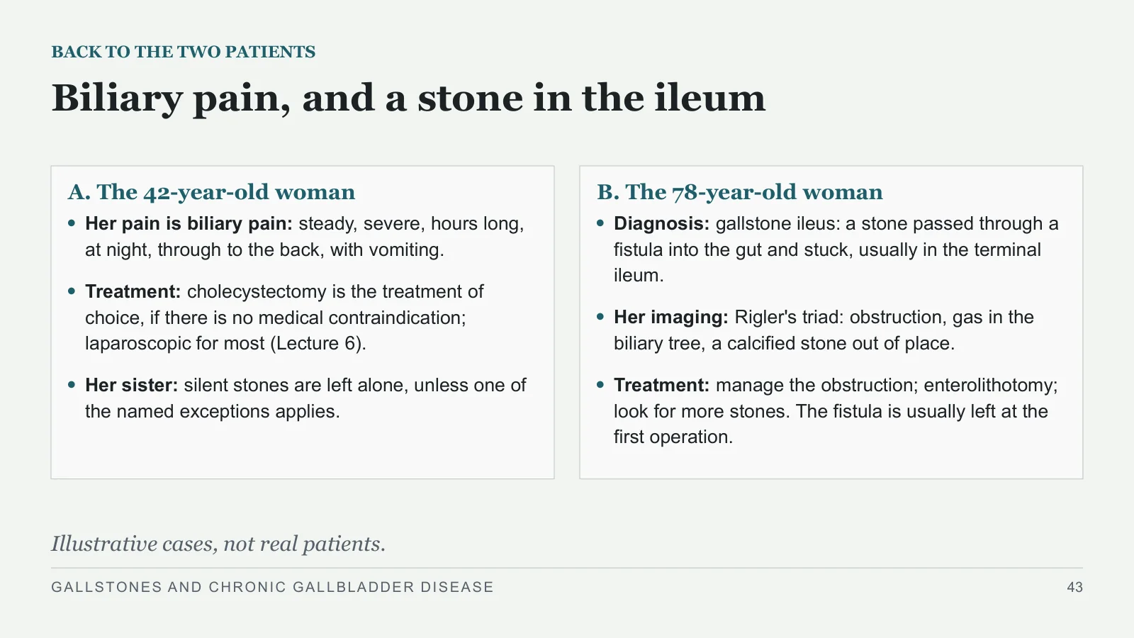
Biliary pain, and a stone in the ileum
BACK TO THE TWO PATIENTS
A. The 42-year-old woman
Her pain is biliary pain: steady, severe, hours long, at night, through to the back, with vomiting.
Treatment: cholecystectomy is the treatment of choice, if there is no medical contraindication; laparoscopic for most (Lecture 6).
Her sister: silent stones are left alone, unless one of the named exceptions applies.
B. The 78-year-old woman
Diagnosis: gallstone ileus: a stone passed through a fistula into the gut and stuck, usually in the terminal ileum.
Her imaging: Rigler's triad: obstruction, gas in the biliary tree, a calcified stone out of place.
Treatment: manage the obstruction; enterolithotomy; look for more stones. The fistula is usually left at the first operation.
Illustrative cases, not real patients.
GALLSTONES AND CHRONIC GALLBLADDER DISEASE
43
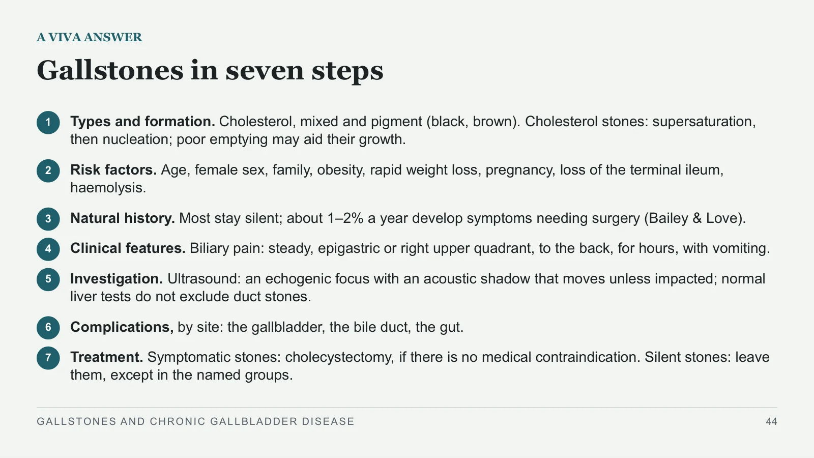
Gallstones in seven steps
A VIVA ANSWER
1
Types and formation. Cholesterol, mixed and pigment (black, brown). Cholesterol stones: supersaturation, then nucleation; poor emptying may aid their growth.
2
Risk factors. Age, female sex, family, obesity, rapid weight loss, pregnancy, loss of the terminal ileum, haemolysis.
3
Natural history. Most stay silent; about 1–2% a year develop symptoms needing surgery (Bailey & Love).
4
Clinical features. Biliary pain: steady, epigastric or right upper quadrant, to the back, for hours, with vomiting.
5
Investigation. Ultrasound: an echogenic focus with an acoustic shadow that moves unless impacted; normal liver tests do not exclude duct stones.
6
Complications, by site: the gallbladder, the bile duct, the gut.
7
Treatment. Symptomatic stones: cholecystectomy, if there is no medical contraindication. Silent stones: leave them, except in the named groups.
GALLSTONES AND CHRONIC GALLBLADDER DISEASE
44
What to carry out of this lecture
TAKE-HOME
1
Cholesterol, mixed or pigment. Cholesterol stones start in supersaturated bile.
2
Biliary pain comes from the outlet. Steady, for hours; fever means more than colic.
3
Most stones stay silent. Treat symptoms; operate on silent stones only for named reasons.
4
Stones at the outlet do more harm. Repeated obstruction can cause chronic cholecystitis; complete cystic duct block, a mucocele; an impacted stone pressing on the bile duct, Mirizzi syndrome.
5
Gallstone ileus. A stone passes through a fistula and blocks the bowel; imaging supports the diagnosis; enterolithotomy, then a search for more stones.
GALLSTONES AND CHRONIC GALLBLADDER DISEASE
45
References
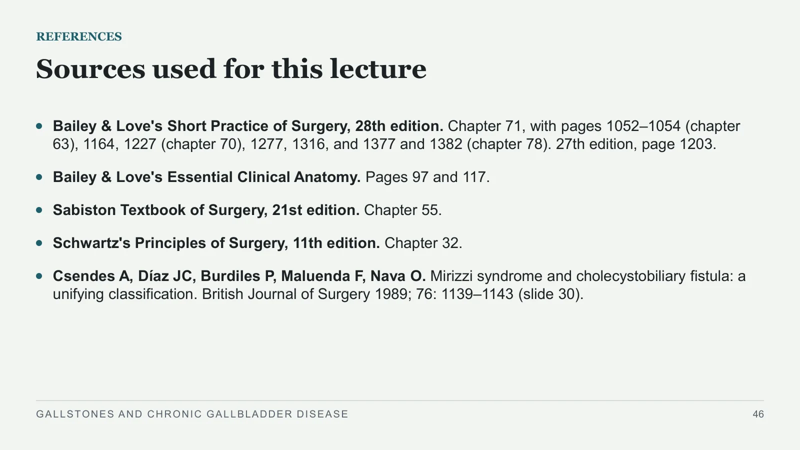
Sources used for this lecture
REFERENCES
Bailey & Love's Short Practice of Surgery, 28th edition. Chapter 71, with pages 1052–1054 (chapter 63), 1164, 1227 (chapter 70), 1277, 1316, and 1377 and 1382 (chapter 78). 27th edition, page 1203.
Bailey & Love's Essential Clinical Anatomy. Pages 97 and 117.
Sabiston Textbook of Surgery, 21st edition. Chapter 55.
Schwartz's Principles of Surgery, 11th edition. Chapter 32.
Csendes A, Díaz JC, Burdiles P, Maluenda F, Nava O. Mirizzi syndrome and cholecystobiliary fistula: a unifying classification. British Journal of Surgery 1989; 76: 1139–1143 (slide 30).
GALLSTONES AND CHRONIC GALLBLADDER DISEASE
46
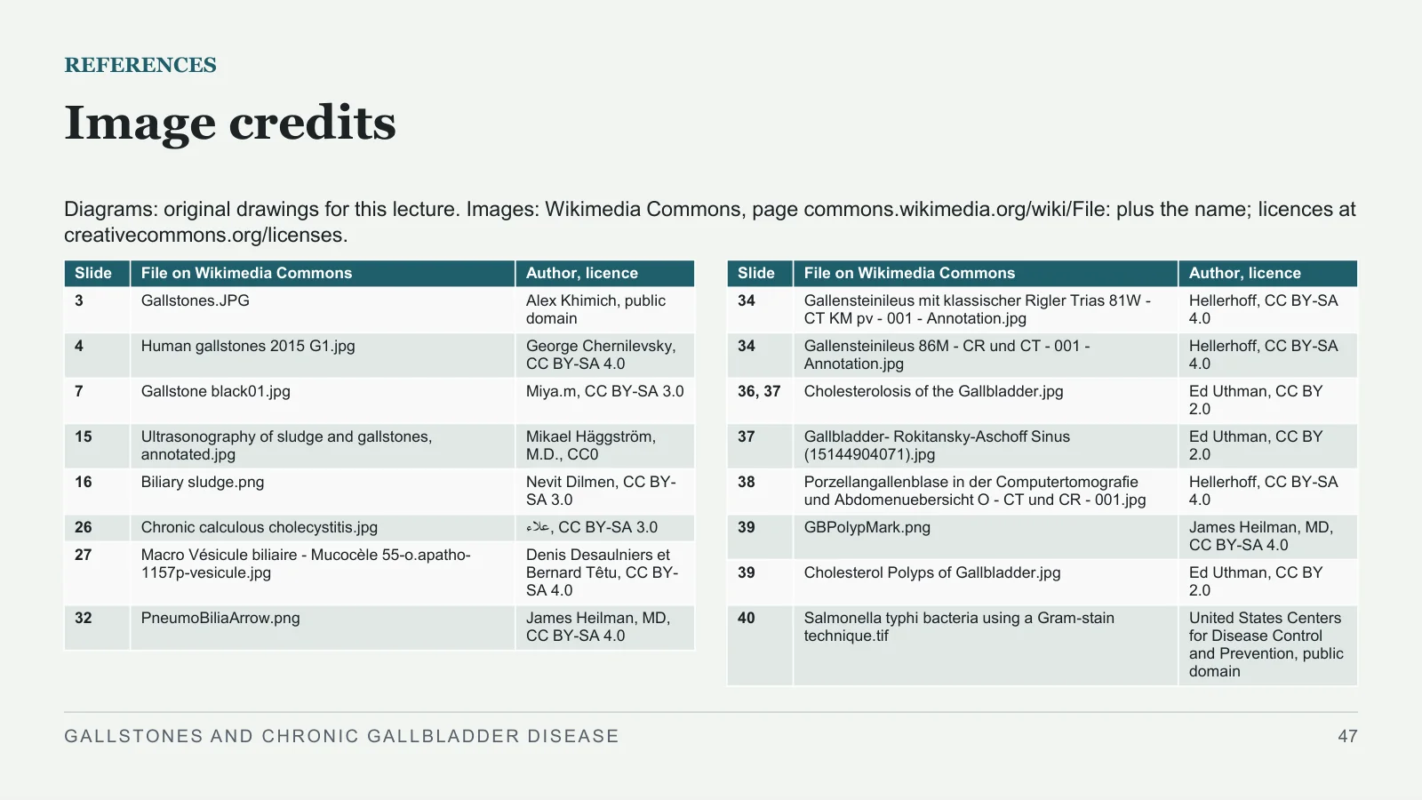
Image credits
REFERENCES
Diagrams: original drawings for this lecture. Images: Wikimedia Commons, page commons.wikimedia.org/wiki/File: plus the name; licences at creativecommons.org/licenses.
GALLSTONES AND CHRONIC GALLBLADDER DISEASE
47
| Slide | File on Wikimedia Commons | Author, licence |
| 3 | Gallstones.JPG | Alex Khimich, public domain |
| 4 | Human gallstones 2015 G1.jpg | George Chernilevsky, CC BY-SA 4.0 |
| 7 | Gallstone black01.jpg | Miya.m, CC BY-SA 3.0 |
| 15 | Ultrasonography of sludge and gallstones, annotated.jpg | Mikael Häggström, M.D., CC0 |
| 16 | Biliary sludge.png | Nevit Dilmen, CC BY-SA 3.0 |
| 26 | Chronic calculous cholecystitis.jpg | علاء, CC BY-SA 3.0 |
| 27 | Macro Vésicule biliaire - Mucocèle 55-o.apatho-1157p-vesicule.jpg | Denis Desaulniers et Bernard Têtu, CC BY-SA 4.0 |
| 32 | PneumoBiliaArrow.png | James Heilman, MD, CC BY-SA 4.0 |
| Slide | File on Wikimedia Commons | Author, licence |
| 34 | Gallensteinileus mit klassischer Rigler Trias 81W - CT KM pv - 001 - Annotation.jpg | Hellerhoff, CC BY-SA 4.0 |
| 34 | Gallensteinileus 86M - CR und CT - 001 - Annotation.jpg | Hellerhoff, CC BY-SA 4.0 |
| 36, 37 | Cholesterolosis of the Gallbladder.jpg | Ed Uthman, CC BY 2.0 |
| 37 | Gallbladder- Rokitansky-Aschoff Sinus (15144904071).jpg | Ed Uthman, CC BY 2.0 |
| 38 | Porzellangallenblase in der Computertomografie und Abdomenuebersicht O - CT und CR - 001.jpg | Hellerhoff, CC BY-SA 4.0 |
| 39 | GBPolypMark.png | James Heilman, MD, CC BY-SA 4.0 |
| 39 | Cholesterol Polyps of Gallbladder.jpg | Ed Uthman, CC BY 2.0 |
| 40 | Salmonella typhi bacteria using a Gram-stain technique.tif | United States Centers for Disease Control and Prevention, public domain |
Sources and image credits
Original lecture diagrams and source/image credits are preserved from the PowerPoint. Each photograph or scan below links to its source and applicable licence. Changes are described in the slide captions; adapted ShareAlike images retain that licence.
Images
- Slide 3: Gallstones.JPG · Alex Khimich, public domain. Slide caption: Photograph: Alex Khimich, public domain, via Wikimedia Commons.
- Slide 4: Human gallstones 2015 G1.jpg · George Chernilevsky, CC BY-SA 4.0. Slide caption: Photograph: George Chernilevsky, CC BY-SA 4.0, via Wikimedia Commons. Adaptations of this image retain the same Creative Commons ShareAlike licence.
- Licence — Human gallstones 2015 G1.jpg · CC BY-SA 4.0
- Slide 7: Gallstone black01.jpg · Miya.m, CC BY-SA 3.0. Slide caption: Photograph: Miya.m, CC BY-SA 3.0, via Wikimedia Commons. Adaptations of this image retain the same Creative Commons ShareAlike licence.
- Licence — Gallstone black01.jpg · CC BY-SA 3.0
- Slide 15: Ultrasonography of sludge and gallstones, annotated.jpg · Mikael Häggström, M.D., CC0. Slide caption: Ultrasound: Mikael Häggström, M.D., CC0, via Wikimedia Commons; the author's labels.
- Licence — Ultrasonography of sludge and gallstones, annotated.jpg · CC0
- Slide 16: Biliary sludge.png · Nevit Dilmen, CC BY-SA 3.0. Slide caption: Ultrasound: Nevit Dilmen, CC BY-SA 3.0, via Wikimedia Commons; the author's label. Adaptations of this image retain the same Creative Commons ShareAlike licence.
- Licence — Biliary sludge.png · CC BY-SA 3.0
- Slide 26: Chronic calculous cholecystitis.jpg · علاء, CC BY-SA 3.0. Slide caption: Specimen: علاء, CC BY-SA 3.0, via Wikimedia Commons. Adaptations of this image retain the same Creative Commons ShareAlike licence.
- Licence — Chronic calculous cholecystitis.jpg · CC BY-SA 3.0
- Slide 27: Macro Vésicule biliaire - Mucocèle 55-o.apatho-1157p-vesicule.jpg · Denis Desaulniers et Bernard Têtu, CC BY-SA 4.0. Source collection: Bibliothèque de l'Université Laval. Slide caption: Specimen: Denis Desaulniers et Bernard Têtu, CC BY-SA 4.0, via Wikimedia Commons. Adaptations of this image retain the same Creative Commons ShareAlike licence.
- Licence — Macro Vésicule biliaire - Mucocèle 55-o.apatho-1157p-vesicule.jpg · CC BY-SA 4.0
- Slide 32: PneumoBiliaArrow.png · James Heilman, MD, CC BY-SA 4.0. Slide caption: CT: James Heilman, MD, CC BY-SA 4.0, via Wikimedia Commons; the author's arrow. Adaptations of this image retain the same Creative Commons ShareAlike licence.
- Licence — PneumoBiliaArrow.png · CC BY-SA 4.0
- Slide 34: Gallensteinileus mit klassischer Rigler Trias 81W - CT KM pv - 001 - Annotation.jpg · Hellerhoff, CC BY-SA 4.0. Slide caption: CT and X-ray: Hellerhoff, CC BY-SA 4.0, via Wikimedia Commons; cropped from two figures, with the author's arrow and circles, two rings redrawn. Adaptations of this image retain the same Creative Commons ShareAlike licence.
- Licence — Gallensteinileus mit klassischer Rigler Trias 81W - CT KM pv - 001 - Annotation.jpg · CC BY-SA 4.0
- Slide 34: Gallensteinileus 86M - CR und CT - 001 - Annotation.jpg · Hellerhoff, CC BY-SA 4.0. Slide caption: CT and X-ray: Hellerhoff, CC BY-SA 4.0, via Wikimedia Commons; cropped from two figures, with the author's arrow and circles, two rings redrawn. Adaptations of this image retain the same Creative Commons ShareAlike licence.
- Licence — Gallensteinileus 86M - CR und CT - 001 - Annotation.jpg · CC BY-SA 4.0
- Slide 36, 37: Cholesterolosis of the Gallbladder.jpg · Ed Uthman, CC BY 2.0. Slide caption: Photograph: Ed Uthman, CC BY 2.0, via Wikimedia Commons. Micrograph: Ed Uthman, CC BY 2.0, via Wikimedia Commons.
- Licence — Cholesterolosis of the Gallbladder.jpg · CC BY 2.0
- Slide 37: Gallbladder- Rokitansky-Aschoff Sinus (15144904071).jpg · Ed Uthman, CC BY 2.0. Slide caption: Photograph: Ed Uthman, CC BY 2.0, via Wikimedia Commons. Micrograph: Ed Uthman, CC BY 2.0, via Wikimedia Commons.
- Licence — Gallbladder- Rokitansky-Aschoff Sinus (15144904071).jpg · CC BY 2.0
- Slide 38: Porzellangallenblase in der Computertomografie und Abdomenuebersicht O - CT und CR - 001.jpg · Hellerhoff, CC BY-SA 4.0. Slide caption: CT and X-ray: Hellerhoff, CC BY-SA 4.0, via Wikimedia Commons. Adaptations of this image retain the same Creative Commons ShareAlike licence.
- Licence — Porzellangallenblase in der Computertomografie und Abdomenuebersicht O - CT und CR - 001.jpg · CC BY-SA 4.0
- Slide 39: GBPolypMark.png · James Heilman, MD, CC BY-SA 4.0. Slide caption: Ultrasound (left): James Heilman, MD, CC BY-SA 4.0, the author's arrow. Specimen (right): Ed Uthman, CC BY 2.0. Both via Wikimedia Commons. Adaptations of this image retain the same Creative Commons ShareAlike licence.
- Licence — GBPolypMark.png · CC BY-SA 4.0
- Slide 39: Cholesterol Polyps of Gallbladder.jpg · Ed Uthman, CC BY 2.0. Slide caption: Ultrasound (left): James Heilman, MD, CC BY-SA 4.0, the author's arrow. Specimen (right): Ed Uthman, CC BY 2.0. Both via Wikimedia Commons.
- Licence — Cholesterol Polyps of Gallbladder.jpg · CC BY 2.0
- Slide 40: Salmonella typhi bacteria using a Gram-stain technique.tif · United States Centers for Disease Control and Prevention, public domain. Slide caption: Micrograph: United States Centers for Disease Control and Prevention, public domain, via Wikimedia Commons; cropped to its round field.


Sources
- [1] REFERENCES
- [2] Sources used for this lecture
- [3] Bailey & Love's Short Practice of Surgery, 28th edition. Chapter 71, with pages 1052–1054 (chapter 63), 1164, 1227 (chapter 70), 1277, 1316, and 1377 and 1382 (chapter 78). 27th edition, page 1203.
- [4] Bailey & Love's Essential Clinical Anatomy. Pages 97 and 117.
- [5] Sabiston Textbook of Surgery, 21st edition. Chapter 55.
- [6] Schwartz's Principles of Surgery, 11th edition. Chapter 32.
- [7] Csendes A, Díaz JC, Burdiles P, Maluenda F, Nava O. Mirizzi syndrome and cholecystobiliary fistula: a unifying classification. British Journal of Surgery 1989; 76: 1139–1143 (slide 30).