Hepatobiliary surgery · Lecture 10
Bile duct cancer · 56 slides
Cholangiocarcinoma.
Follow two cases of malignant obstruction to see how the tumour's location shapes staging, drainage and treatment.
About the downloads
PowerPoint without speaker notes. SHA-256 checksums verify the downloaded files.
Contents · 11 parts
Opening cases
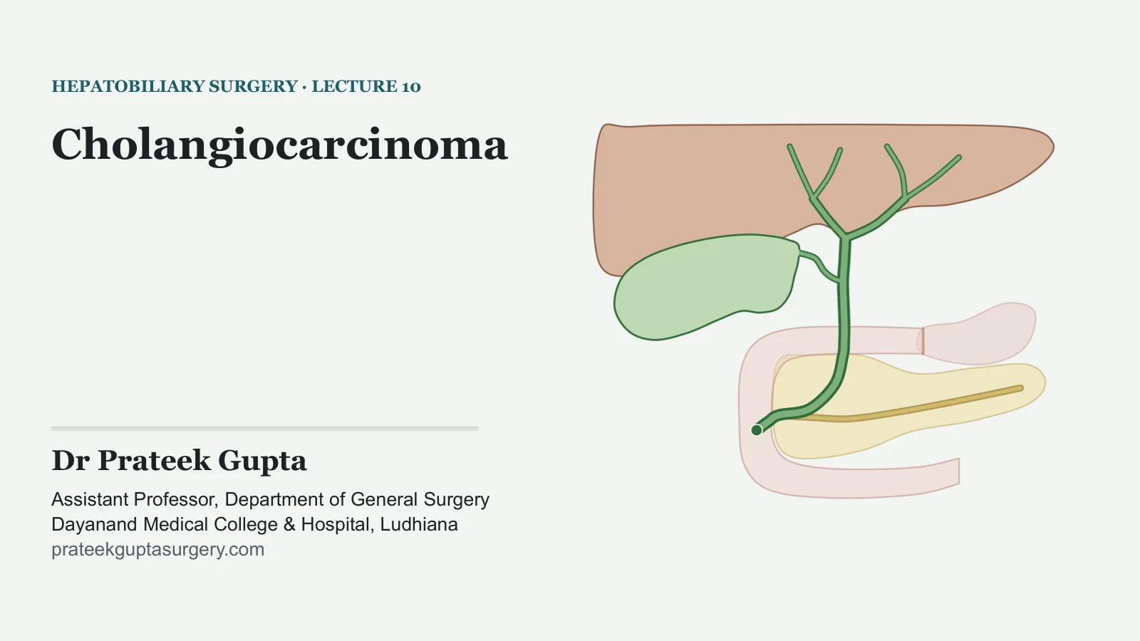
Cholangiocarcinoma
HEPATOBILIARY SURGERY · LECTURE 10
Dr Prateek Gupta
Assistant Professor, Department of General Surgery Dayanand Medical College & Hospital, Ludhiana prateekguptasurgery.com
Two patients, one cancer
TWO PATIENTS
A · A man in his sixties
Painless jaundice, itching and weight loss. The scan shows dilated ducts inside the liver on both sides and a collapsed gallbladder.
B · A woman in her seventies
Painless jaundice, dark urine and pale stools. Her gallbladder can be felt. The scan shows dilated ducts inside and outside the liver, a distended gallbladder, and no stone or pancreatic mass.
Where is each block, and what comes next?
Illustrative patients, not real ones.
CHOLANGIOCARCINOMA
2
What and where
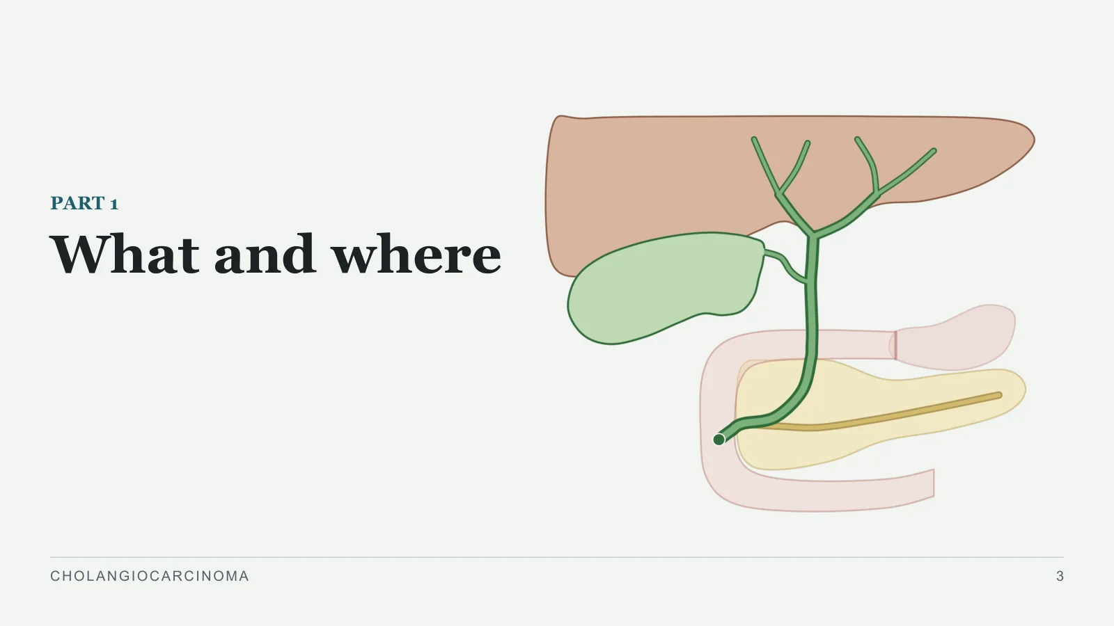
What and where
PART 1
CHOLANGIOCARCINOMA
3
A cancer of the bile duct lining
WHAT AND WHERE
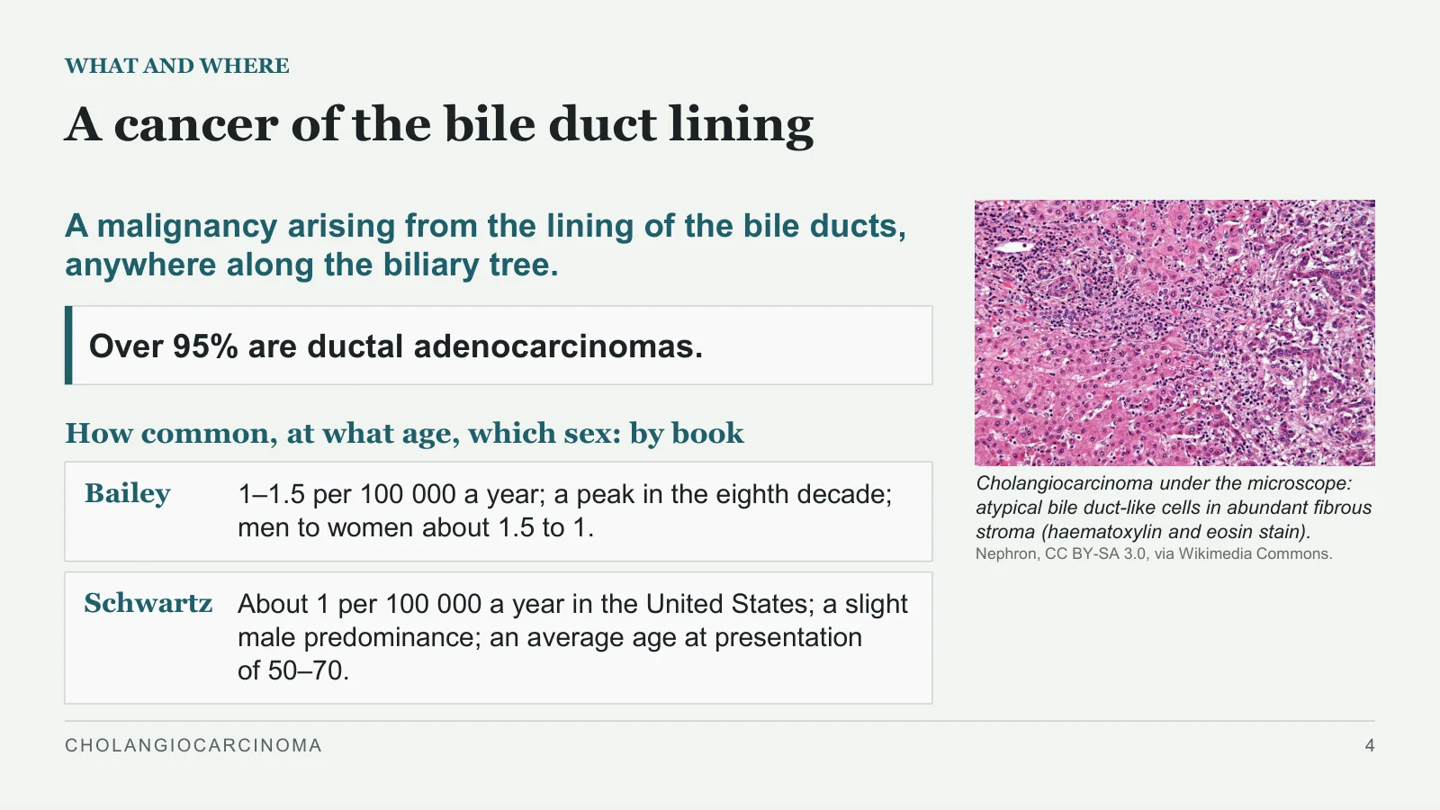
Cholangiocarcinoma under the microscope: atypical bile duct-like cells in abundant fibrous stroma (haematoxylin and eosin stain).
Nephron, CC BY-SA 3.0, via Wikimedia Commons.
A malignancy arising from the lining of the bile ducts, anywhere along the biliary tree.
Over 95% are ductal adenocarcinomas.
How common, at what age, which sex: by book
Bailey
1–1.5 per 100 000 a year; a peak in the eighth decade; men to women about 1.5 to 1.
Schwartz
About 1 per 100 000 a year in the United States; a slight male predominance; an average age at presentation of 50–70.
CHOLANGIOCARCINOMA
4
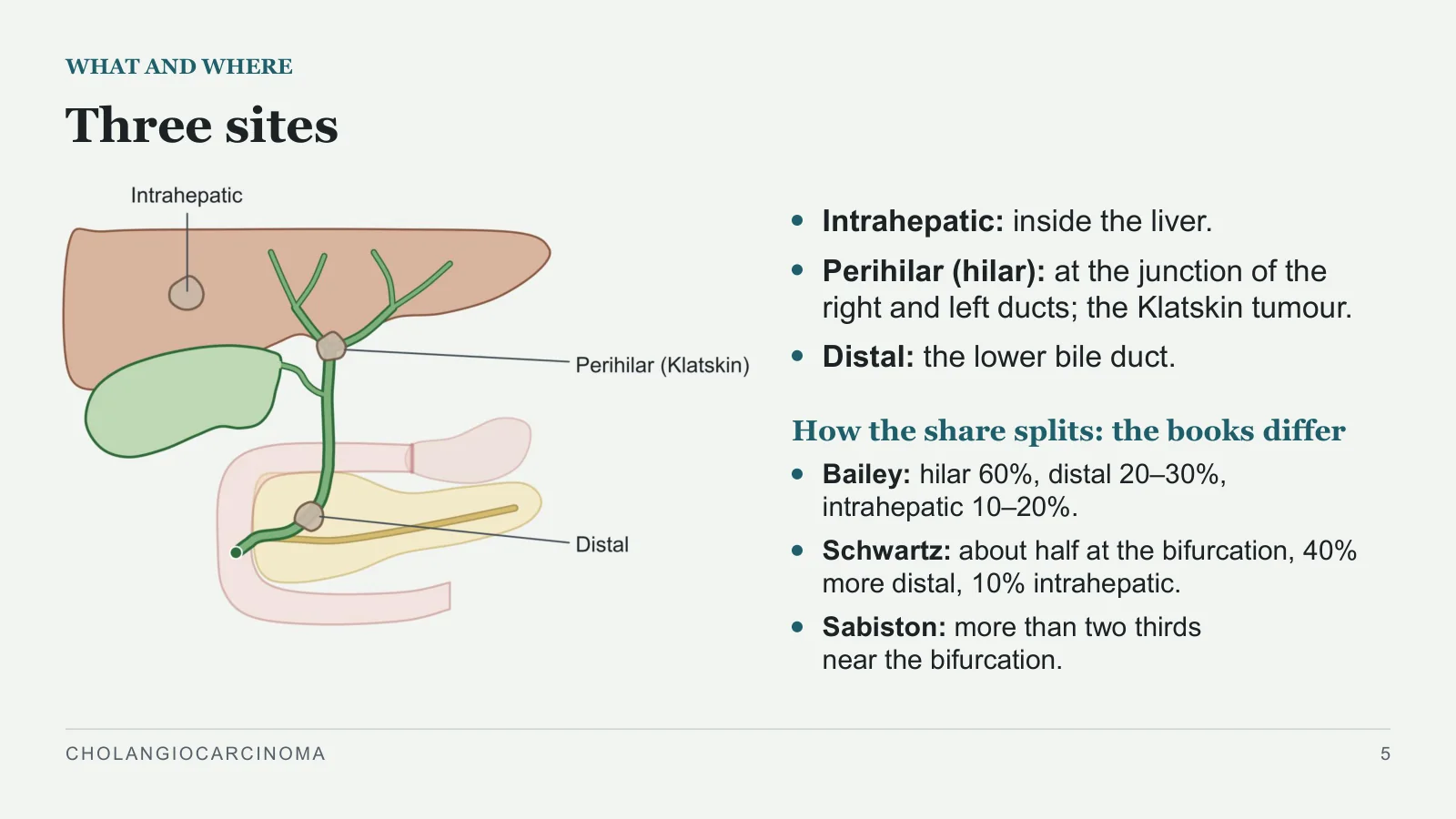
Three sites
WHAT AND WHERE
Intrahepatic: inside the liver.
Perihilar (hilar): at the junction of the right and left ducts; the Klatskin tumour.
Distal: the lower bile duct.
How the share splits: the books differ
Bailey: hilar 60%, distal 20–30%, intrahepatic 10–20%.
Schwartz: about half at the bifurcation, 40% more distal, 10% intrahepatic.
Sabiston: more than two thirds near the bifurcation.
CHOLANGIOCARCINOMA
5
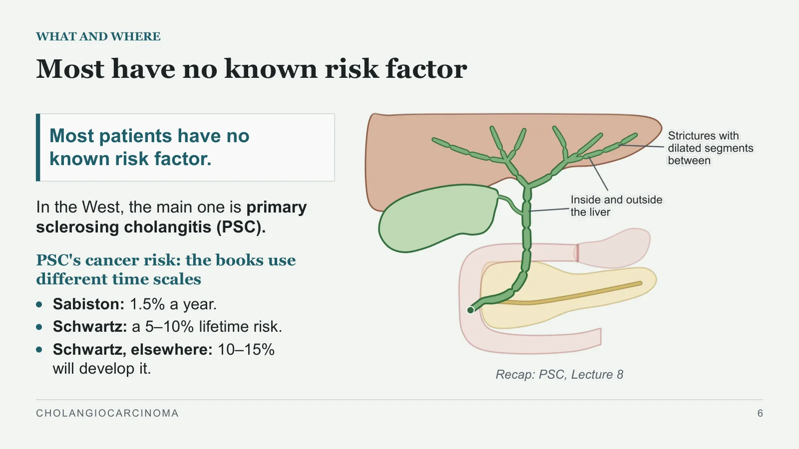
Most have no known risk factor
WHAT AND WHERE
Recap: PSC, Lecture 8
Most patients have no known risk factor.
In the West, the main one is primary sclerosing cholangitis (PSC).
PSC's cancer risk: the books use different time scales
Sabiston: 1.5% a year.
Schwartz: a 5–10% lifetime risk.
Schwartz, elsewhere: 10–15% will develop it.
CHOLANGIOCARCINOMA
6
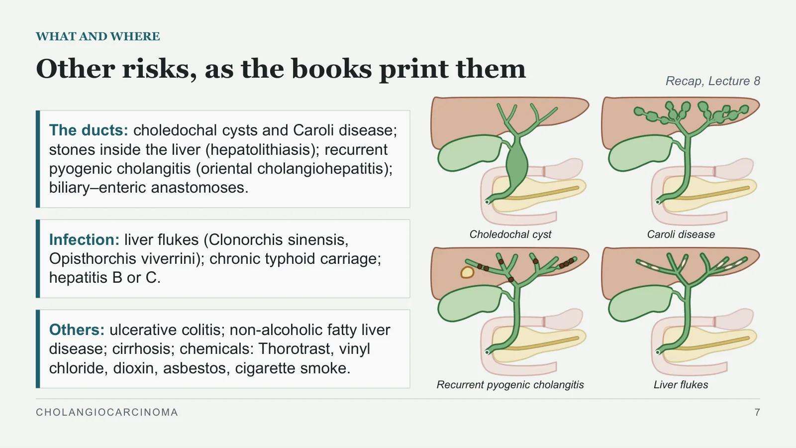
Other risks, as the books print them
WHAT AND WHERE
The ducts: choledochal cysts and Caroli disease; stones inside the liver (hepatolithiasis); recurrent pyogenic cholangitis (oriental cholangiohepatitis); biliary–enteric anastomoses.
Infection: liver flukes (Clonorchis sinensis, Opisthorchis viverrini); chronic typhoid carriage; hepatitis B or C.
Others: ulcerative colitis; non-alcoholic fatty liver disease; cirrhosis; chemicals: Thorotrast, vinyl chloride, dioxin, asbestos, cigarette smoke.
Recap, Lecture 8
Choledochal cyst
Caroli disease
Recurrent pyogenic cholangitis
Liver flukes
CHOLANGIOCARCINOMA
7
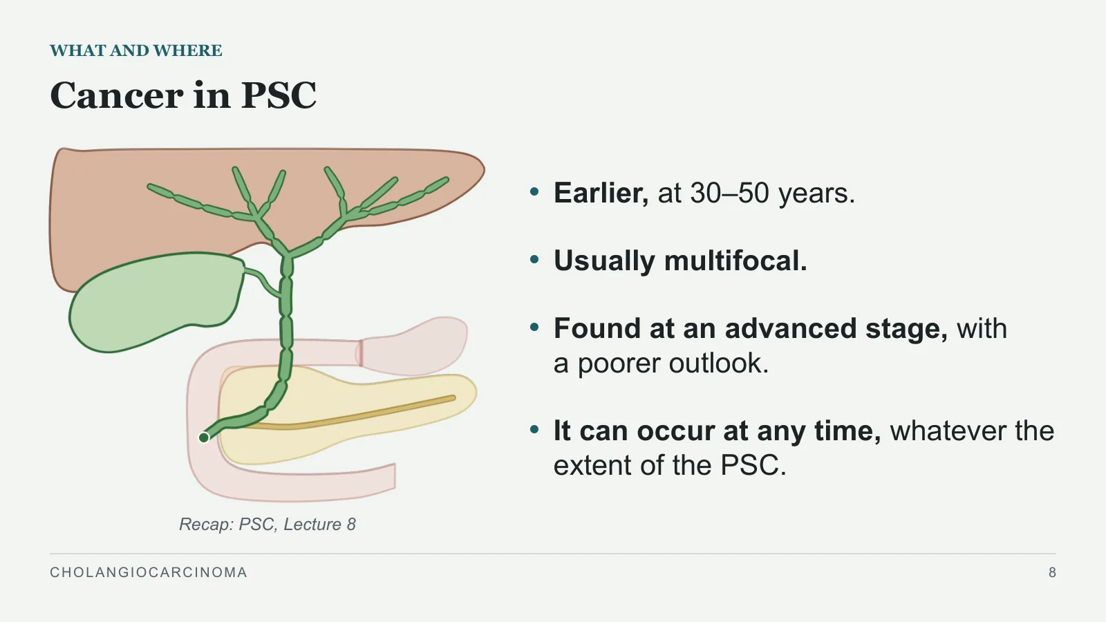
Cancer in PSC
WHAT AND WHERE
Recap: PSC, Lecture 8
Earlier, at 30–50 years.
Usually multifocal.
Found at an advanced stage, with a poorer outlook.
It can occur at any time, whatever the extent of the PSC.
CHOLANGIOCARCINOMA
8
How it grows
How it grows
PART 2
CHOLANGIOCARCINOMA
9
It spreads under the lining
HOW IT GROWS
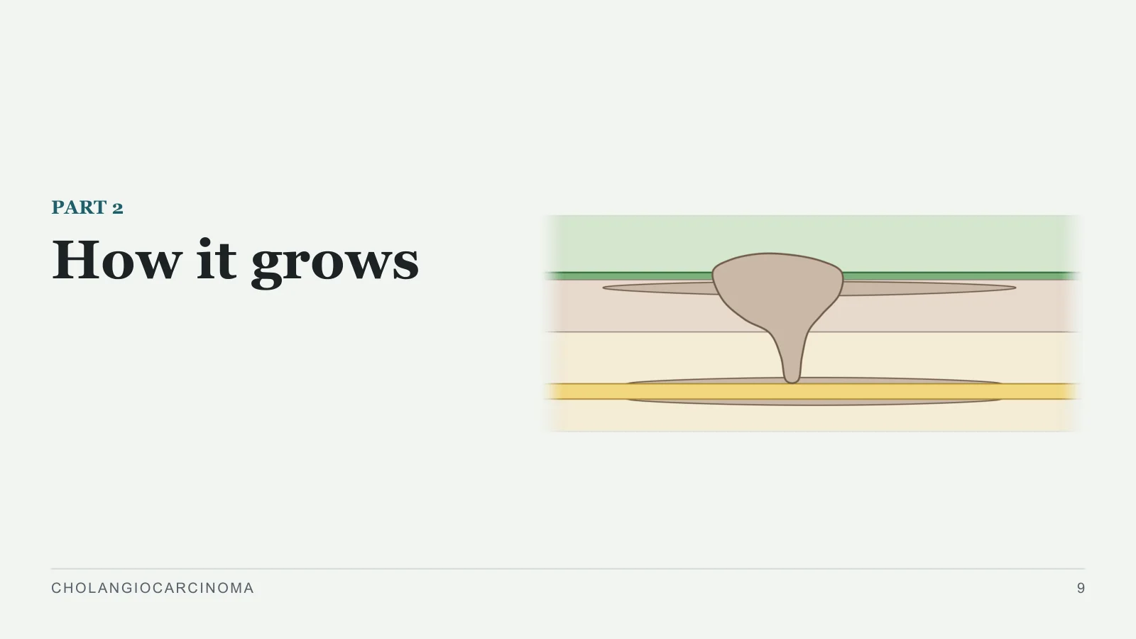
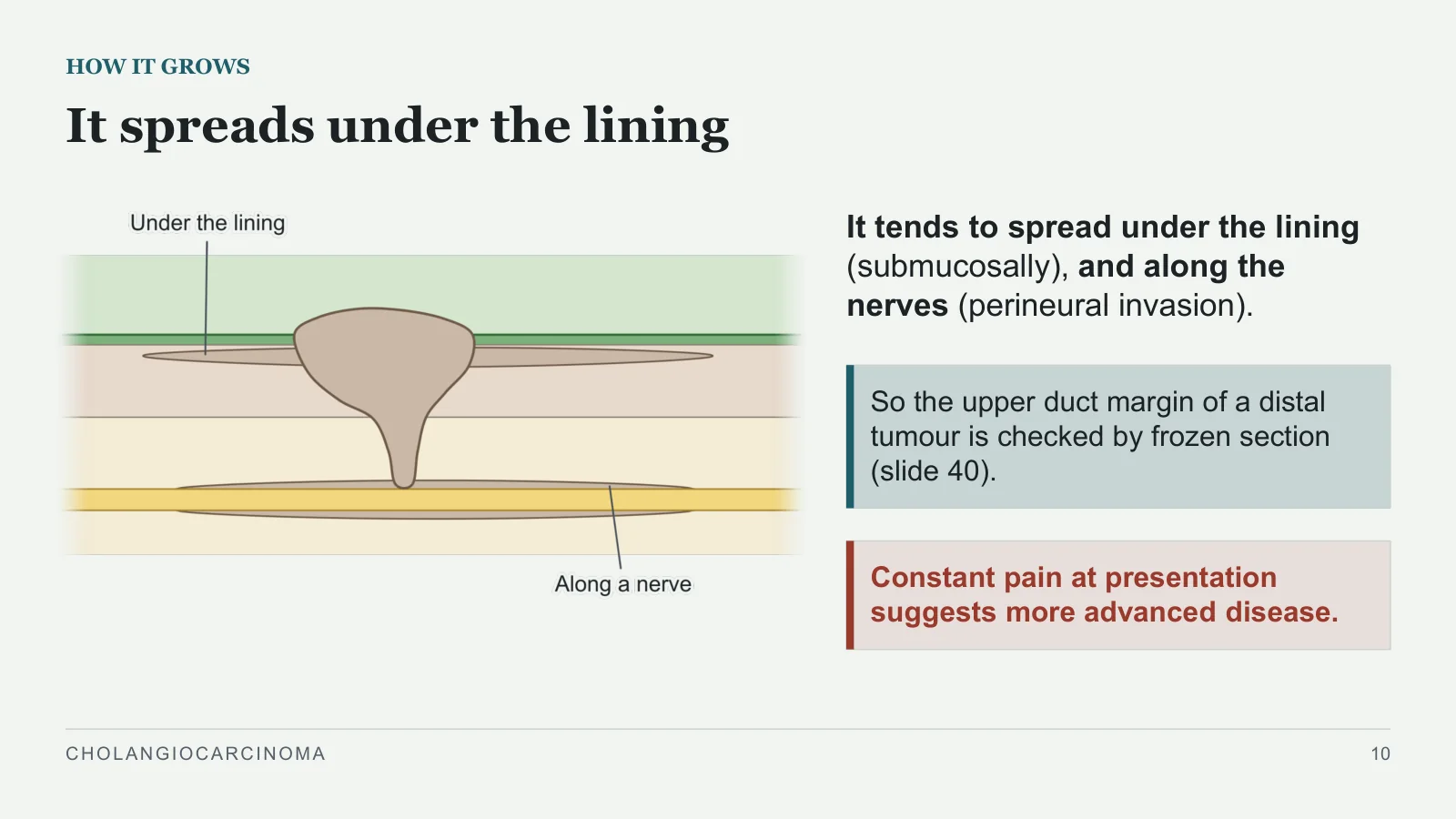
It tends to spread under the lining (submucosally), and along the nerves (perineural invasion).
So the upper duct margin of a distal tumour is checked by frozen section (slide 40).
Constant pain at presentation suggests more advanced disease.
CHOLANGIOCARCINOMA
10
Its types: the books name them differently
HOW IT GROWS
Bailey
28th edition
Three histological types: mass forming, intraductal growing, periductal infiltrating.
Sabiston
Sclerosing, nodular, papillary. Sclerosing tumours tend to be proximal, with fibrosis around the duct and a ring-like narrowing.
Schwartz
Nodular (the commonest), scirrhous, diffusely infiltrating, papillary.
CHOLANGIOCARCINOMA
11
How it presents
How it presents
PART 3
CHOLANGIOCARCINOMA
12
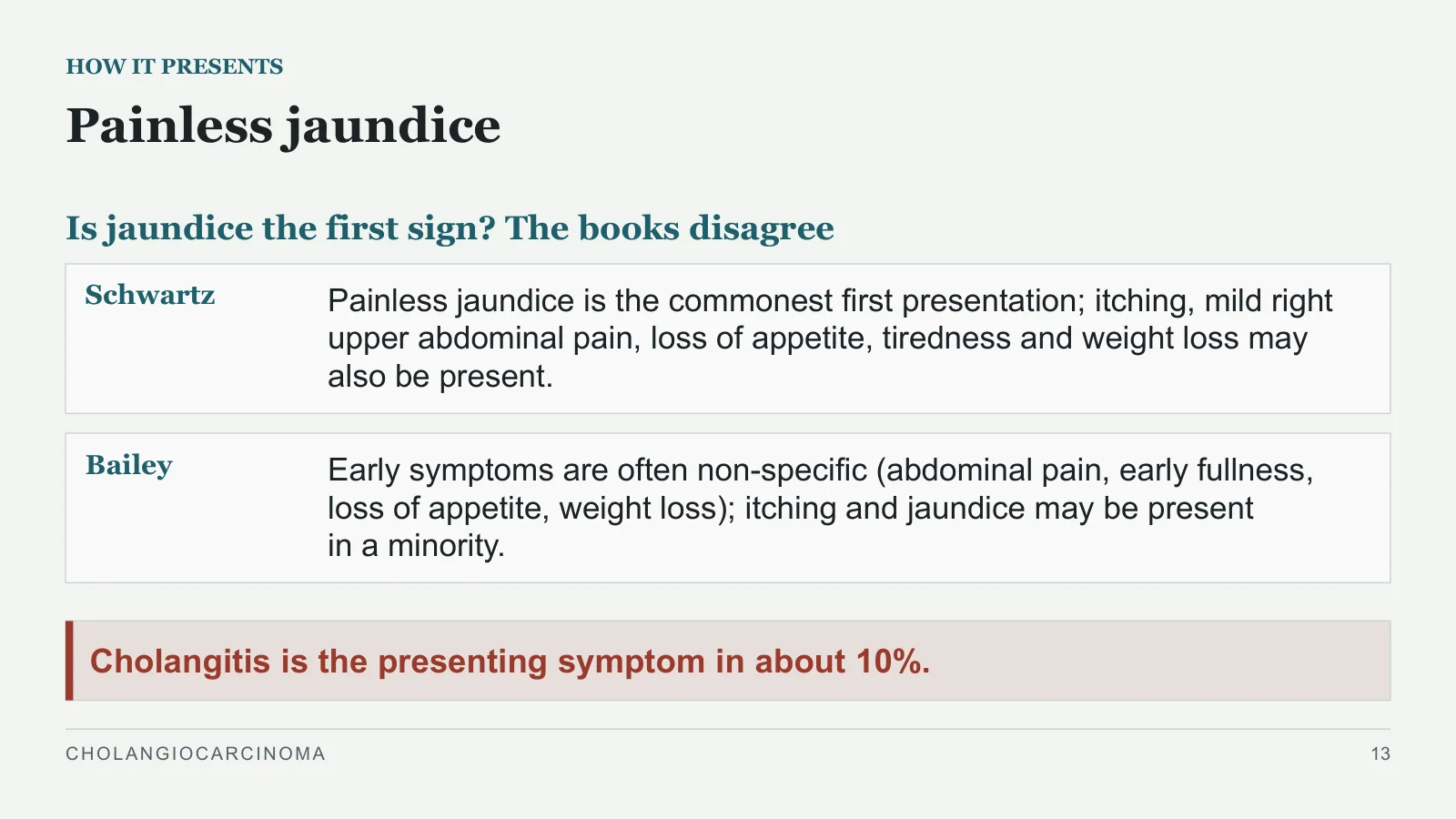
Painless jaundice
HOW IT PRESENTS
Is jaundice the first sign? The books disagree
Schwartz
Painless jaundice is the commonest first presentation; itching, mild right upper abdominal pain, loss of appetite, tiredness and weight loss may also be present.
Bailey
Early symptoms are often non-specific (abdominal pain, early fullness, loss of appetite, weight loss); itching and jaundice may be present in a minority.
Cholangitis is the presenting symptom in about 10%.
CHOLANGIOCARCINOMA
13
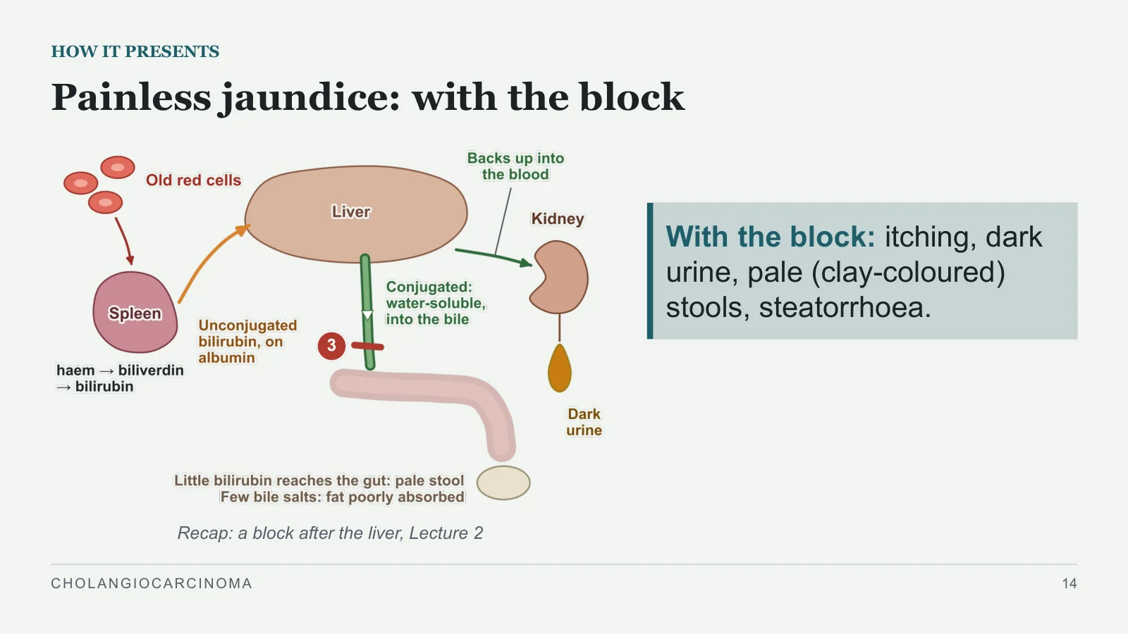
Painless jaundice: with the block
HOW IT PRESENTS
Recap: a block after the liver, Lecture 2
With the block: itching, dark urine, pale (clay-coloured) stools, steatorrhoea.
CHOLANGIOCARCINOMA
14
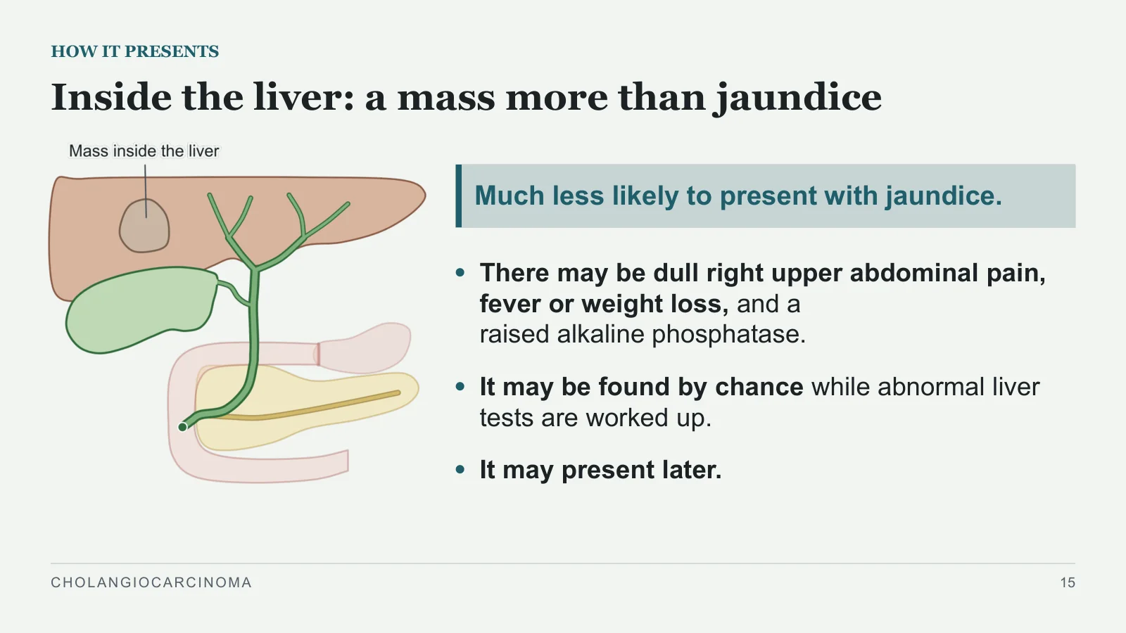
Inside the liver: a mass more than jaundice
HOW IT PRESENTS
Much less likely to present with jaundice.
There may be dull right upper abdominal pain, fever or weight loss, and a raised alkaline phosphatase.
It may be found by chance while abnormal liver tests are worked up.
It may present later.
CHOLANGIOCARCINOMA
15
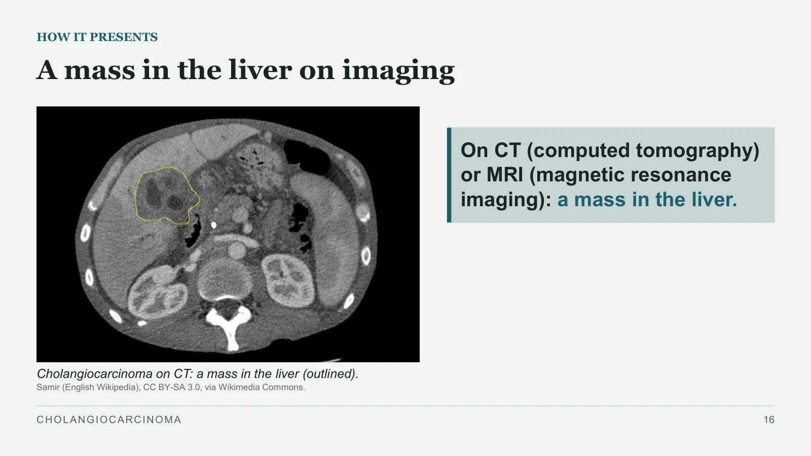
A mass in the liver on imaging
HOW IT PRESENTS
Cholangiocarcinoma on CT: a mass in the liver (outlined).
Samir (English Wikipedia), CC BY-SA 3.0, via Wikimedia Commons.
On CT (computed tomography) or MRI (magnetic resonance imaging): a mass in the liver.
CHOLANGIOCARCINOMA
16
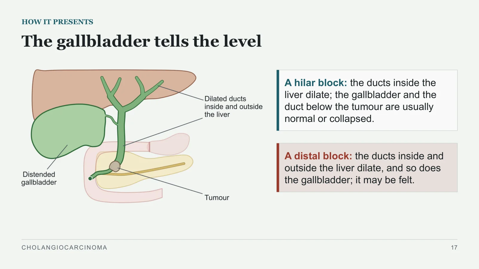
The gallbladder tells the level
HOW IT PRESENTS
A hilar block: the ducts inside the liver dilate; the gallbladder and the duct below the tumour are usually normal or collapsed.
A distal block: the ducts inside and outside the liver dilate, and so does the gallbladder; it may be felt.
CHOLANGIOCARCINOMA
17
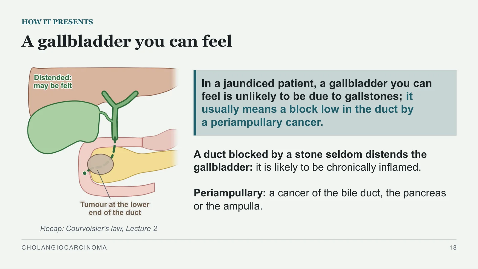
A gallbladder you can feel
HOW IT PRESENTS
Recap: Courvoisier's law, Lecture 2
In a jaundiced patient, a gallbladder you can feel is unlikely to be due to gallstones; it usually means a block low in the duct by a periampullary cancer.
A duct blocked by a stone seldom distends the gallbladder: it is likely to be chronically inflamed.
Periampullary: a cancer of the bile duct, the pancreas or the ampulla.
CHOLANGIOCARCINOMA
18
Investigation
Investigation
PART 4
CHOLANGIOCARCINOMA
19
Ultrasound first
INVESTIGATION
Lecture 2: the tools
Ultrasound comes first: it shows dilated ducts and the level of the block.
For this cancer, how much it shows: the books differ
Sabiston
It may show dilated ducts inside the liver, but does not usually show the site of the block.
Schwartz
The first test is ultrasound or CT; the tumour itself is usually hard to see on ultrasound, CT or even MRCP (magnetic resonance cholangiopancreatography).
Reading the pattern
Dilated ducts in both lobes suggest a hilar tumour.
Dilated ducts inside and outside the liver, with no pancreatic head mass or stones, suggest a distal cholangiocarcinoma.
CHOLANGIOCARCINOMA
20
CT and MRCP: the extent and the vessels
INVESTIGATION
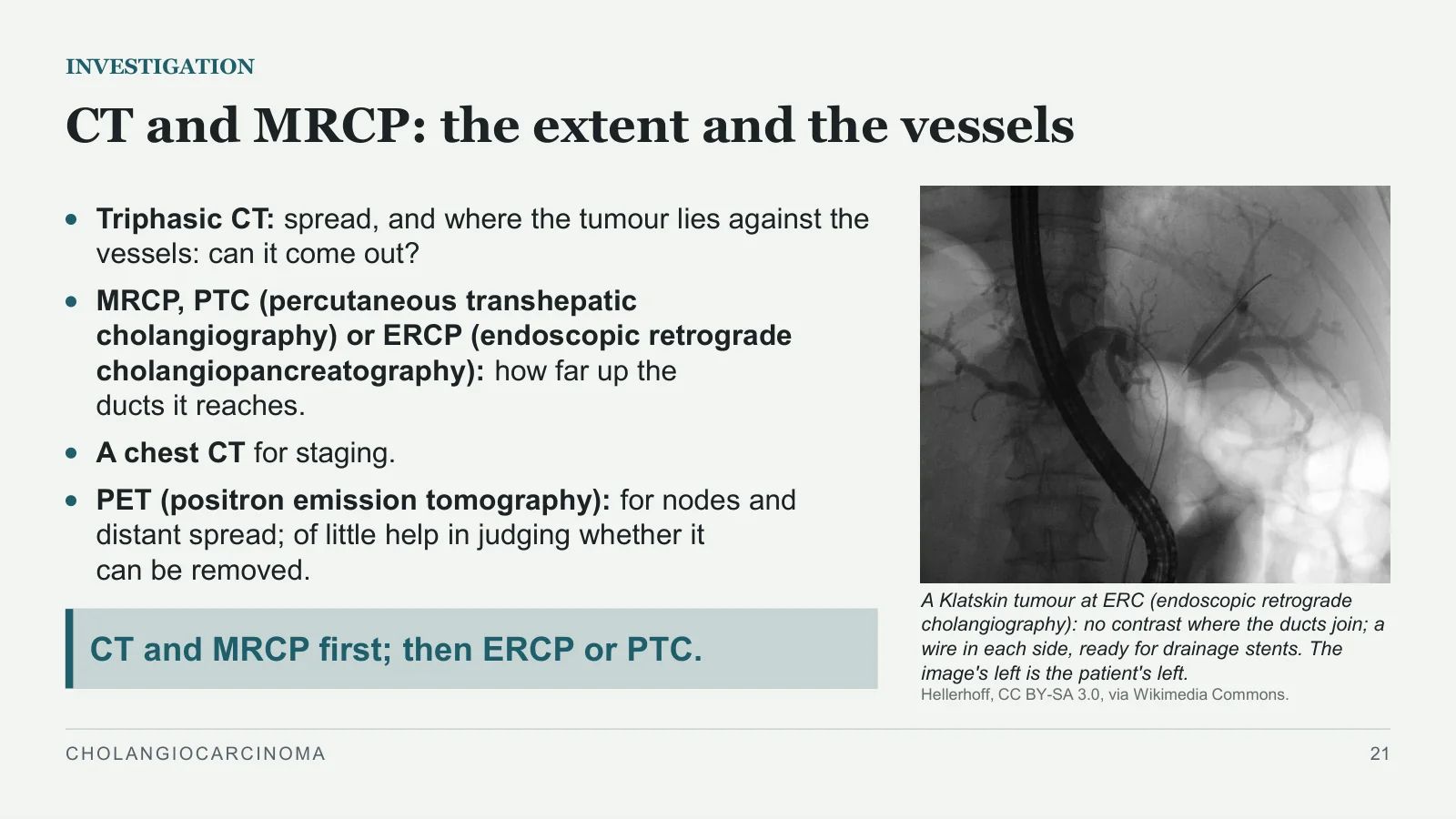
A Klatskin tumour at ERC (endoscopic retrograde cholangiography): no contrast where the ducts join; a wire in each side, ready for drainage stents. The image's left is the patient's left.
Hellerhoff, CC BY-SA 3.0, via Wikimedia Commons.
Triphasic CT: spread, and where the tumour lies against the vessels: can it come out?
MRCP, PTC (percutaneous transhepatic cholangiography) or ERCP (endoscopic retrograde cholangiopancreatography): how far up the ducts it reaches.
A chest CT for staging.
PET (positron emission tomography): for nodes and distant spread; of little help in judging whether it can be removed.
CT and MRCP first; then ERCP or PTC.
CHOLANGIOCARCINOMA
21
One lobe shrinks, the other grows
INVESTIGATION
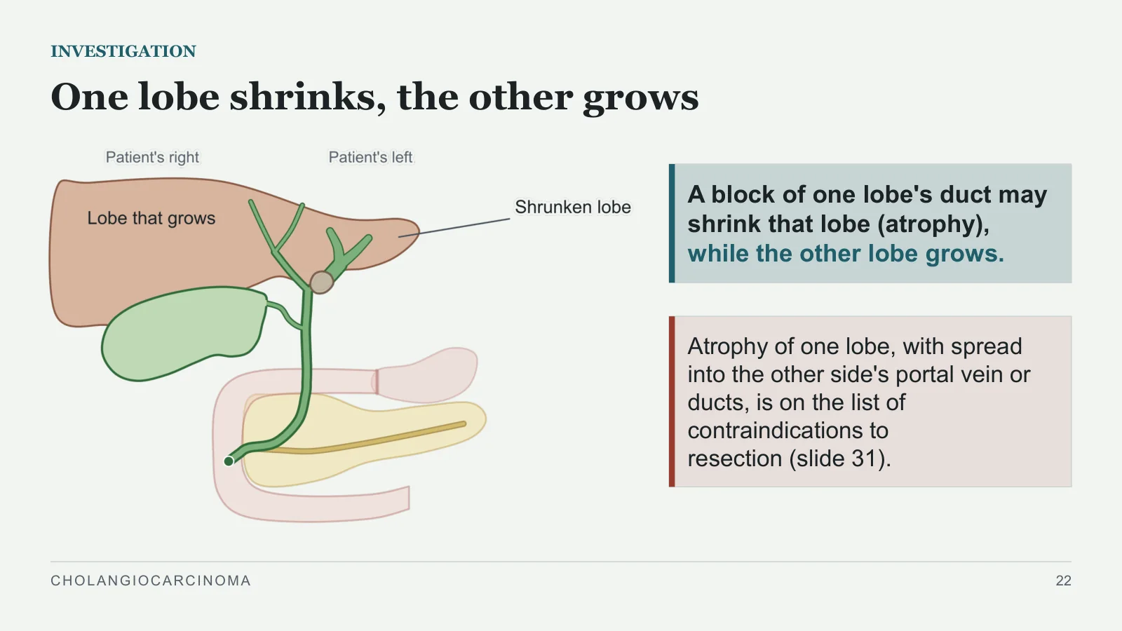
A block of one lobe's duct may shrink that lobe (atrophy), while the other lobe grows.
Atrophy of one lobe, with spread into the other side's portal vein or ducts, is on the list of contraindications to resection (slide 31).
CHOLANGIOCARCINOMA
22
Markers: the books disagree
INVESTIGATION
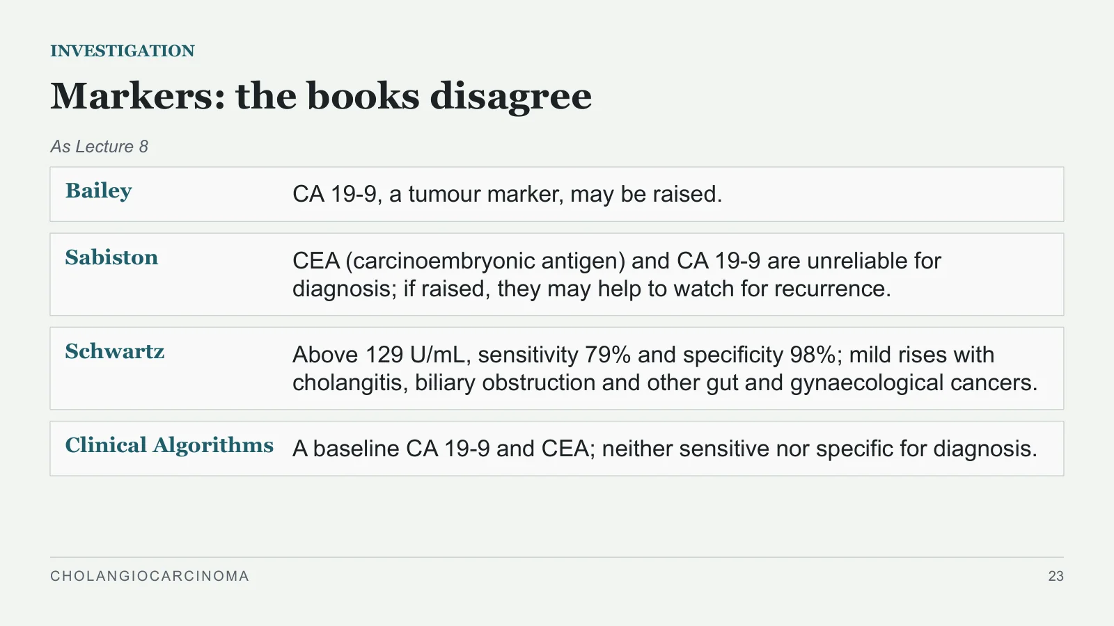
As Lecture 8
Bailey
CA 19-9, a tumour marker, may be raised.
Sabiston
CEA (carcinoembryonic antigen) and CA 19-9 are unreliable for diagnosis; if raised, they may help to watch for recurrence.
Schwartz
Above 129 U/mL, sensitivity 79% and specificity 98%; mild rises with cholangitis, biliary obstruction and other gut and gynaecological cancers.
Clinical Algorithms
A baseline CA 19-9 and CEA; neither sensitive nor specific for diagnosis.
CHOLANGIOCARCINOMA
23
Sampling often misses it
INVESTIGATION
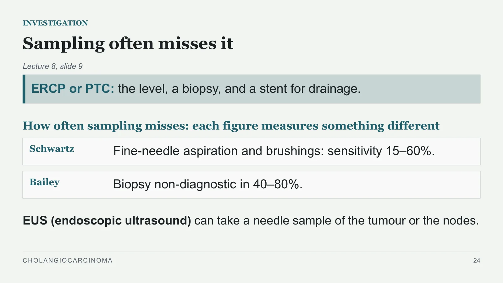
Lecture 8, slide 9
ERCP or PTC: the level, a biopsy, and a stent for drainage.
How often sampling misses: each figure measures something different
Schwartz
Fine-needle aspiration and brushings: sensitivity 15–60%.
Bailey
Biopsy non-diagnostic in 40–80%.
EUS (endoscopic ultrasound) can take a needle sample of the tumour or the nodes.
CHOLANGIOCARCINOMA
24
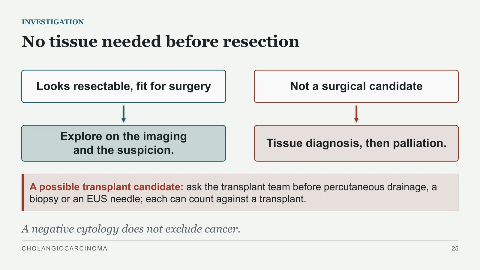
No tissue needed before resection
INVESTIGATION
Looks resectable, fit for surgery
Explore on the imaging and the suspicion.
Not a surgical candidate
Tissue diagnosis, then palliation.
A possible transplant candidate: ask the transplant team before percutaneous drainage, a biopsy or an EUS needle; each can count against a transplant.
A negative cytology does not exclude cancer.
CHOLANGIOCARCINOMA
25
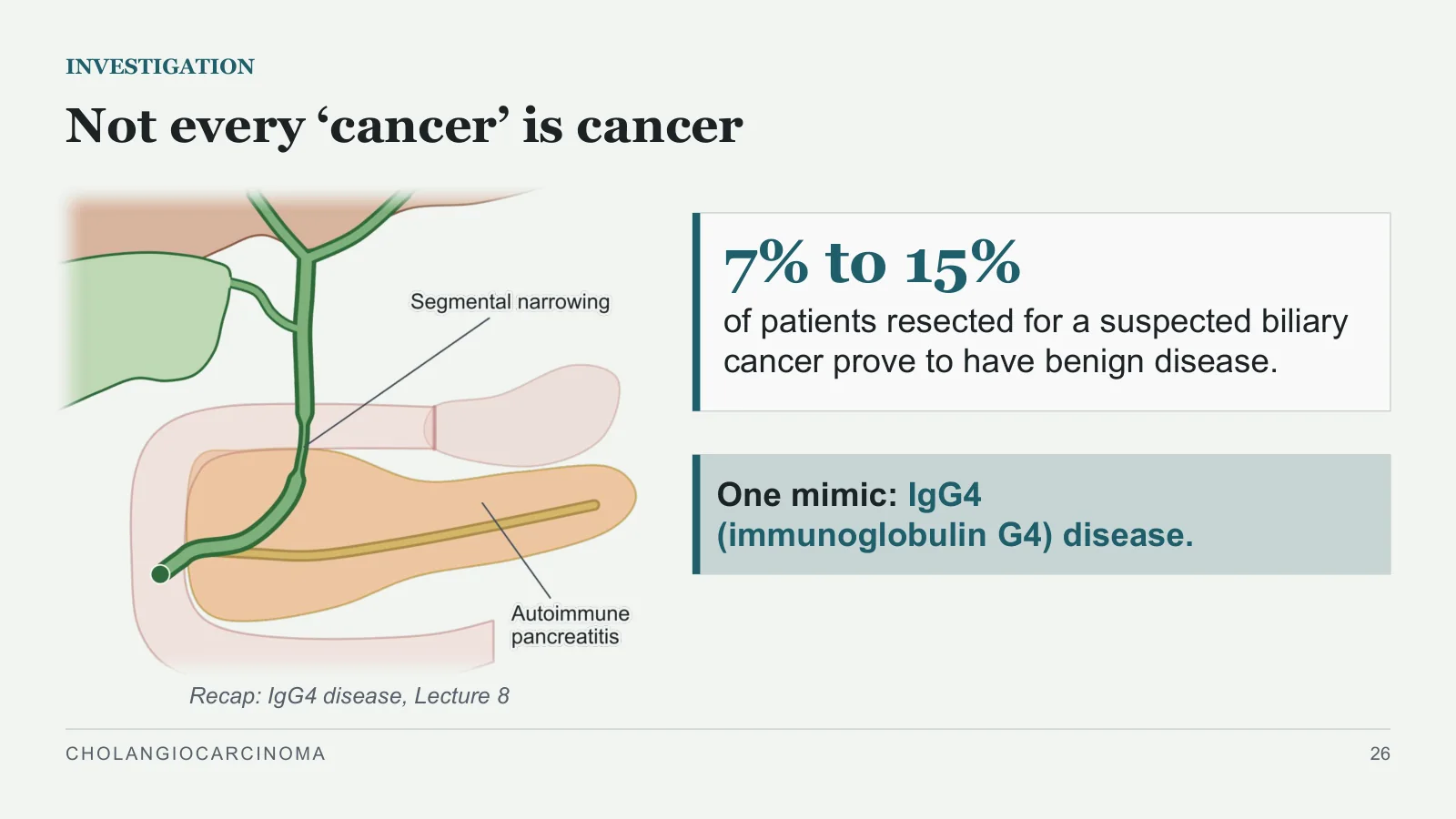
7% to 15%
INVESTIGATION
Not every ‘cancer’ is cancer
Recap: IgG4 disease, Lecture 8
of patients resected for a suspected biliary cancer prove to have benign disease.
One mimic: IgG4 (immunoglobulin G4) disease.
CHOLANGIOCARCINOMA
26
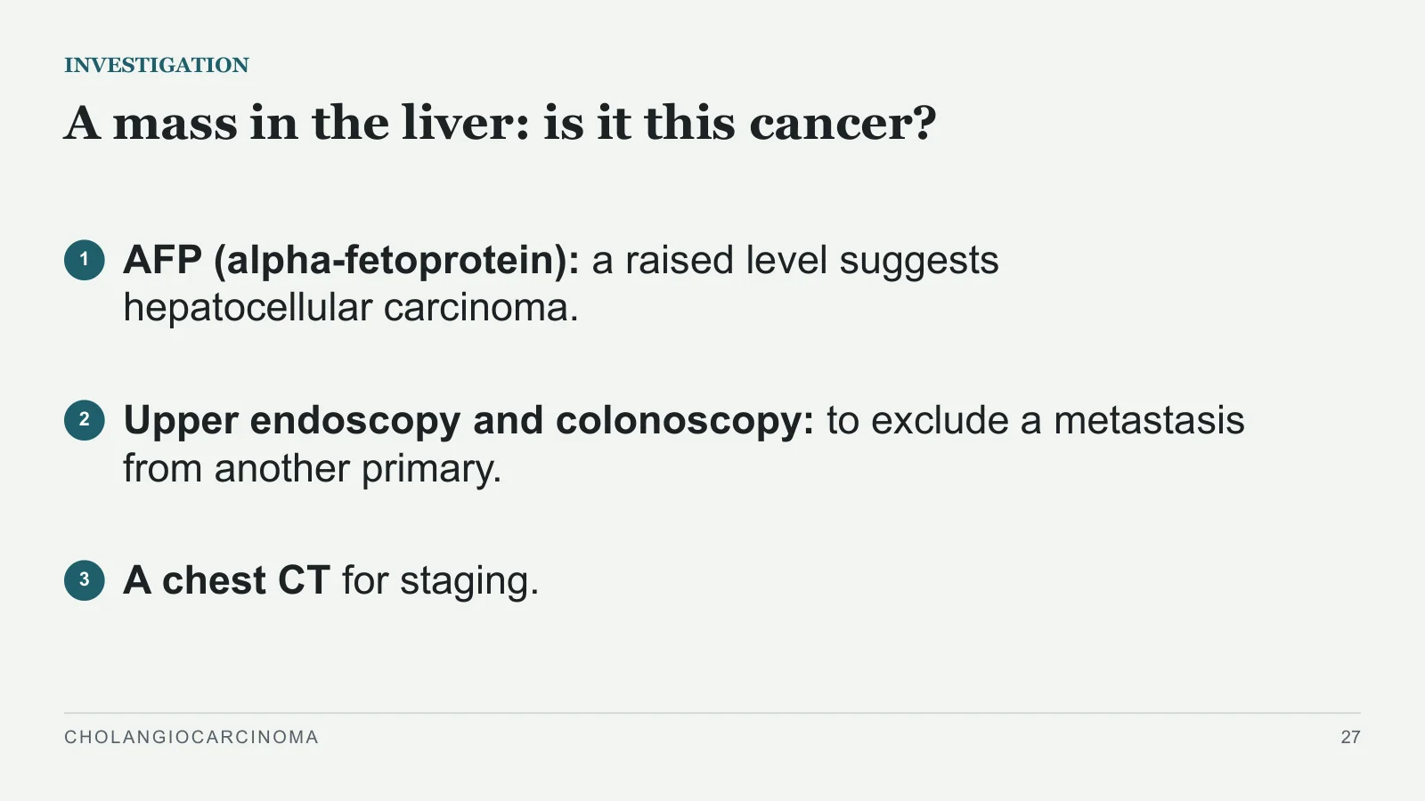
A mass in the liver: is it this cancer?
INVESTIGATION
1
AFP (alpha-fetoprotein): a raised level suggests hepatocellular carcinoma.
2
Upper endoscopy and colonoscopy: to exclude a metastasis from another primary.
3
A chest CT for staging.
CHOLANGIOCARCINOMA
27
Staging and resectability
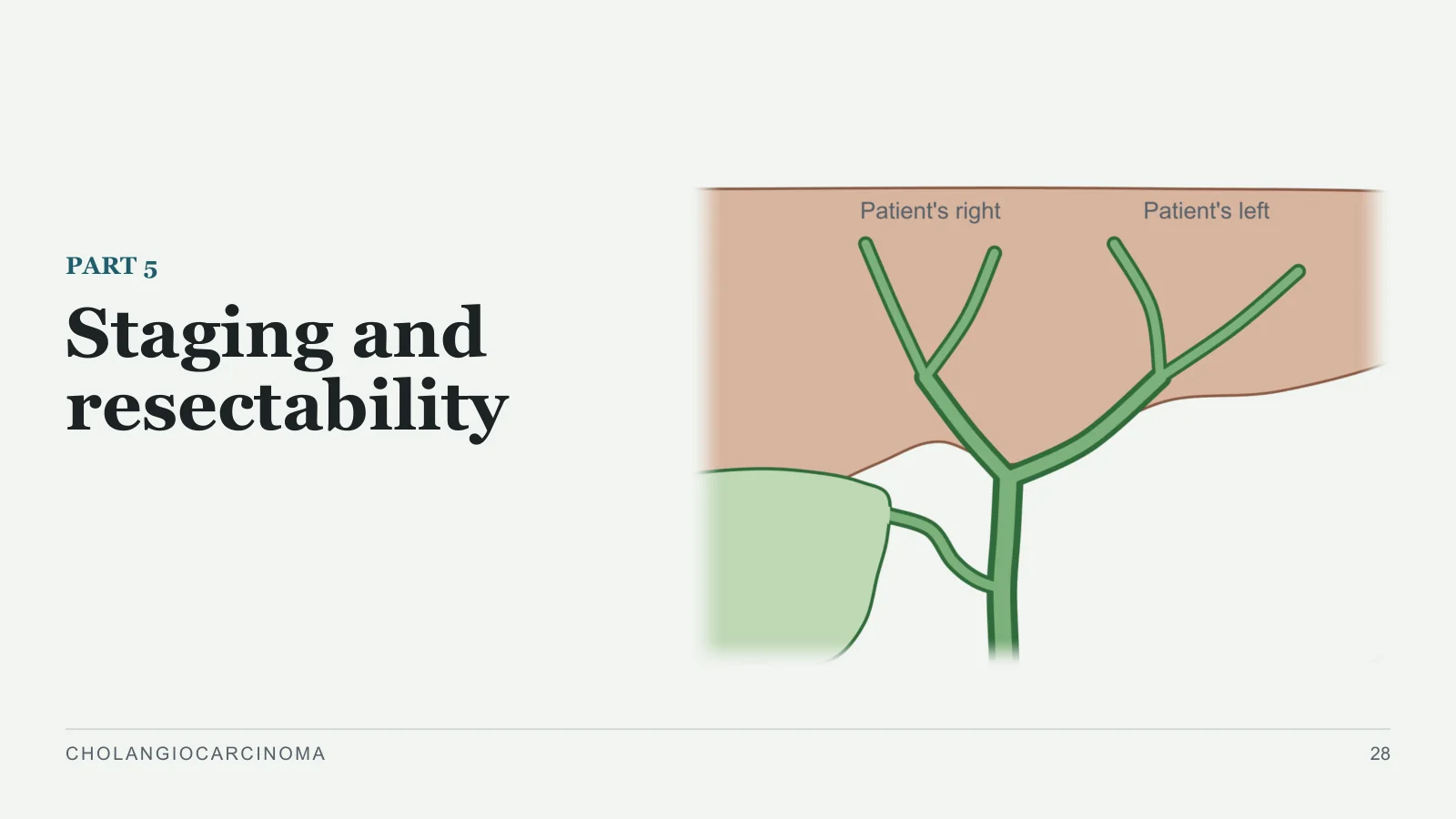
Staging and resectability
PART 5
CHOLANGIOCARCINOMA
28
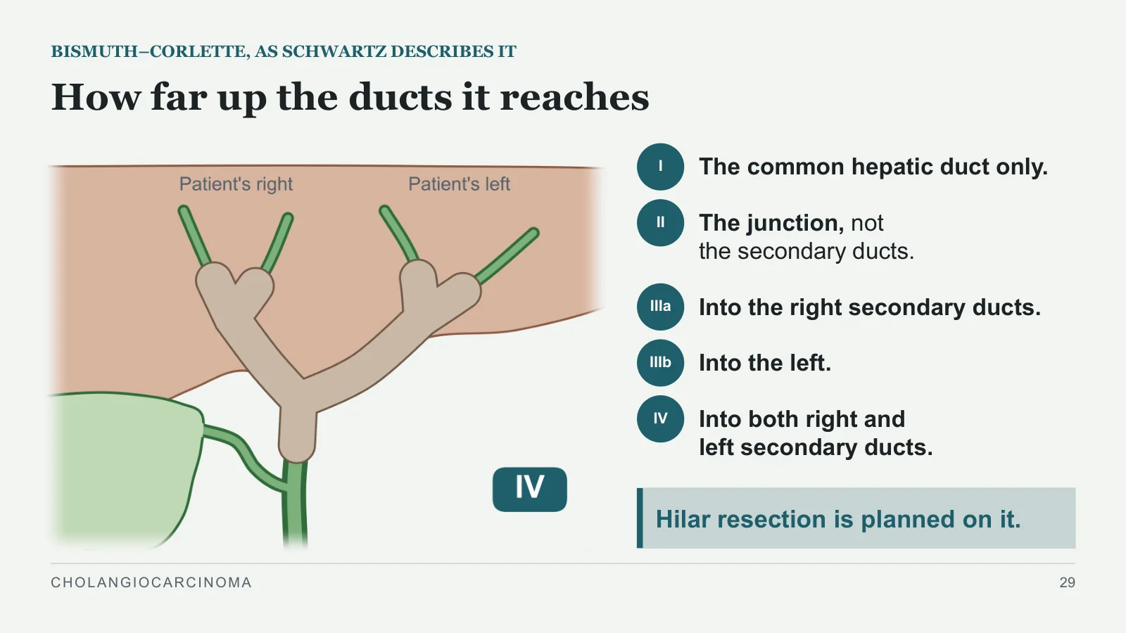
How far up the ducts it reaches
BISMUTH–CORLETTE, AS SCHWARTZ DESCRIBES IT
I
The common hepatic duct only.
II
The junction, not the secondary ducts.
IIIa
Into the right secondary ducts.
IIIb
Into the left.
IV
Into both right and left secondary ducts.
Hilar resection is planned on it.
CHOLANGIOCARCINOMA
29
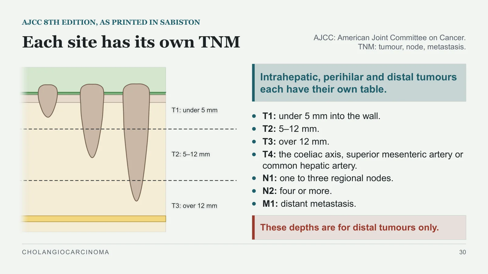
Each site has its own TNM
AJCC 8TH EDITION, AS PRINTED IN SABISTON
AJCC: American Joint Committee on Cancer. TNM: tumour, node, metastasis.
Intrahepatic, perihilar and distal tumours each have their own table.
T1: under 5 mm into the wall.
T2: 5–12 mm.
T3: over 12 mm.
T4: the coeliac axis, superior mesenteric artery or common hepatic artery.
N1: one to three regional nodes.
N2: four or more.
M1: distant metastasis.
These depths are for distal tumours only.
CHOLANGIOCARCINOMA
30
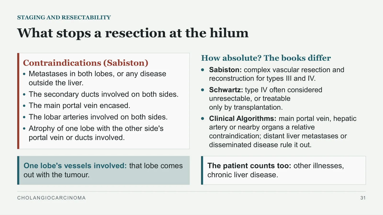
What stops a resection at the hilum
STAGING AND RESECTABILITY
Contraindications (Sabiston)
Metastases in both lobes, or any disease outside the liver.
The secondary ducts involved on both sides.
The main portal vein encased.
The lobar arteries involved on both sides.
Atrophy of one lobe with the other side's portal vein or ducts involved.
How absolute? The books differ
Sabiston: complex vascular resection and reconstruction for types III and IV.
Schwartz: type IV often considered unresectable, or treatable only by transplantation.
Clinical Algorithms: main portal vein, hepatic artery or nearby organs a relative contraindication; distant liver metastases or disseminated disease rule it out.
One lobe's vessels involved: that lobe comes out with the tumour.
The patient counts too: other illnesses, chronic liver disease.
CHOLANGIOCARCINOMA
31
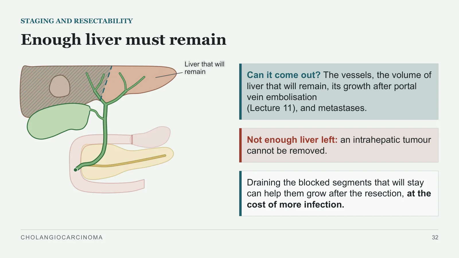
Enough liver must remain
STAGING AND RESECTABILITY
Can it come out? The vessels, the volume of liver that will remain, its growth after portal vein embolisation (Lecture 11), and metastases.
Not enough liver left: an intrahepatic tumour cannot be removed.
Draining the blocked segments that will stay can help them grow after the resection, at the cost of more infection.
CHOLANGIOCARCINOMA
32
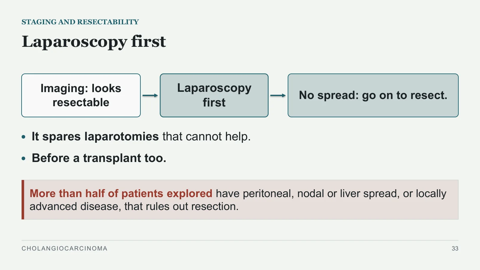
Laparoscopy first
STAGING AND RESECTABILITY
Imaging: looks resectable
Laparoscopy first
No spread: go on to resect.
It spares laparotomies that cannot help.
Before a transplant too.
More than half of patients explored have peritoneal, nodal or liver spread, or locally advanced disease, that rules out resection.
CHOLANGIOCARCINOMA
33
Treatment
Treatment
PART 6
CHOLANGIOCARCINOMA
34
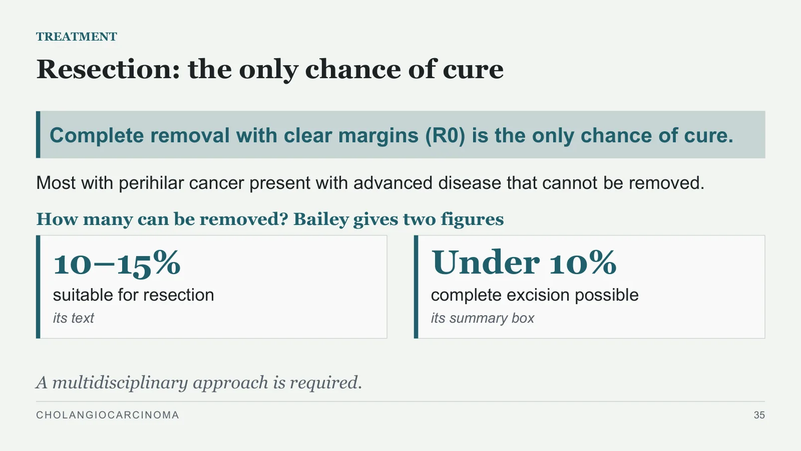
10–15%
TREATMENT
Resection: the only chance of cure
Complete removal with clear margins (R0) is the only chance of cure.
Most with perihilar cancer present with advanced disease that cannot be removed.
How many can be removed? Bailey gives two figures
suitable for resection
its text
Under 10%
complete excision possible
its summary box
A multidisciplinary approach is required.
CHOLANGIOCARCINOMA
35
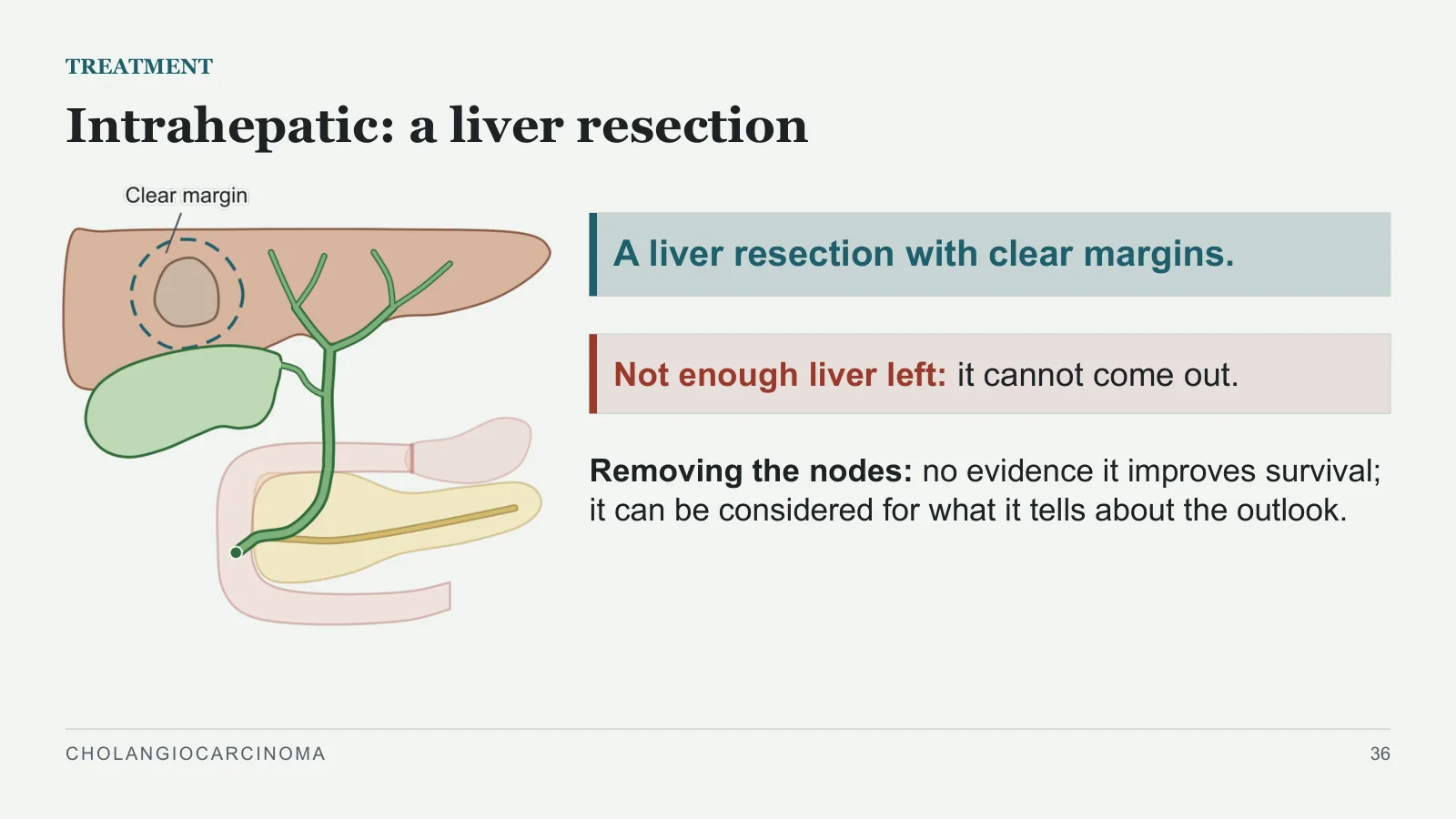
Intrahepatic: a liver resection
TREATMENT
A liver resection with clear margins.
Not enough liver left: it cannot come out.
Removing the nodes: no evidence it improves survival; it can be considered for what it tells about the outlook.
CHOLANGIOCARCINOMA
36
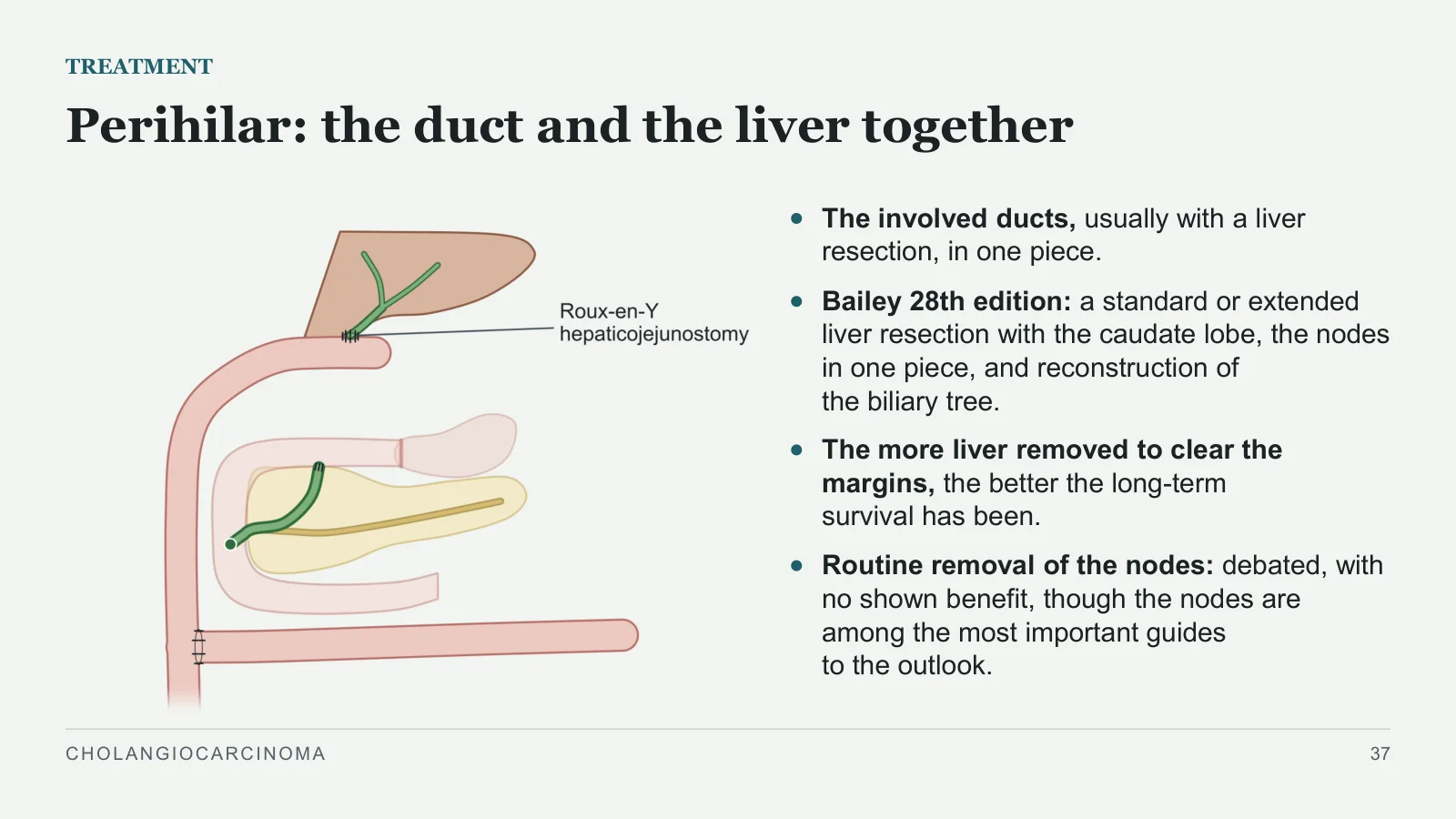
Perihilar: the duct and the liver together
TREATMENT
The involved ducts, usually with a liver resection, in one piece.
Bailey 28th edition: a standard or extended liver resection with the caudate lobe, the nodes in one piece, and reconstruction of the biliary tree.
The more liver removed to clear the margins, the better the long-term survival has been.
Routine removal of the nodes: debated, with no shown benefit, though the nodes are among the most important guides to the outlook.
CHOLANGIOCARCINOMA
37
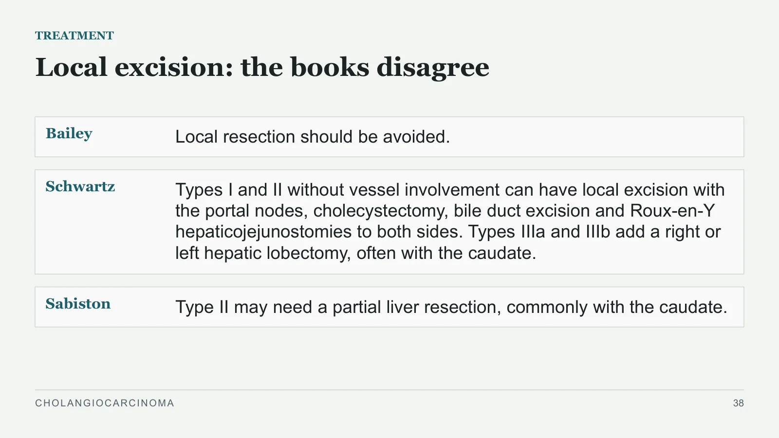
Local excision: the books disagree
TREATMENT
Bailey
Local resection should be avoided.
Schwartz
Types I and II without vessel involvement can have local excision with the portal nodes, cholecystectomy, bile duct excision and Roux-en-Y hepaticojejunostomies to both sides. Types IIIa and IIIb add a right or left hepatic lobectomy, often with the caudate.
Sabiston
Type II may need a partial liver resection, commonly with the caudate.
CHOLANGIOCARCINOMA
38
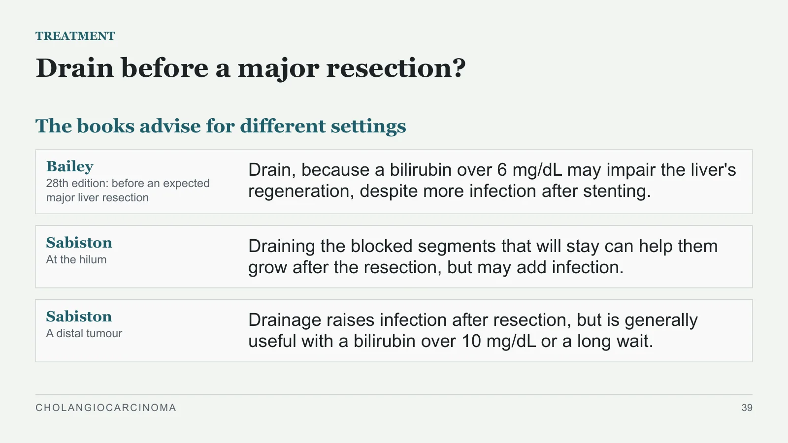
Drain before a major resection?
TREATMENT
The books advise for different settings
Bailey
28th edition: before an expected major liver resection
Drain, because a bilirubin over 6 mg/dL may impair the liver's regeneration, despite more infection after stenting.
Sabiston
At the hilum
Draining the blocked segments that will stay can help them grow after the resection, but may add infection.
Sabiston
A distal tumour
Drainage raises infection after resection, but is generally useful with a bilirubin over 10 mg/dL or a long wait.
CHOLANGIOCARCINOMA
39
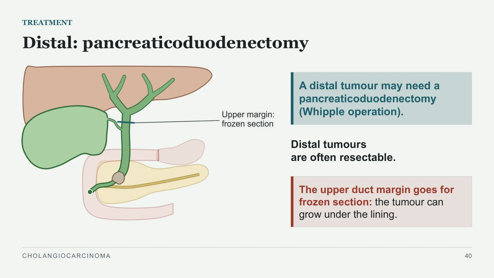
Distal: pancreaticoduodenectomy
TREATMENT
A distal tumour may need a pancreaticoduodenectomy (Whipple operation).
Distal tumours are often resectable.
The upper duct margin goes for frozen section: the tumour can grow under the lining.
CHOLANGIOCARCINOMA
40
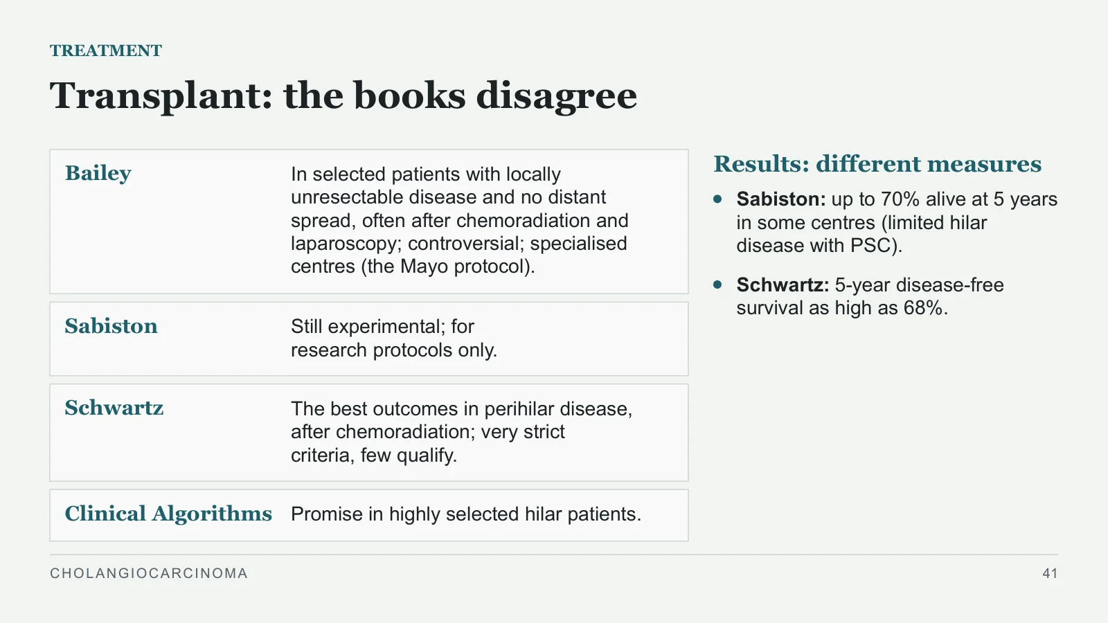
Transplant: the books disagree
TREATMENT
Bailey
In selected patients with locally unresectable disease and no distant spread, often after chemoradiation and laparoscopy; controversial; specialised centres (the Mayo protocol).
Sabiston
Still experimental; for research protocols only.
Schwartz
The best outcomes in perihilar disease, after chemoradiation; very strict criteria, few qualify.
Clinical Algorithms
Promise in highly selected hilar patients.
Results: different measures
Sabiston: up to 70% alive at 5 years in some centres (limited hilar disease with PSC).
Schwartz: 5-year disease-free survival as high as 68%.
CHOLANGIOCARCINOMA
41
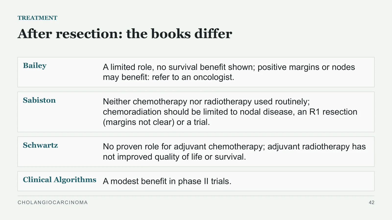
After resection: the books differ
TREATMENT
Bailey
A limited role, no survival benefit shown; positive margins or nodes may benefit: refer to an oncologist.
Sabiston
Neither chemotherapy nor radiotherapy used routinely; chemoradiation should be limited to nodal disease, an R1 resection (margins not clear) or a trial.
Schwartz
No proven role for adjuvant chemotherapy; adjuvant radiotherapy has not improved quality of life or survival.
Clinical Algorithms
A modest benefit in phase II trials.
CHOLANGIOCARCINOMA
42
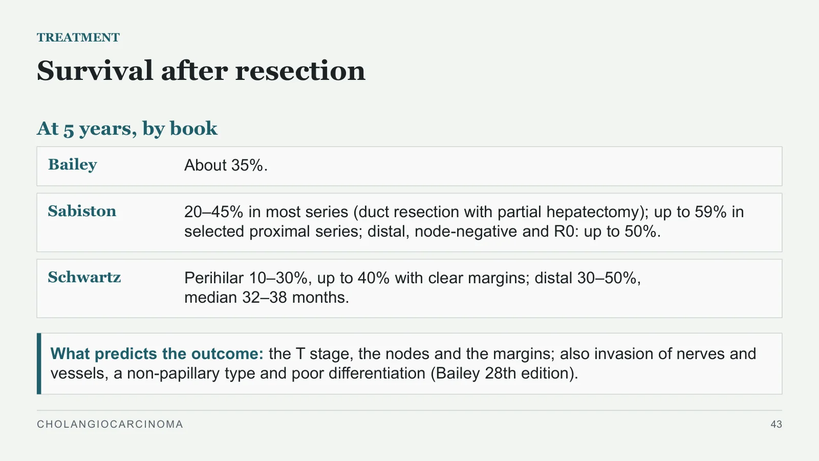
Survival after resection
TREATMENT
At 5 years, by book
Bailey
About 35%.
Sabiston
20–45% in most series (duct resection with partial hepatectomy); up to 59% in selected proximal series; distal, node-negative and R0: up to 50%.
Schwartz
Perihilar 10–30%, up to 40% with clear margins; distal 30–50%, median 32–38 months.
What predicts the outcome: the T stage, the nodes and the margins; also invasion of nerves and vessels, a non-papillary type and poor differentiation (Bailey 28th edition).
CHOLANGIOCARCINOMA
43
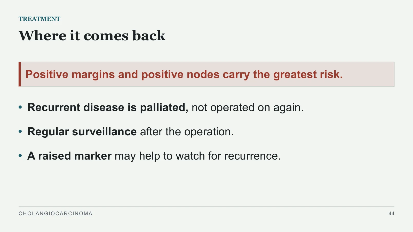
Where it comes back
TREATMENT
Positive margins and positive nodes carry the greatest risk.
Recurrent disease is palliated, not operated on again.
Regular surveillance after the operation.
A raised marker may help to watch for recurrence.
CHOLANGIOCARCINOMA
44
Palliation
Palliation
PART 7
CHOLANGIOCARCINOMA
45
5 to 8 months
PALLIATION
Most need palliation
The majority receive palliative care only.
The aims: relieve the symptoms (jaundice, itching, pain, a blocked duodenum), and prevent liver failure and cholangitis.
Without resection, median survival.
CHOLANGIOCARCINOMA
46
Endoscopic or through the liver
PALLIATION
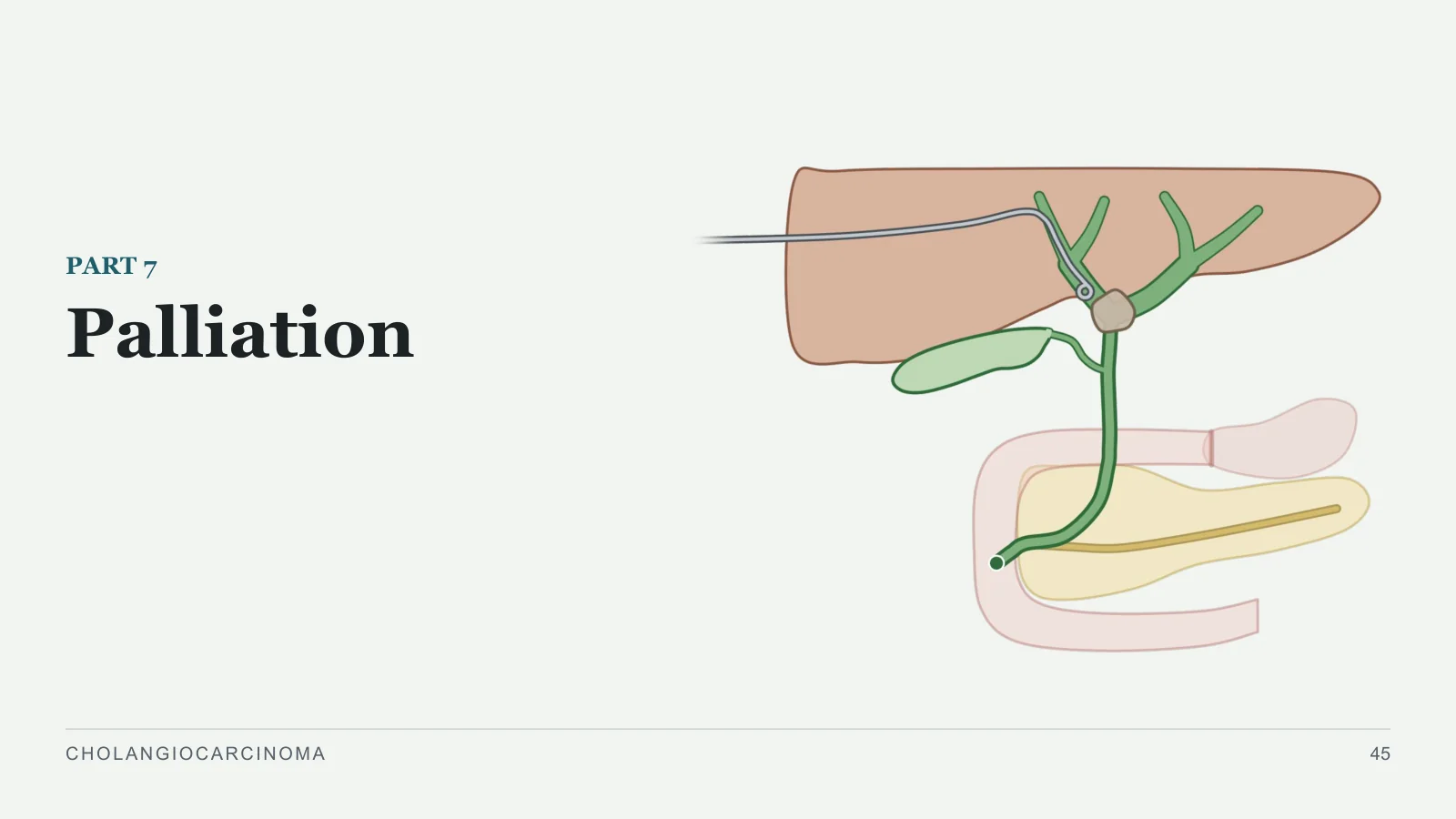
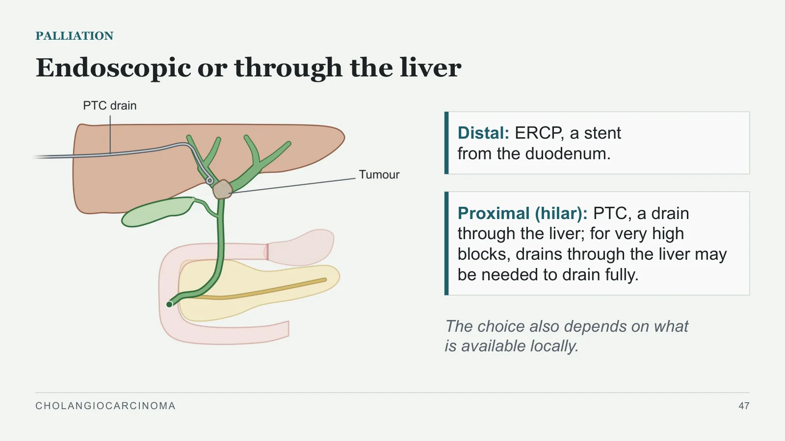
Distal: ERCP, a stent from the duodenum.
Proximal (hilar): PTC, a drain through the liver; for very high blocks, drains through the liver may be needed to drain fully.
The choice also depends on what is available locally.
CHOLANGIOCARCINOMA
47
Which stent, which segments
PALLIATION
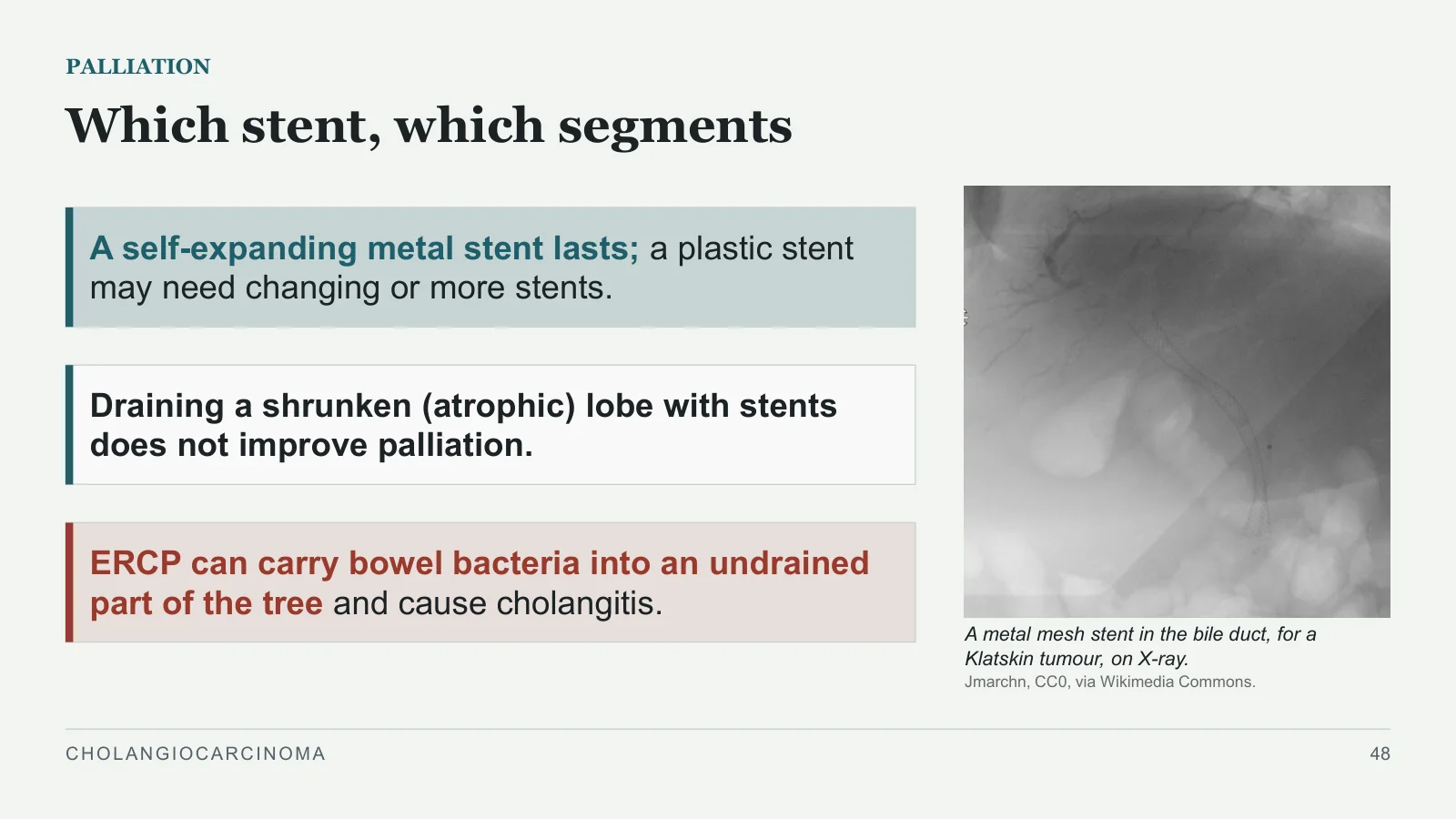
A metal mesh stent in the bile duct, for a Klatskin tumour, on X-ray.
Jmarchn, CC0, via Wikimedia Commons.
A self-expanding metal stent lasts; a plastic stent may need changing or more stents.
Draining a shrunken (atrophic) lobe with stents does not improve palliation.
ERCP can carry bowel bacteria into an undrained part of the tree and cause cholangitis.
CHOLANGIOCARCINOMA
48
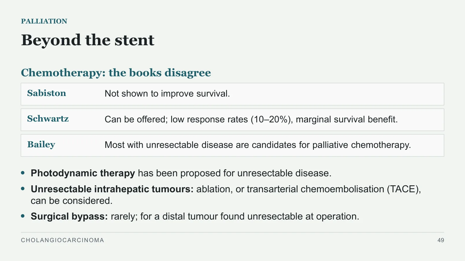
Beyond the stent
PALLIATION
Chemotherapy: the books disagree
Sabiston
Not shown to improve survival.
Schwartz
Can be offered; low response rates (10–20%), marginal survival benefit.
Bailey
Most with unresectable disease are candidates for palliative chemotherapy.
Photodynamic therapy has been proposed for unresectable disease.
Unresectable intrahepatic tumours: ablation, or transarterial chemoembolisation (TACE), can be considered.
Surgical bypass: rarely; for a distal tumour found unresectable at operation.
CHOLANGIOCARCINOMA
49
Early detection
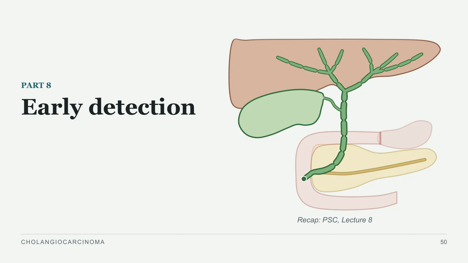
Early detection
PART 8
Recap: PSC, Lecture 8
CHOLANGIOCARCINOMA
50
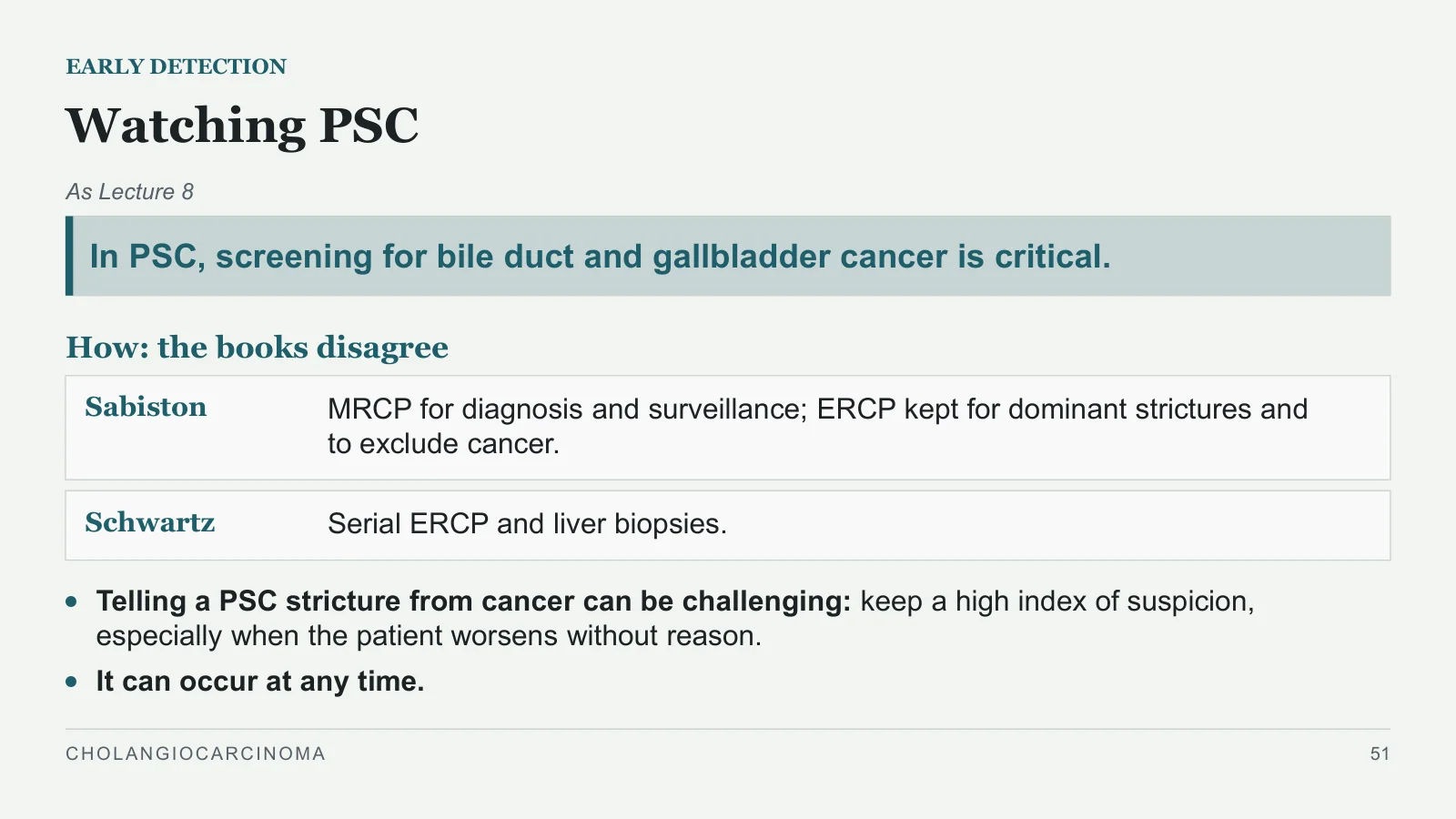
Watching PSC
EARLY DETECTION
As Lecture 8
In PSC, screening for bile duct and gallbladder cancer is critical.
How: the books disagree
Sabiston
MRCP for diagnosis and surveillance; ERCP kept for dominant strictures and to exclude cancer.
Schwartz
Serial ERCP and liver biopsies.
Telling a PSC stricture from cancer can be challenging: keep a high index of suspicion, especially when the patient worsens without reason.
It can occur at any time.
CHOLANGIOCARCINOMA
51
Case recap and take-home
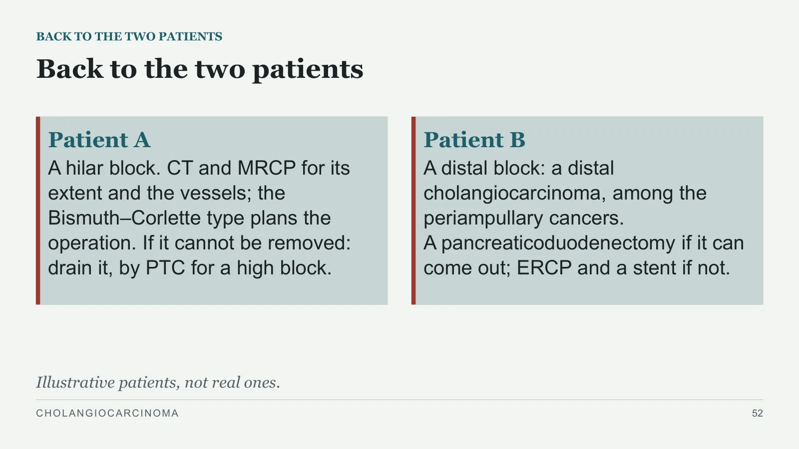
Back to the two patients
BACK TO THE TWO PATIENTS
Patient A
A hilar block. CT and MRCP for its extent and the vessels; the Bismuth–Corlette type plans the operation. If it cannot be removed: drain it, by PTC for a high block.
Patient B
A distal block: a distal cholangiocarcinoma, among the periampullary cancers. A pancreaticoduodenectomy if it can come out; ERCP and a stent if not.
Illustrative patients, not real ones.
CHOLANGIOCARCINOMA
52
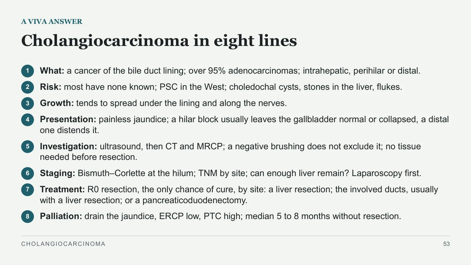
Cholangiocarcinoma in eight lines
A VIVA ANSWER
1
What: a cancer of the bile duct lining; over 95% adenocarcinomas; intrahepatic, perihilar or distal.
2
Risk: most have none known; PSC in the West; choledochal cysts, stones in the liver, flukes.
3
Growth: tends to spread under the lining and along the nerves.
4
Presentation: painless jaundice; a hilar block usually leaves the gallbladder normal or collapsed, a distal one distends it.
5
Investigation: ultrasound, then CT and MRCP; a negative brushing does not exclude it; no tissue needed before resection.
6
Staging: Bismuth–Corlette at the hilum; TNM by site; can enough liver remain? Laparoscopy first.
7
Treatment: R0 resection, the only chance of cure, by site: a liver resection; the involved ducts, usually with a liver resection; or a pancreaticoduodenectomy.
8
Palliation: drain the jaundice, ERCP low, PTC high; median 5 to 8 months without resection.
CHOLANGIOCARCINOMA
53
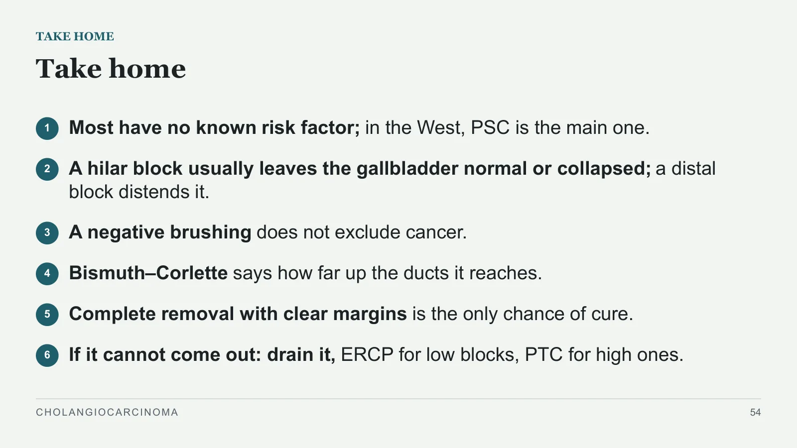
Take home
TAKE HOME
1
Most have no known risk factor; in the West, PSC is the main one.
2
A hilar block usually leaves the gallbladder normal or collapsed; a distal block distends it.
3
A negative brushing does not exclude cancer.
4
Bismuth–Corlette says how far up the ducts it reaches.
5
Complete removal with clear margins is the only chance of cure.
6
If it cannot come out: drain it, ERCP for low blocks, PTC for high ones.
CHOLANGIOCARCINOMA
54
Sources and image credits
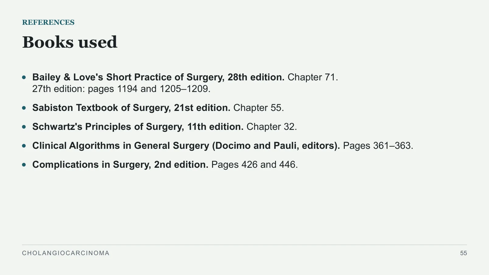
Books used
REFERENCES
Bailey & Love's Short Practice of Surgery, 28th edition. Chapter 71. 27th edition: pages 1194 and 1205–1209.
Sabiston Textbook of Surgery, 21st edition. Chapter 55.
Schwartz's Principles of Surgery, 11th edition. Chapter 32.
Clinical Algorithms in General Surgery (Docimo and Pauli, editors). Pages 361–363.
Complications in Surgery, 2nd edition. Pages 426 and 446.
CHOLANGIOCARCINOMA
55
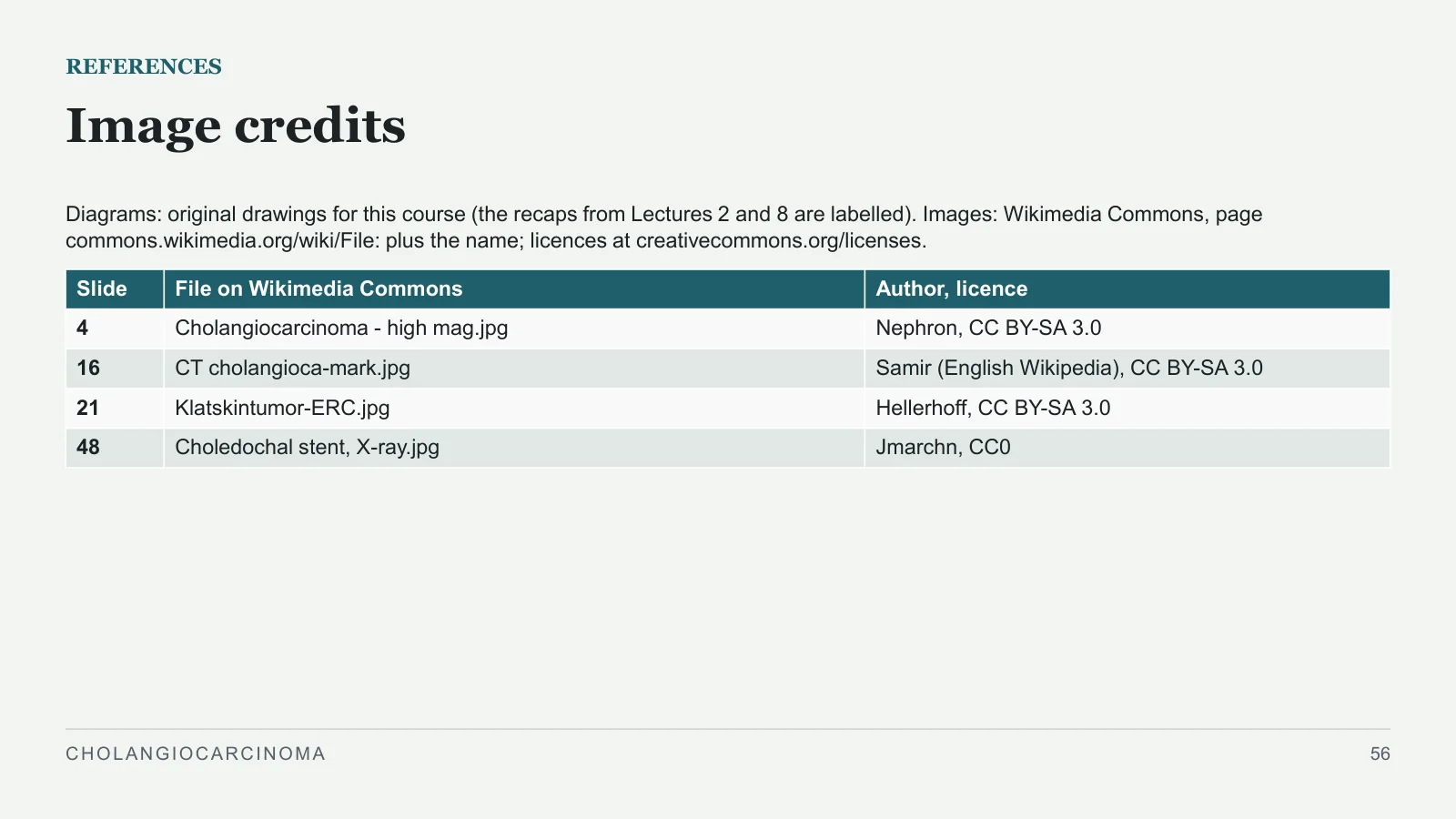
Image credits
REFERENCES
Diagrams: original drawings for this course (the recaps from Lectures 2 and 8 are labelled). Images: Wikimedia Commons, page commons.wikimedia.org/wiki/File: plus the name; licences at creativecommons.org/licenses.
CHOLANGIOCARCINOMA
56
| Slide | File on Wikimedia Commons | Author, licence |
| 4 | Cholangiocarcinoma - high mag.jpg | Nephron, CC BY-SA 3.0 |
| 16 | CT cholangioca-mark.jpg | Samir (English Wikipedia), CC BY-SA 3.0 |
| 21 | Klatskintumor-ERC.jpg | Hellerhoff, CC BY-SA 3.0 |
| 48 | Choledochal stent, X-ray.jpg | Jmarchn, CC0 |
Sources and image credits
Sources and image credits are reproduced from the designated slides.
Images
- Slide 4 · Cholangiocarcinoma - high mag.jpg · Nephron, CC BY-SA 3.0 · CC BY-SA 3.0
- Slide 16 · CT cholangioca-mark.jpg · Samir (English Wikipedia), CC BY-SA 3.0 · CC BY-SA 3.0
- Slide 21 · Klatskintumor-ERC.jpg · Hellerhoff, CC BY-SA 3.0 · CC BY-SA 3.0
- Slide 48 · Choledochal stent, X-ray.jpg · Jmarchn, CC0 · CC0


Sources
- [1] REFERENCES
- [2] Books used
- [3] Bailey & Love's Short Practice of Surgery, 28th edition. Chapter 71. 27th edition: pages 1194 and 1205–1209.
- [4] Sabiston Textbook of Surgery, 21st edition. Chapter 55.
- [5] Schwartz's Principles of Surgery, 11th edition. Chapter 32.
- [6] Clinical Algorithms in General Surgery (Docimo and Pauli, editors). Pages 361–363.
- [7] Complications in Surgery, 2nd edition. Pages 426 and 446.