Hepatobiliary surgery · Lecture 9
Biliary oncology · 58 slides
Gallbladder cancer.
Two cases frame how gallbladder cancer is found and how investigation and staging guide the next decision.
About the downloads
PowerPoint without speaker notes. SHA-256 checksums verify the downloaded files.
Contents · 11 parts
Opening cases
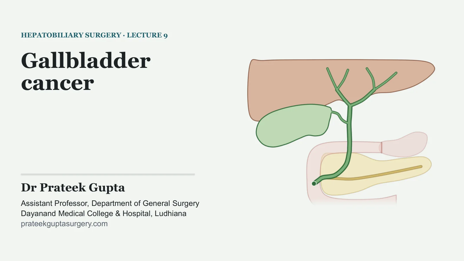
Gallbladder cancer
HEPATOBILIARY SURGERY · LECTURE 9
Gallbladder cancer
Dr Prateek Gupta
Assistant Professor, Department of General Surgery Dayanand Medical College & Hospital, Ludhiana prateekguptasurgery.com
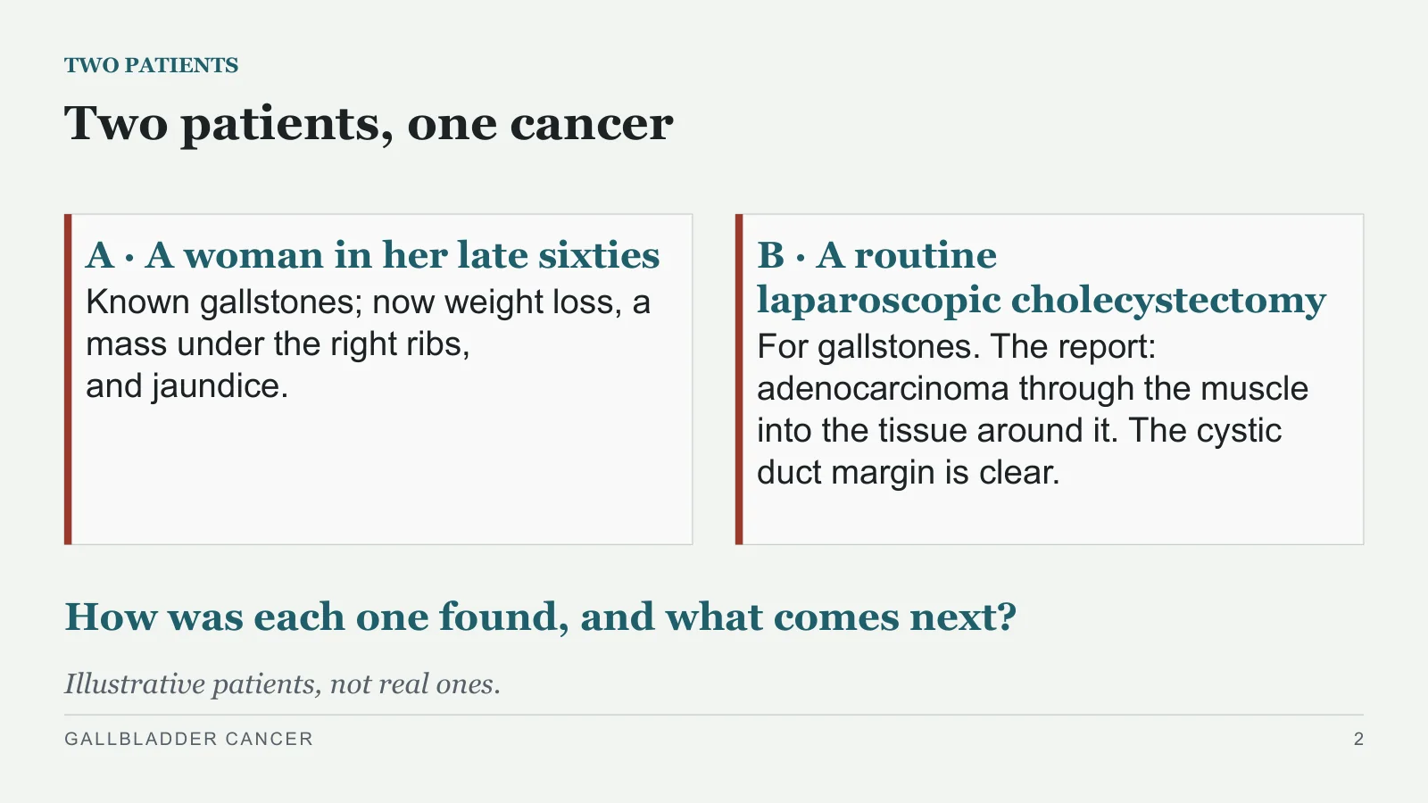
Two patients, one cancer
TWO PATIENTS
A · A woman in her late sixties
Known gallstones; now weight loss, a mass under the right ribs, and jaundice.
B · A routine laparoscopic cholecystectomy
For gallstones. The report: adenocarcinoma through the muscle into the tissue around it. The cystic duct margin is clear.
How was each one found, and what comes next?
Illustrative patients, not real ones.
GALLBLADDER CANCER
2
Who gets it
Who gets it
PART 1
GALLBLADDER CANCER
3
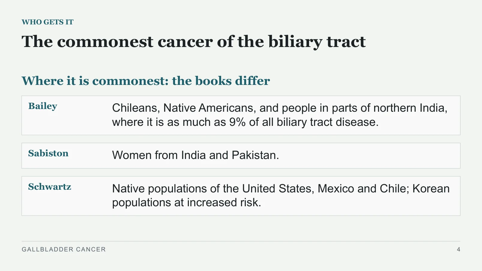
The commonest cancer of the biliary tract
WHO GETS IT
Where it is commonest: the books differ
Bailey
Chileans, Native Americans, and people in parts of northern India, where it is as much as 9% of all biliary tract disease.
Sabiston
Women from India and Pakistan.
Schwartz
Native populations of the United States, Mexico and Chile; Korean populations at increased risk.
GALLBLADDER CANCER
4
Mostly older women
WHO GETS IT
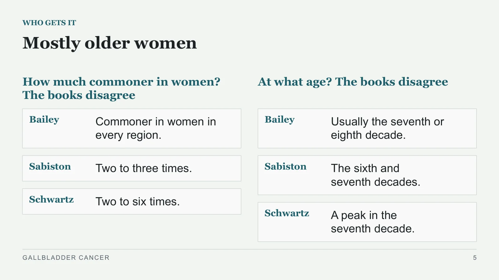
How much commoner in women? The books disagree
Bailey
Commoner in women in every region.
Sabiston
Two to three times.
Schwartz
Two to six times.
At what age? The books disagree
Bailey
Usually the seventh or eighth decade.
Sabiston
The sixth and seventh decades.
Schwartz
A peak in the seventh decade.
GALLBLADDER CANCER
5
Most patients have gallstones
WHO GETS IT
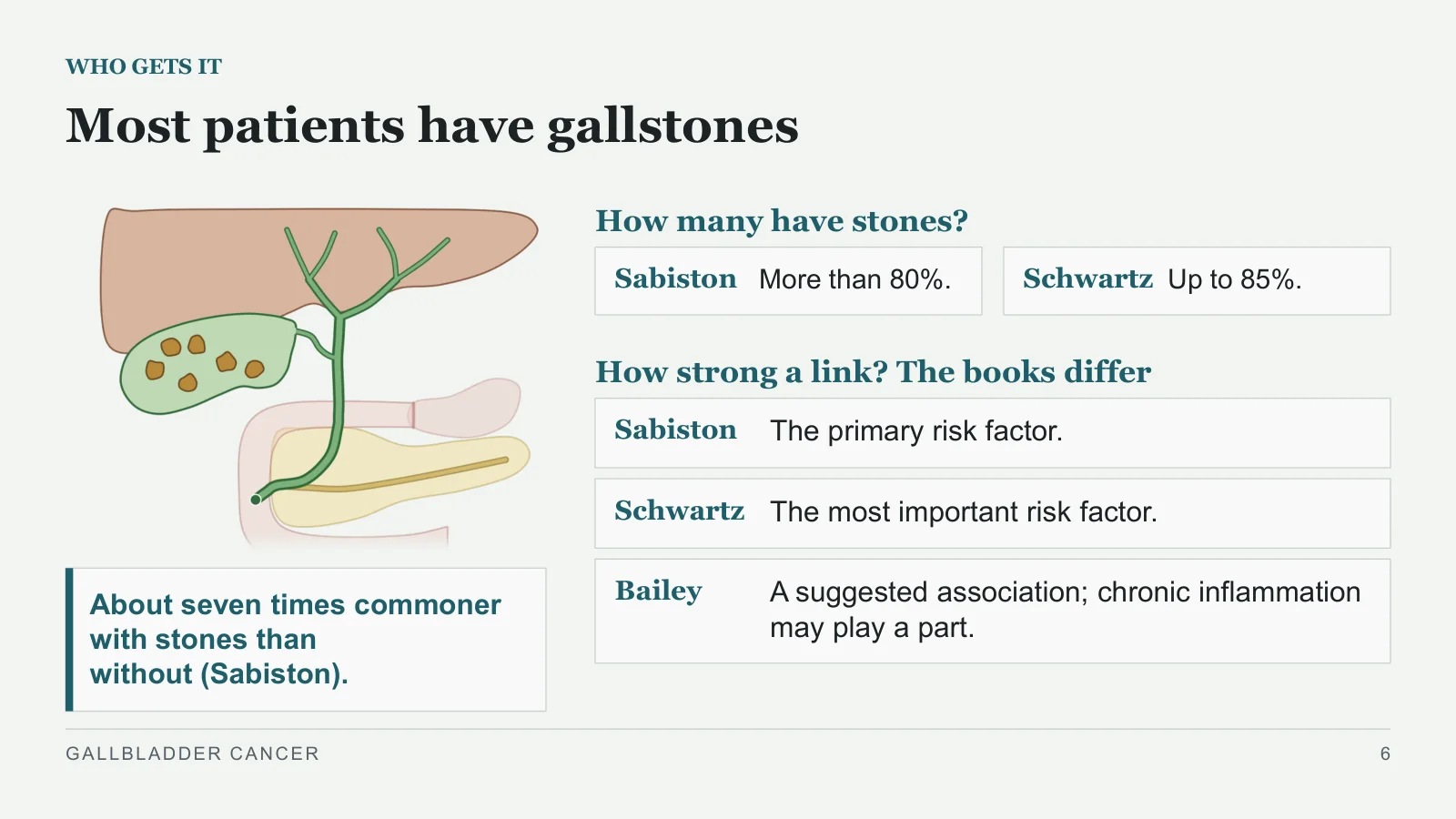
About seven times commoner with stones than without (Sabiston).
How many have stones?
Sabiston
More than 80%.
Schwartz
Up to 85%.
How strong a link? The books differ
Sabiston
The primary risk factor.
Schwartz
The most important risk factor.
Bailey
A suggested association; chronic inflammation may play a part.
GALLBLADDER CANCER
6
Few people with gallstones have it
WHO GETS IT
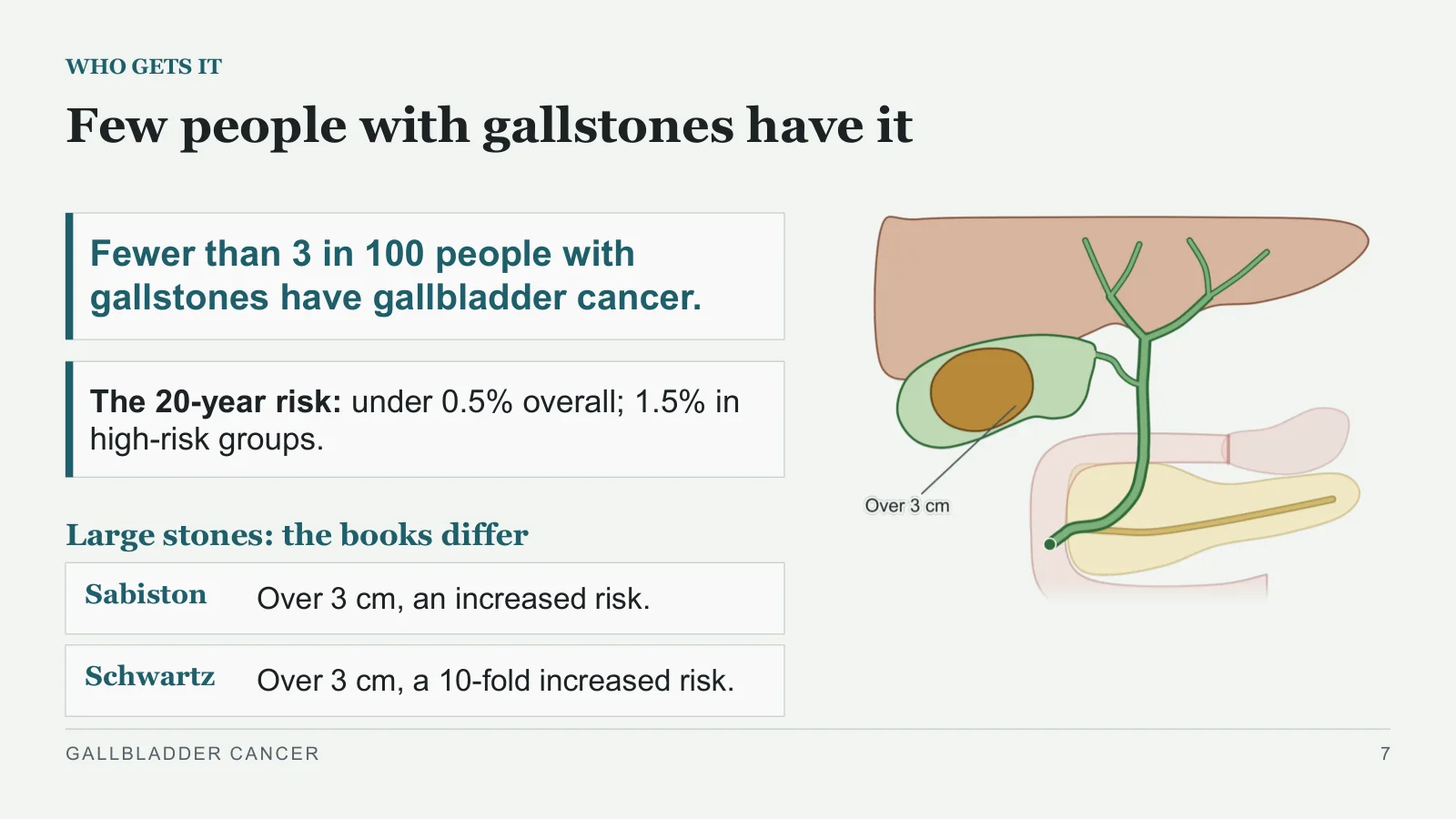
Fewer than 3 in 100 people with gallstones have gallbladder cancer.
The 20-year risk: under 0.5% overall; 1.5% in high-risk groups.
Large stones: the books differ
Sabiston
Over 3 cm, an increased risk.
Schwartz
Over 3 cm, a 10-fold increased risk.
GALLBLADDER CANCER
7
A calcified wall
WHO GETS IT
Recap: Lecture 3
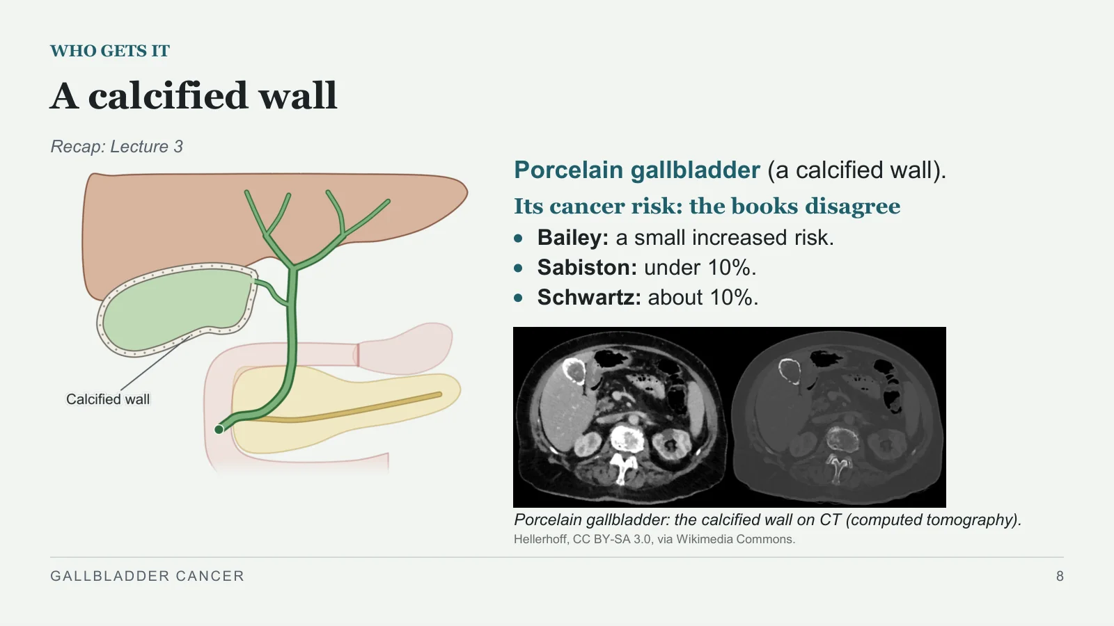
Porcelain gallbladder (a calcified wall).
Its cancer risk: the books disagree
Bailey: a small increased risk.
Sabiston: under 10%.
Schwartz: about 10%.
Porcelain gallbladder: the calcified wall on CT (computed tomography).
Hellerhoff, CC BY-SA 3.0, via Wikimedia Commons.
GALLBLADDER CANCER
8
A large polyp
WHO GETS IT
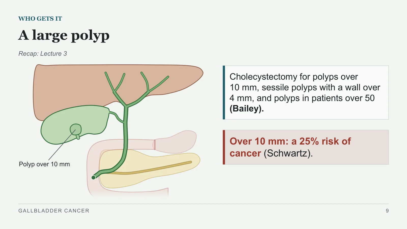
Recap: Lecture 3
Cholecystectomy for polyps over 10 mm, sessile polyps with a wall over 4 mm, and polyps in patients over 50 (Bailey).
Over 10 mm: a 25% risk of cancer (Schwartz).
GALLBLADDER CANCER
9
Risks beyond the stones
WHO GETS IT
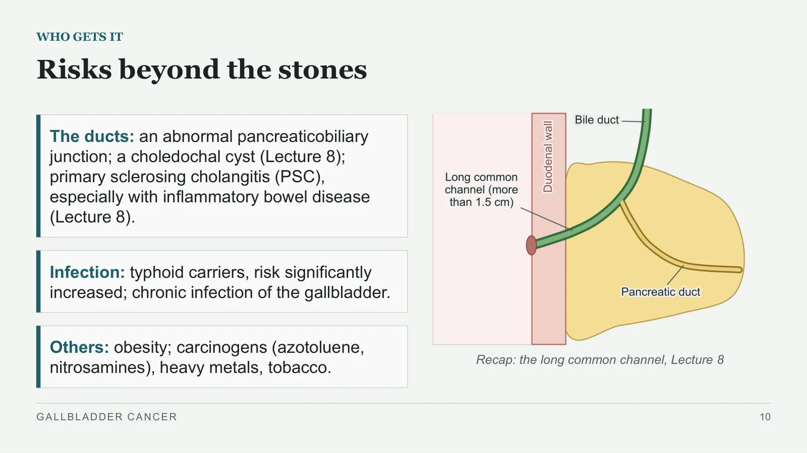
Recap: the long common channel, Lecture 8
The ducts: an abnormal pancreaticobiliary junction; a choledochal cyst (Lecture 8); primary sclerosing cholangitis (PSC), especially with inflammatory bowel disease (Lecture 8).
Infection: typhoid carriers, risk significantly increased; chronic infection of the gallbladder.
Others: obesity; carcinogens (azotoluene, nitrosamines), heavy metals, tobacco.
GALLBLADDER CANCER
10
Pathology and spread
Pathology and spread
PART 2
GALLBLADDER CANCER
11
Adenocarcinoma, from the fundus or body
PATHOLOGY AND SPREAD
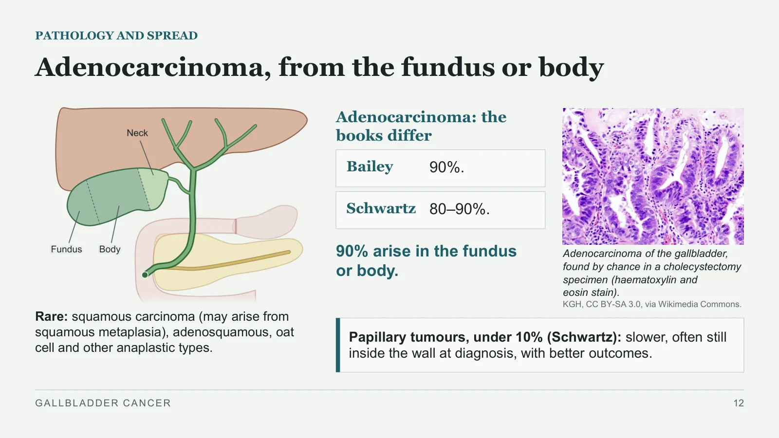
Rare: squamous carcinoma (may arise from squamous metaplasia), adenosquamous, oat cell and other anaplastic types.
Adenocarcinoma of the gallbladder, found by chance in a cholecystectomy specimen (haematoxylin and eosin stain).
KGH, CC BY-SA 3.0, via Wikimedia Commons.
Adenocarcinoma: the books differ
Bailey
90%.
Schwartz
80–90%.
90% arise in the fundus or body.
Papillary tumours, under 10% (Schwartz): slower, often still inside the wall at diagnosis, with better outcomes.
GALLBLADDER CANCER
12
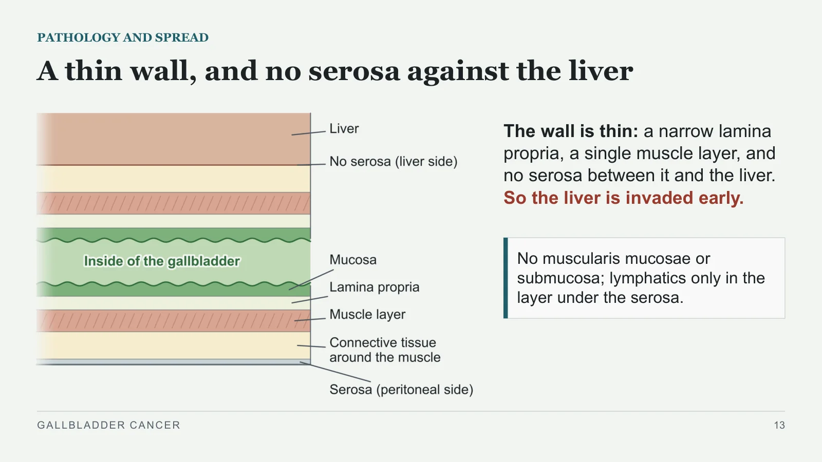
A thin wall, and no serosa against the liver
PATHOLOGY AND SPREAD
The wall is thin: a narrow lamina propria, a single muscle layer, and no serosa between it and the liver. So the liver is invaded early.
No muscularis mucosae or submucosa; lymphatics only in the layer under the serosa.
GALLBLADDER CANCER
13
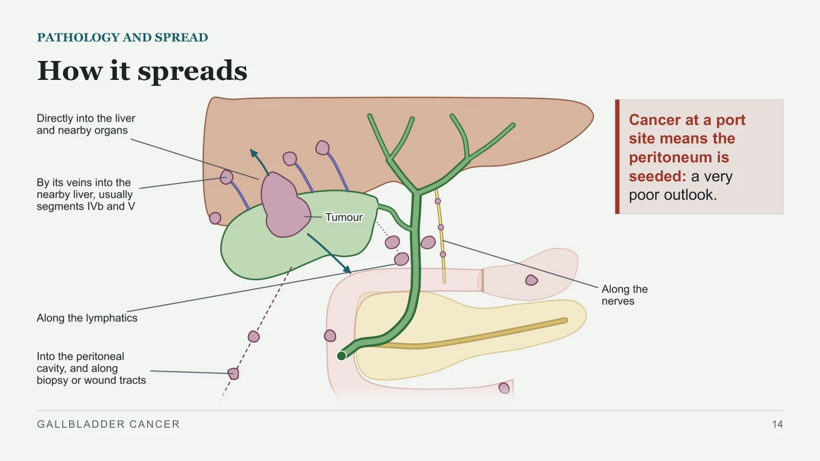
How it spreads
PATHOLOGY AND SPREAD
Cancer at a port site means the peritoneum is seeded: a very poor outlook.
GALLBLADDER CANCER
14
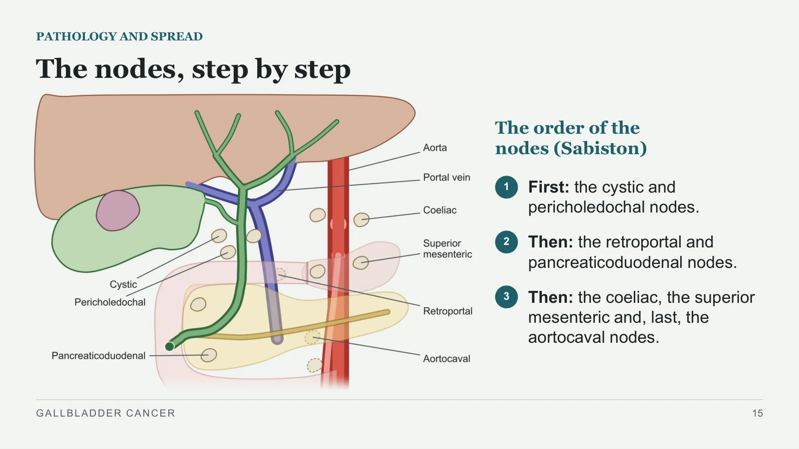
The nodes, step by step
PATHOLOGY AND SPREAD
The order of the nodes (Sabiston)
1
First: the cystic and pericholedochal nodes.
2
Then: the retroportal and pancreaticoduodenal nodes.
3
Then: the coeliac, the superior mesenteric and, last, the aortocaval nodes.
GALLBLADDER CANCER
15
Why it is found late
PATHOLOGY AND SPREAD
Few or no early symptoms; when there are some, they usually look like biliary colic or cholecystitis.
Most arise in the fundus or body, so most give no symptoms until late.
How late? The books measure it differently
Bailey
Resected in less than 10%.
Schwartz
Only 10–25% found while still inside the gallbladder.
Sabiston
At presentation, nodes in 35% and distant metastases in 40%.
GALLBLADDER CANCER
16
How it presents
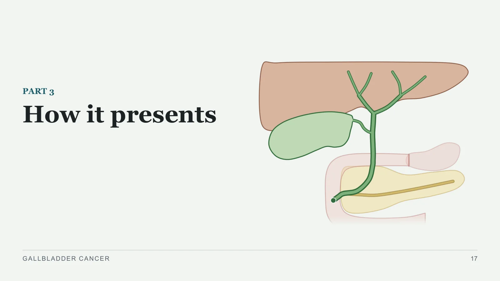
How it presents
PART 3
GALLBLADDER CANCER
17
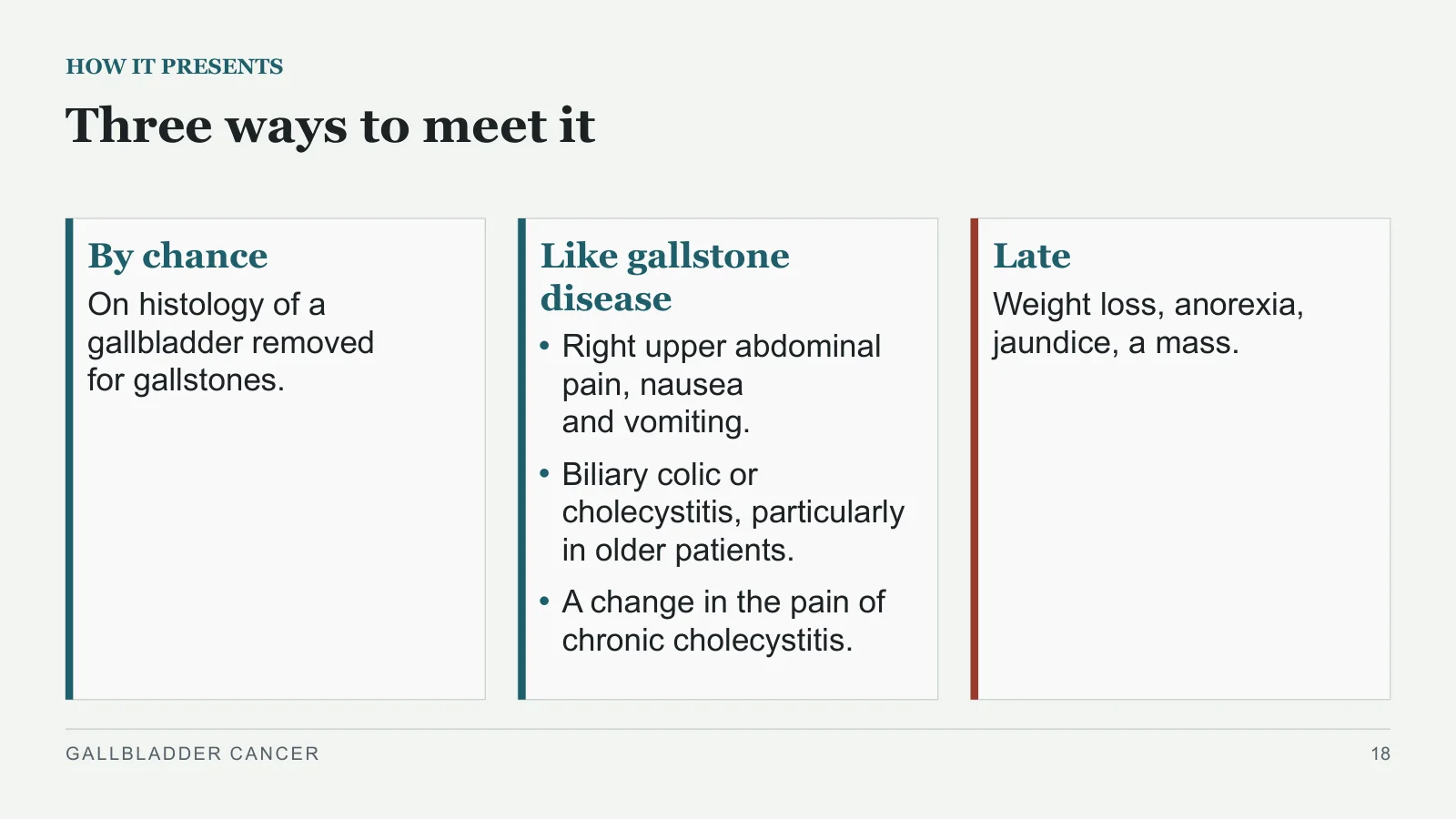
Three ways to meet it
HOW IT PRESENTS
By chance
On histology of a gallbladder removed for gallstones.
Like gallstone disease
Right upper abdominal pain, nausea and vomiting.
Biliary colic or cholecystitis, particularly in older patients.
A change in the pain of chronic cholecystitis.
Late
Weight loss, anorexia, jaundice, a mass.
GALLBLADDER CANCER
18
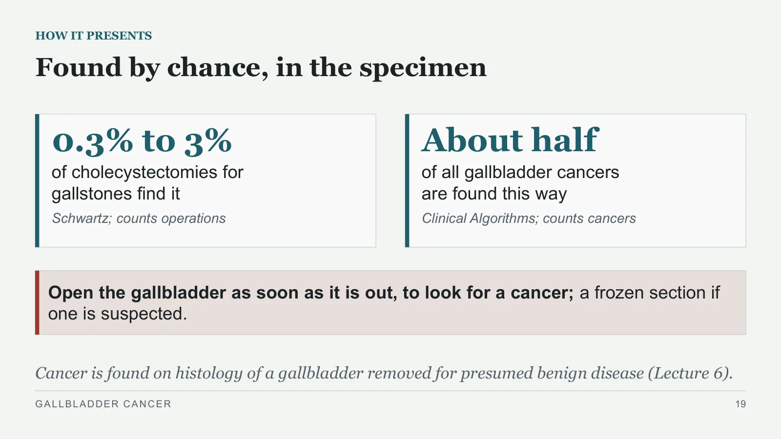
0.3% to 3%
HOW IT PRESENTS
Found by chance, in the specimen
of cholecystectomies for gallstones find it
Schwartz; counts operations
About half
of all gallbladder cancers are found this way
Clinical Algorithms; counts cancers
Open the gallbladder as soon as it is out, to look for a cancer; a frozen section if one is suspected.
Cancer is found on histology of a gallbladder removed for presumed benign disease (Lecture 6).
GALLBLADDER CANCER
19
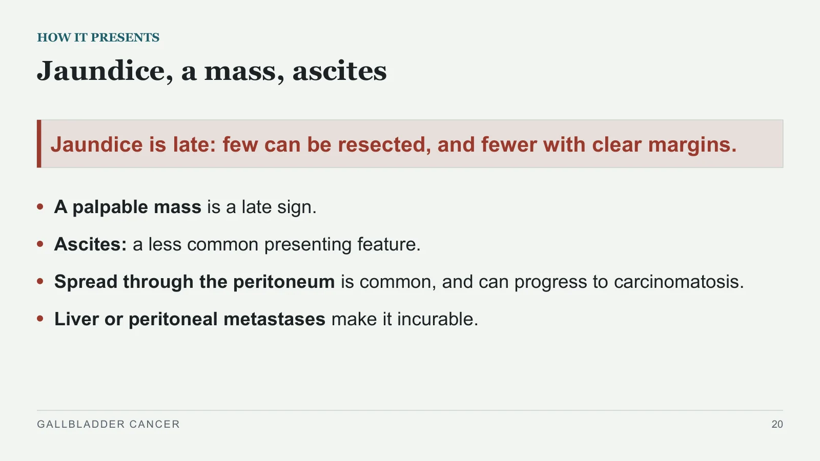
Jaundice, a mass, ascites
HOW IT PRESENTS
Jaundice is late: few can be resected, and fewer with clear margins.
A palpable mass is a late sign.
Ascites: a less common presenting feature.
Spread through the peritoneum is common, and can progress to carcinomatosis.
Liver or peritoneal metastases make it incurable.
GALLBLADDER CANCER
20
Investigation
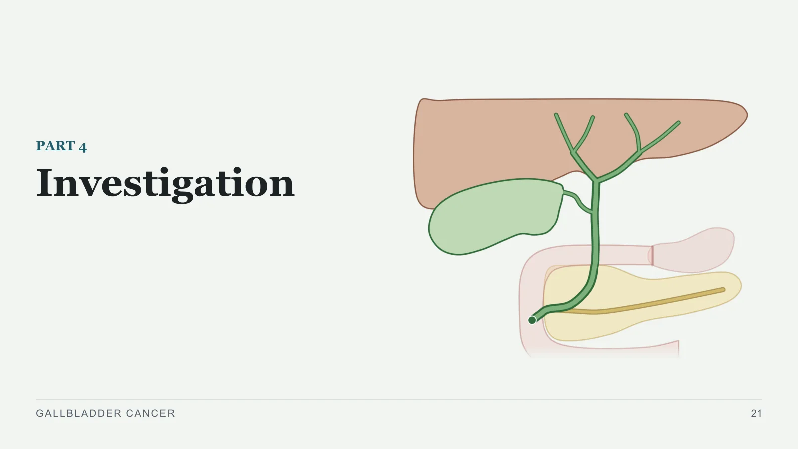
Investigation
PART 4
GALLBLADDER CANCER
21
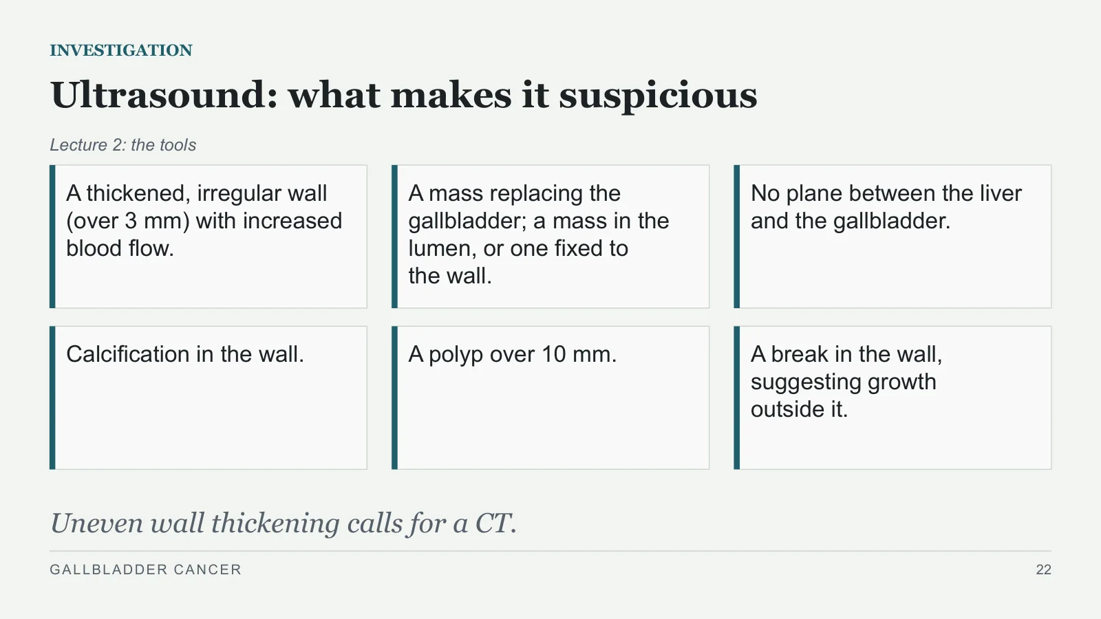
Ultrasound: what makes it suspicious
INVESTIGATION
Lecture 2: the tools
A thickened, irregular wall (over 3 mm) with increased blood flow.
A mass replacing the gallbladder; a mass in the lumen, or one fixed to the wall.
No plane between the liver and the gallbladder.
Calcification in the wall.
A polyp over 10 mm.
A break in the wall, suggesting growth outside it.
Uneven wall thickening calls for a CT.
GALLBLADDER CANCER
22
CT and MRI: how far it has gone
INVESTIGATION
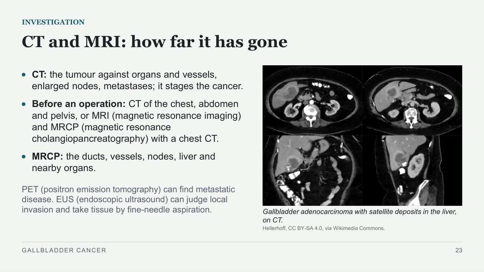
Gallbladder adenocarcinoma with satellite deposits in the liver, on CT.
Hellerhoff, CC BY-SA 4.0, via Wikimedia Commons.
CT: the tumour against organs and vessels, enlarged nodes, metastases; it stages the cancer.
Before an operation: CT of the chest, abdomen and pelvis, or MRI (magnetic resonance imaging) and MRCP (magnetic resonance cholangiopancreatography) with a chest CT.
MRCP: the ducts, vessels, nodes, liver and nearby organs.
PET (positron emission tomography) can find metastatic disease. EUS (endoscopic ultrasound) can judge local invasion and take tissue by fine-needle aspiration.
GALLBLADDER CANCER
23
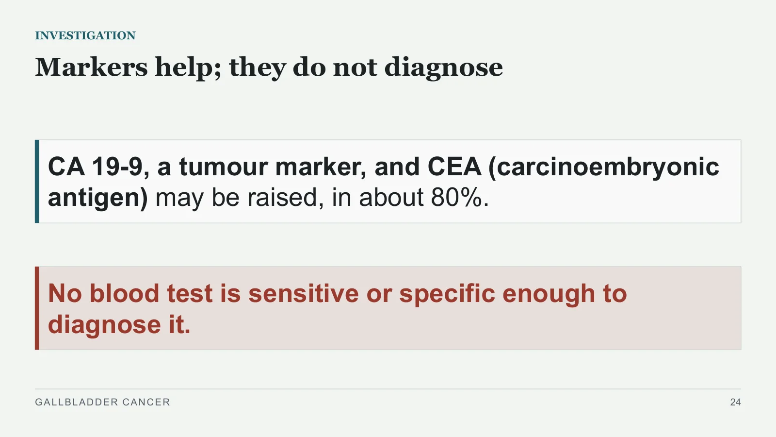
Markers help; they do not diagnose
INVESTIGATION
CA 19-9, a tumour marker, and CEA (carcinoembryonic antigen) may be raised, in about 80%.
No blood test is sensitive or specific enough to diagnose it.
GALLBLADDER CANCER
24
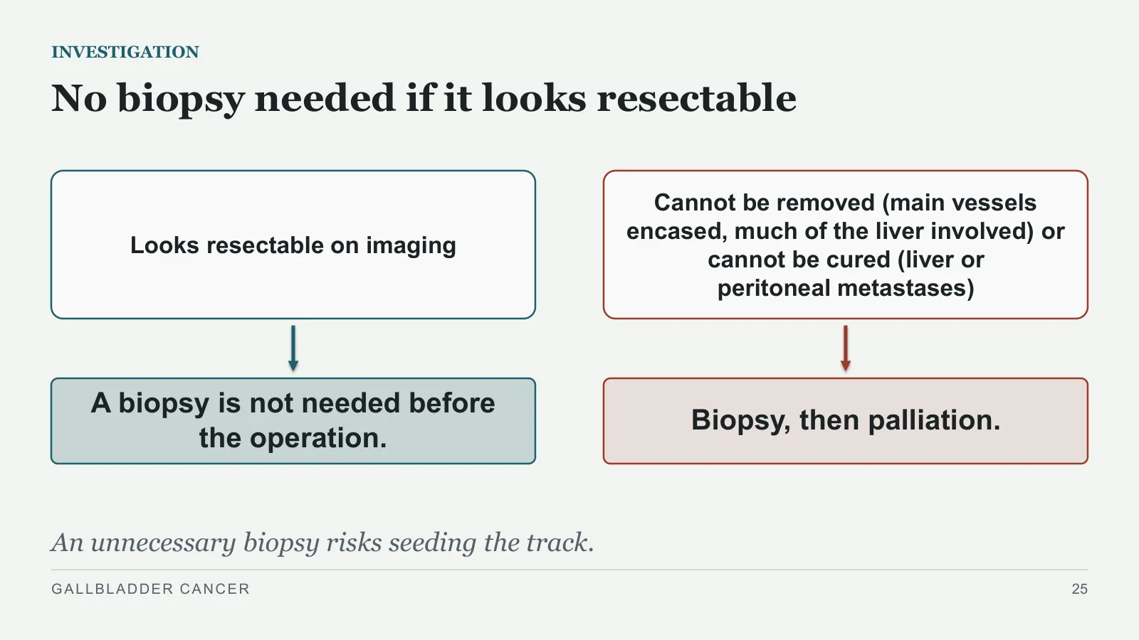
No biopsy needed if it looks resectable
INVESTIGATION
Looks resectable on imaging
A biopsy is not needed before the operation.
Cannot be removed (main vessels encased, much of the liver involved) or cannot be cured (liver or peritoneal metastases)
Biopsy, then palliation.
An unnecessary biopsy risks seeding the track.
GALLBLADDER CANCER
25
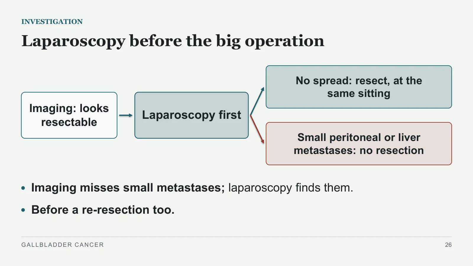
Laparoscopy before the big operation
INVESTIGATION
Imaging: looks resectable
Laparoscopy first
No spread: resect, at the same sitting
Small peritoneal or liver metastases: no resection
Imaging misses small metastases; laparoscopy finds them.
Before a re-resection too.
GALLBLADDER CANCER
26
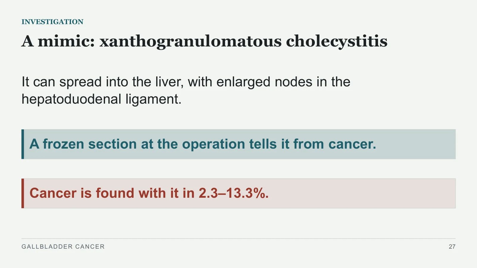
A mimic: xanthogranulomatous cholecystitis
INVESTIGATION
It can spread into the liver, with enlarged nodes in the hepatoduodenal ligament.
A frozen section at the operation tells it from cancer.
Cancer is found with it in 2.3–13.3%.
GALLBLADDER CANCER
27
Staging
Staging
PART 5
GALLBLADDER CANCER
28
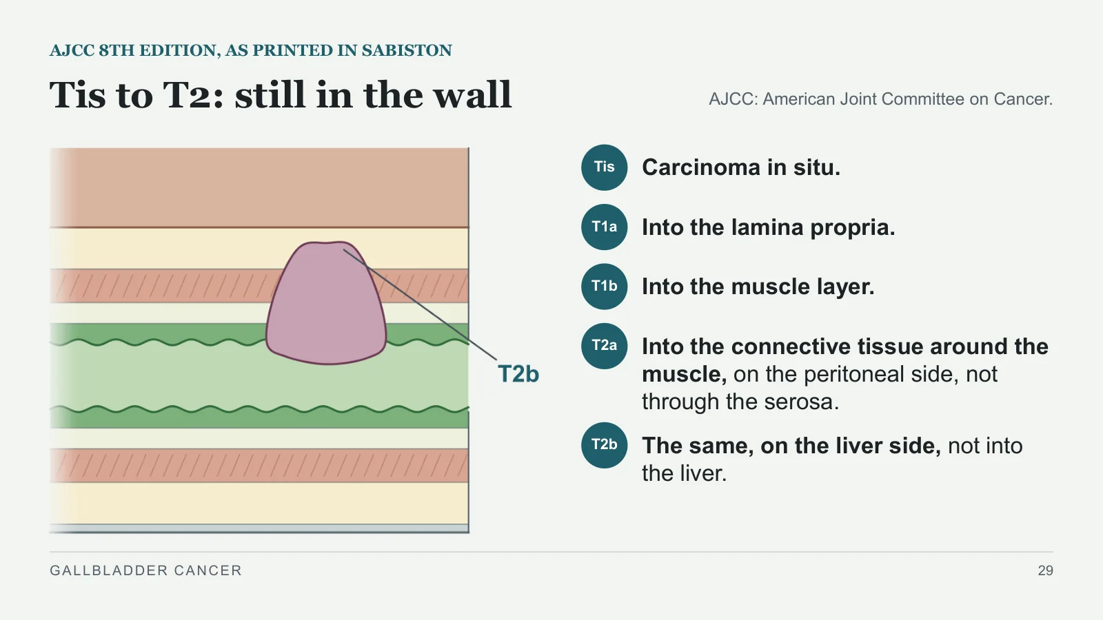
Tis to T2: still in the wall
AJCC 8TH EDITION, AS PRINTED IN SABISTON
AJCC: American Joint Committee on Cancer.
Tis
Carcinoma in situ.
T1a
Into the lamina propria.
T1b
Into the muscle layer.
T2a
Into the connective tissue around the muscle, on the peritoneal side, not through the serosa.
T2b
The same, on the liver side, not into the liver.
GALLBLADDER CANCER
29
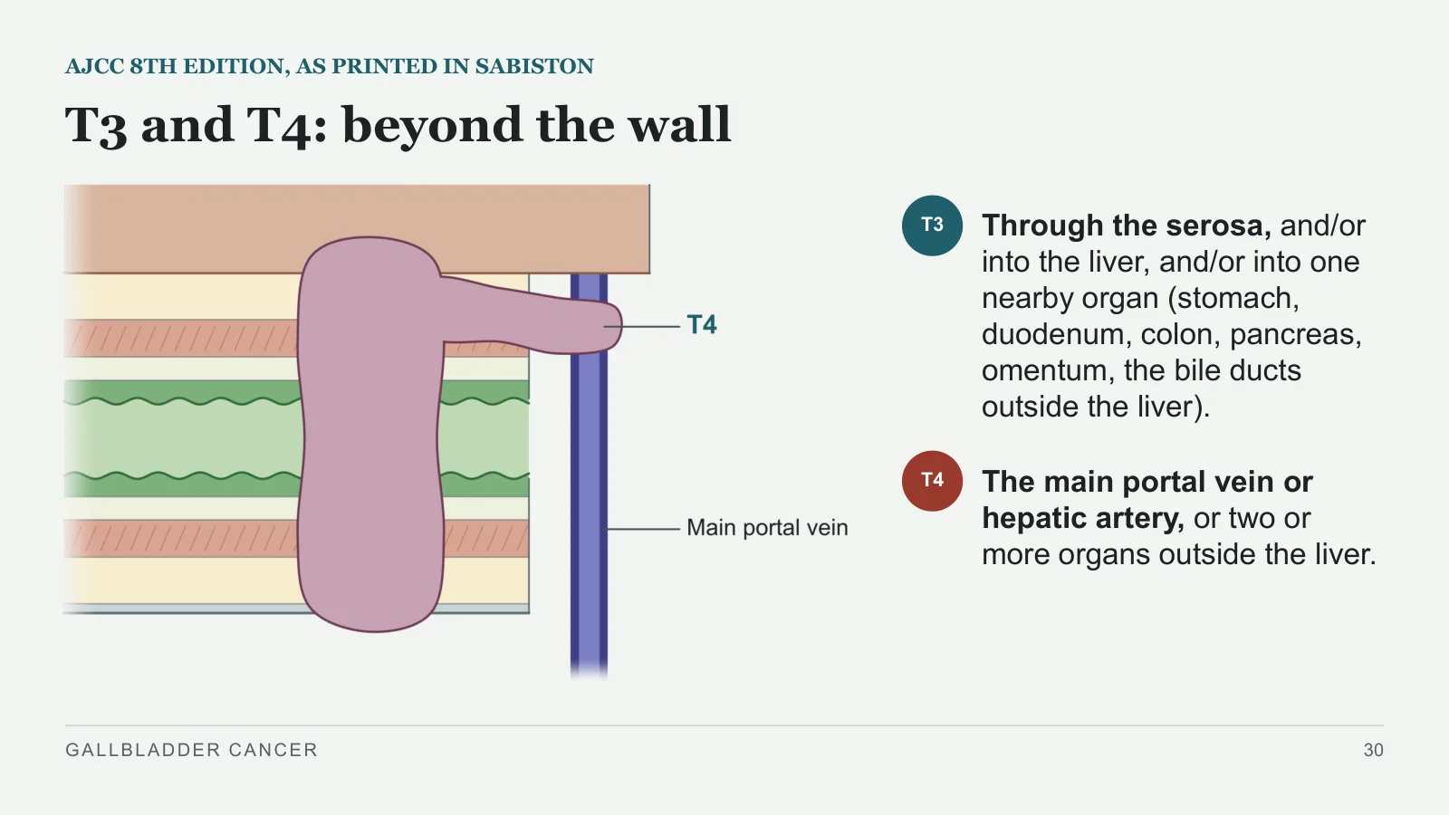
T3 and T4: beyond the wall
AJCC 8TH EDITION, AS PRINTED IN SABISTON
T3
Through the serosa, and/or into the liver, and/or into one nearby organ (stomach, duodenum, colon, pancreas, omentum, the bile ducts outside the liver).
T4
The main portal vein or hepatic artery, or two or more organs outside the liver.
GALLBLADDER CANCER
30
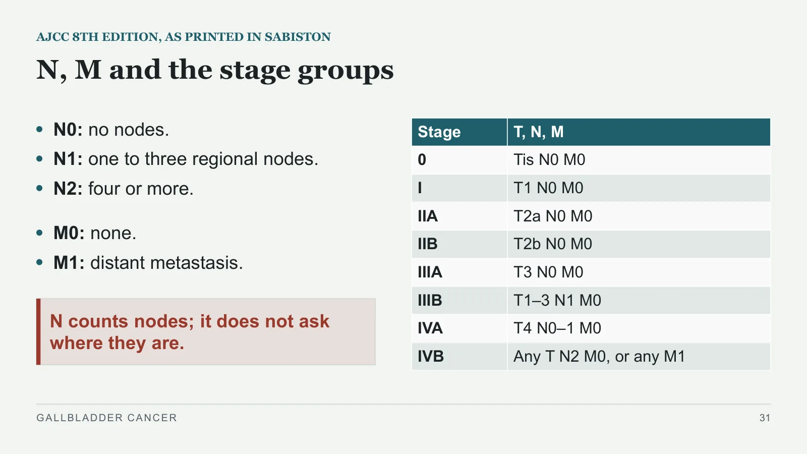
N, M and the stage groups
AJCC 8TH EDITION, AS PRINTED IN SABISTON
N0: no nodes.
N1: one to three regional nodes.
N2: four or more.
M0: none.
M1: distant metastasis.
N counts nodes; it does not ask where they are.
GALLBLADDER CANCER
31
| Stage | T, N, M |
| 0 | Tis N0 M0 |
| I | T1 N0 M0 |
| IIA | T2a N0 M0 |
| IIB | T2b N0 M0 |
| IIIA | T3 N0 M0 |
| IIIB | T1–3 N1 M0 |
| IVA | T4 N0–1 M0 |
| IVB | Any T N2 M0, or any M1 |
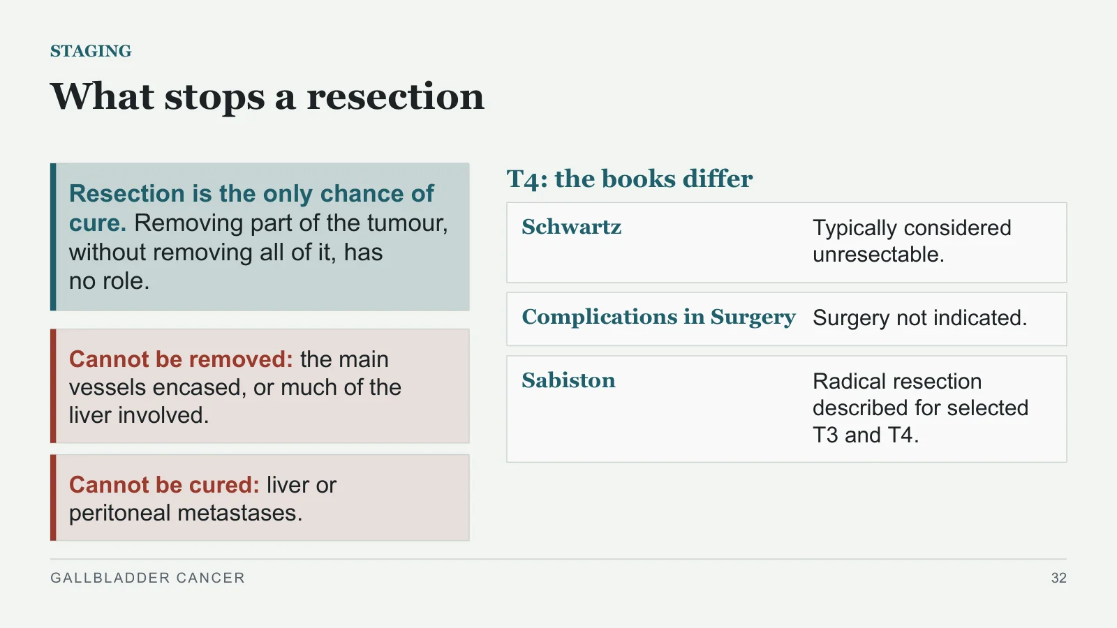
What stops a resection
STAGING
Resection is the only chance of cure. Removing part of the tumour, without removing all of it, has no role.
Cannot be removed: the main vessels encased, or much of the liver involved.
Cannot be cured: liver or peritoneal metastases.
T4: the books differ
Schwartz
Typically considered unresectable.
Complications in Surgery
Surgery not indicated.
Sabiston
Radical resection described for selected T3 and T4.
GALLBLADDER CANCER
32
Treatment
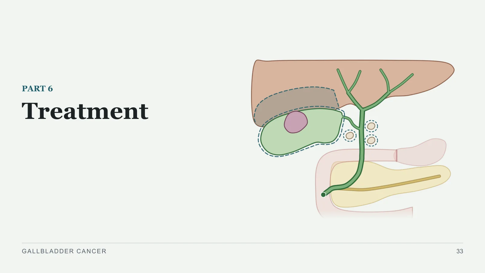
Treatment
PART 6
GALLBLADDER CANCER
33
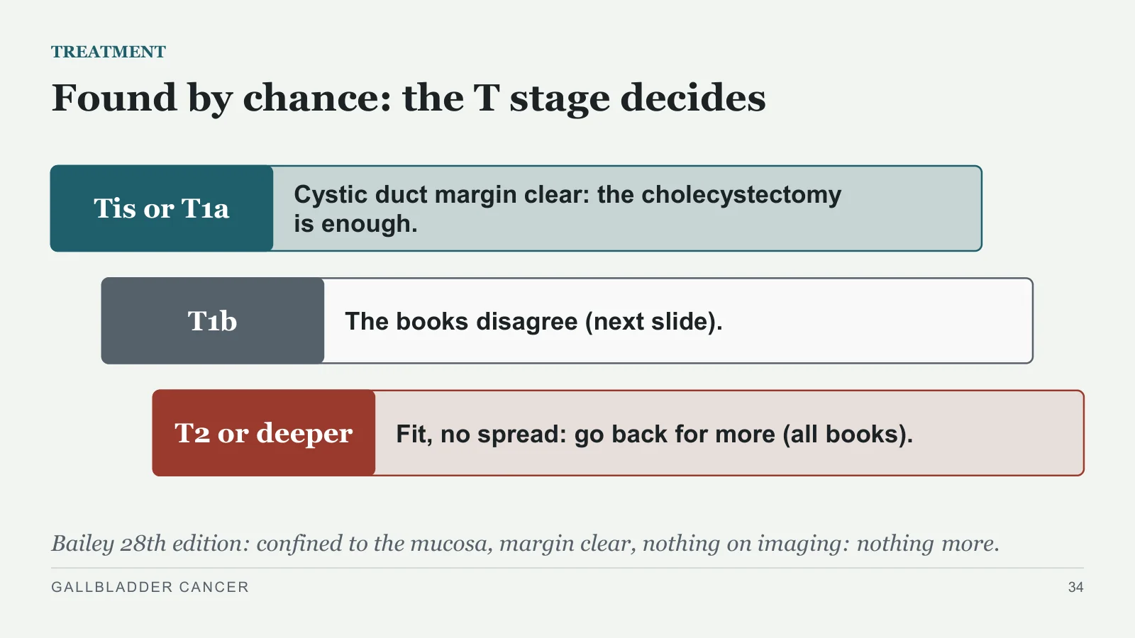
Found by chance: the T stage decides
TREATMENT
Tis or T1a
Cystic duct margin clear: the cholecystectomy is enough.
T1b
The books disagree (next slide).
T2 or deeper
Fit, no spread: go back for more (all books).
Bailey 28th edition: confined to the mucosa, margin clear, nothing on imaging: nothing more.
GALLBLADDER CANCER
34
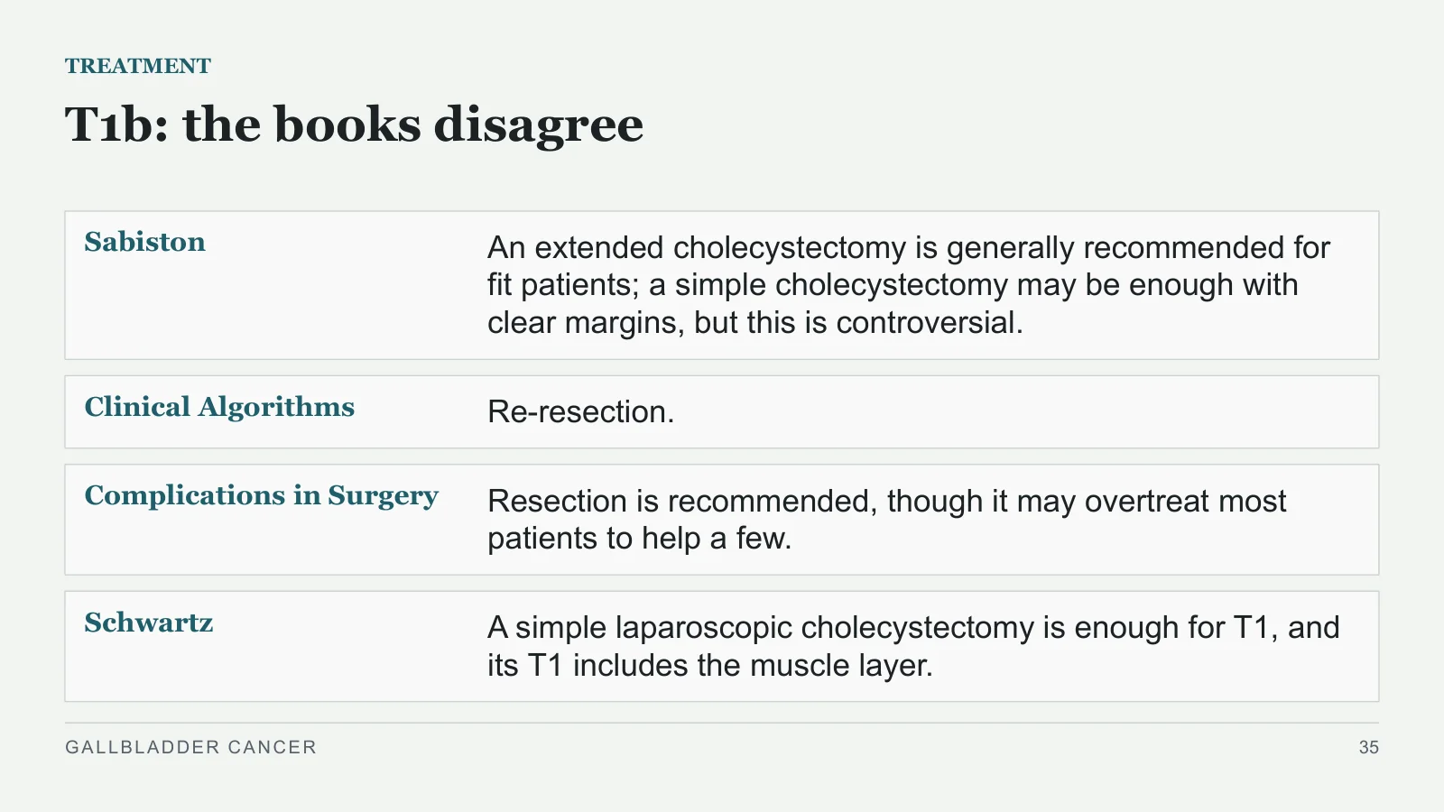
T1b: the books disagree
TREATMENT
Sabiston
An extended cholecystectomy is generally recommended for fit patients; a simple cholecystectomy may be enough with clear margins, but this is controversial.
Clinical Algorithms
Re-resection.
Complications in Surgery
Resection is recommended, though it may overtreat most patients to help a few.
Schwartz
A simple laparoscopic cholecystectomy is enough for T1, and its T1 includes the muscle layer.
GALLBLADDER CANCER
35
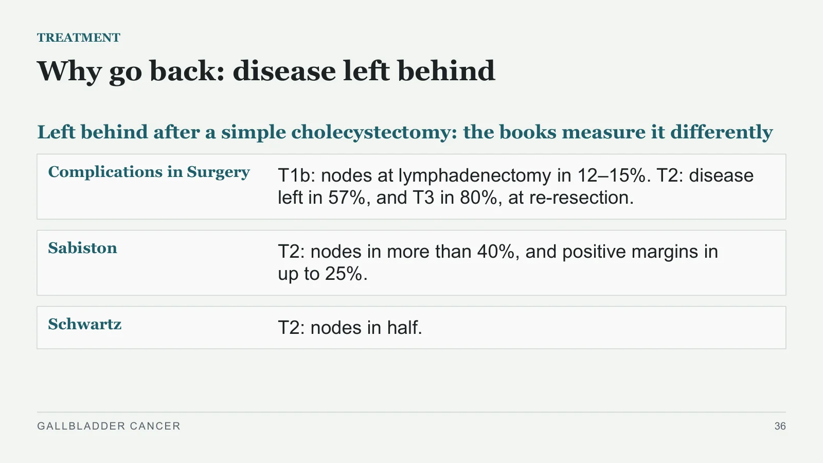
Why go back: disease left behind
TREATMENT
Left behind after a simple cholecystectomy: the books measure it differently
Complications in Surgery
T1b: nodes at lymphadenectomy in 12–15%. T2: disease left in 57%, and T3 in 80%, at re-resection.
Sabiston
T2: nodes in more than 40%, and positive margins in up to 25%.
Schwartz
T2: nodes in half.
GALLBLADDER CANCER
36
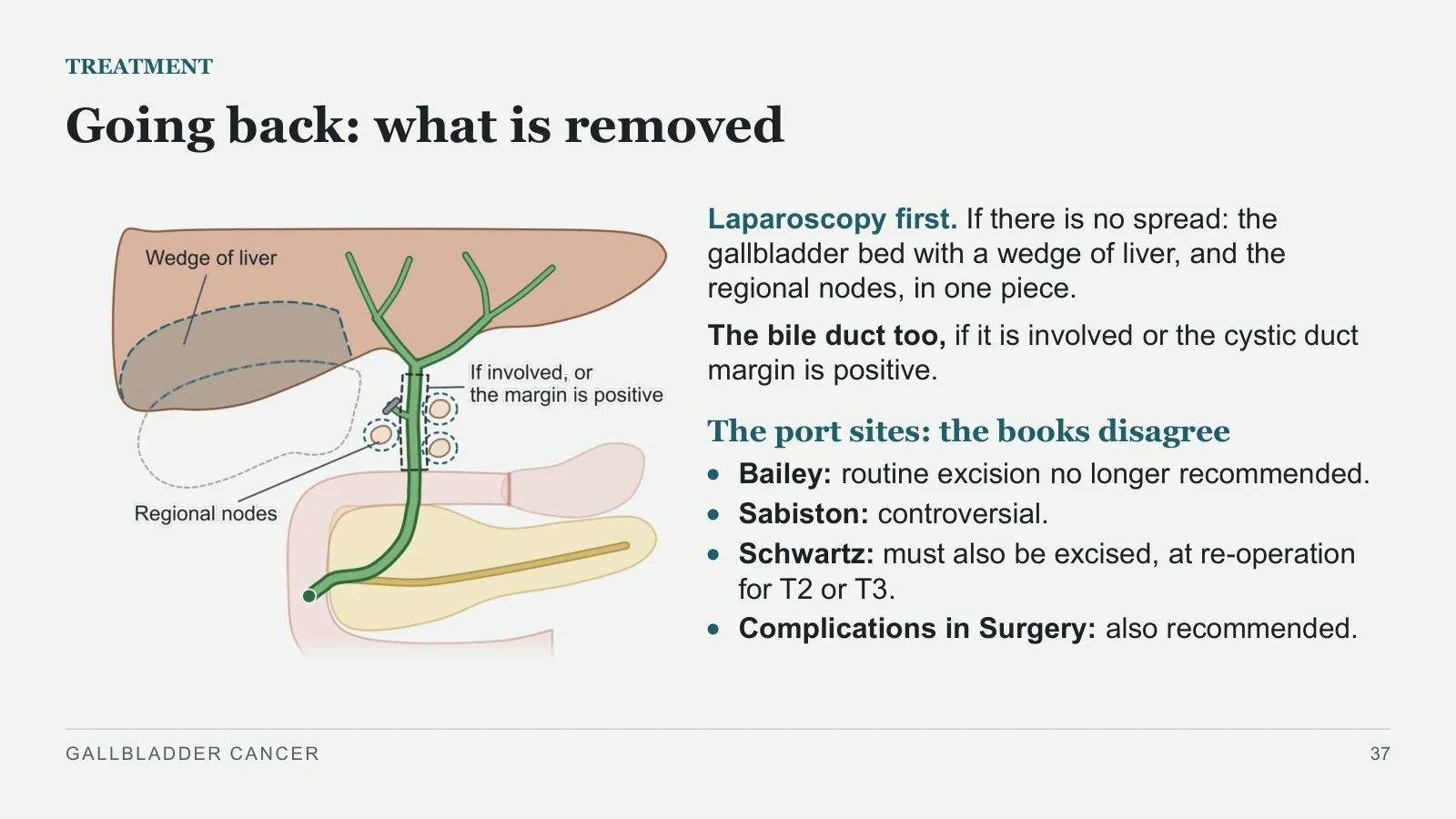
Going back: what is removed
TREATMENT
Laparoscopy first. If there is no spread: the gallbladder bed with a wedge of liver, and the regional nodes, in one piece.
The bile duct too, if it is involved or the cystic duct margin is positive.
The port sites: the books disagree
Bailey: routine excision no longer recommended.
Sabiston: controversial.
Schwartz: must also be excised, at re-operation for T2 or T3.
Complications in Surgery: also recommended.
GALLBLADDER CANCER
37
Suspected before the operation
TREATMENT
Take care not to enter the gallbladder or spill bile.
Laparoscopic cholecystectomy? The books differ in scope
Fischer
Not for a suspected or confirmed cancer.
Complications in Surgery
Cancer is the exception to the laparoscopic standard.
Clinical Algorithms
For a 1–2 cm mass or polyp without invasion, laparoscopic cholecystectomy with a frozen section is appropriate.
The extended operation is done open as standard (Schwartz).
GALLBLADDER CANCER
38
Suspected during the operation
TREATMENT
Convert to open, or stop and refer.
A positive frozen section: the books disagree
Clinical Algorithms
Go on to an extended cholecystectomy and lymphadenectomy, if the surgeon is comfortable.
Complications in Surgery
No further surgery until the final report, because depth is hard to judge on a frozen section.
GALLBLADDER CANCER
39
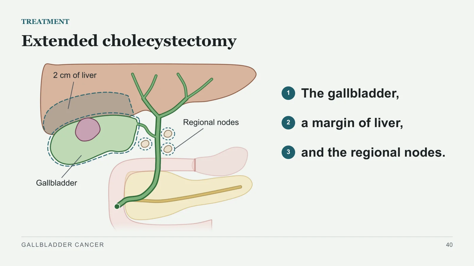
Extended cholecystectomy
TREATMENT
1
The gallbladder,
2
a margin of liver,
3
and the regional nodes.
GALLBLADDER CANCER
40
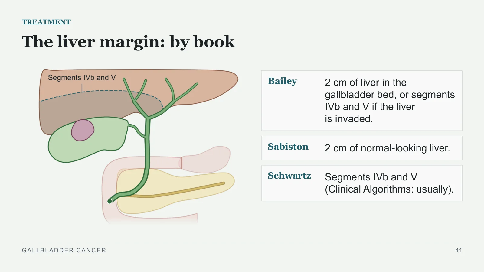
The liver margin: by book
TREATMENT
Bailey
2 cm of liver in the gallbladder bed, or segments IVb and V if the liver is invaded.
Sabiston
2 cm of normal-looking liver.
Schwartz
Segments IVb and V (Clinical Algorithms: usually).
GALLBLADDER CANCER
41
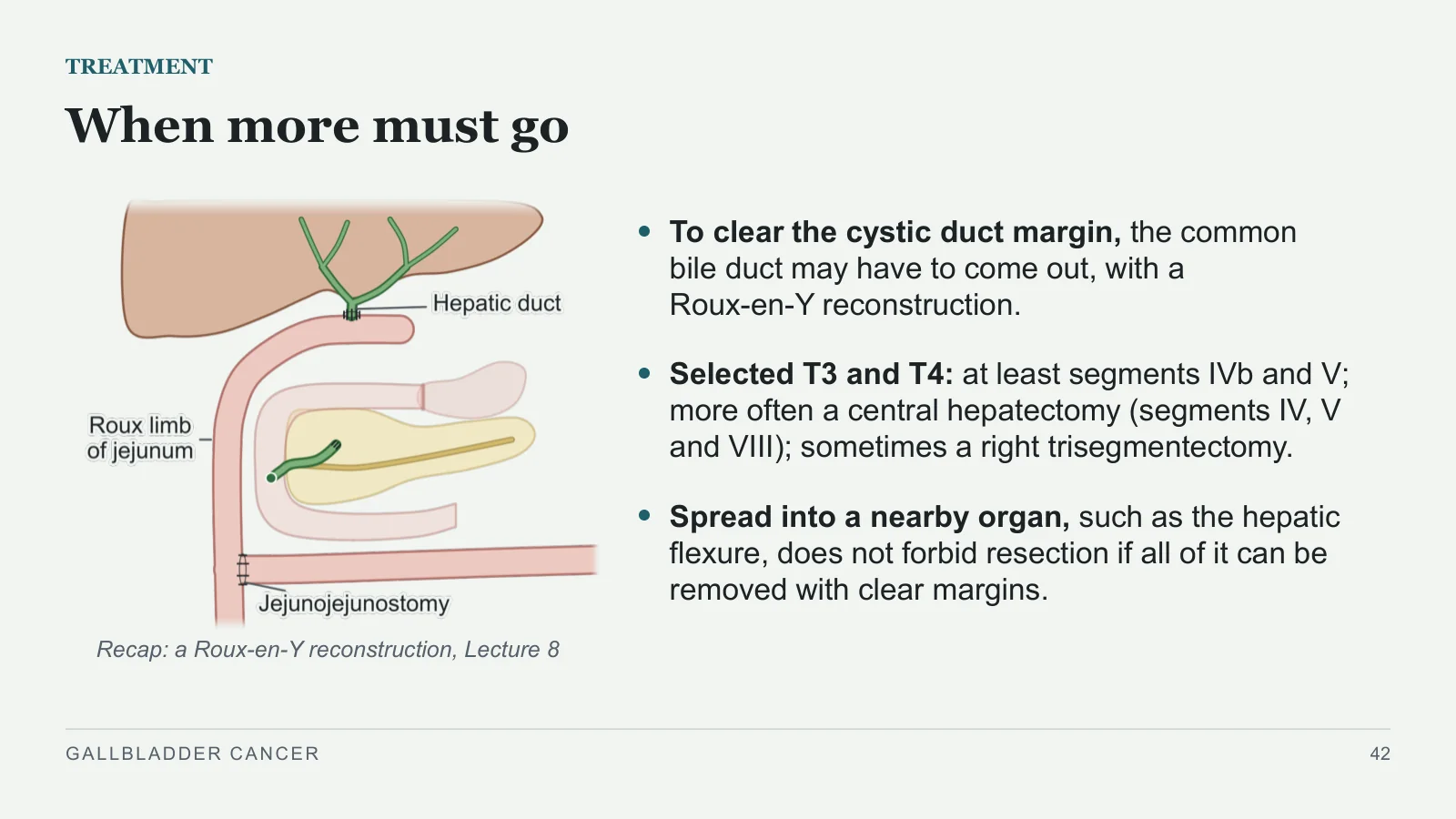
When more must go
TREATMENT
Recap: a Roux-en-Y reconstruction, Lecture 8
To clear the cystic duct margin, the common bile duct may have to come out, with a Roux-en-Y reconstruction.
Selected T3 and T4: at least segments IVb and V; more often a central hepatectomy (segments IV, V and VIII); sometimes a right trisegmentectomy.
Spread into a nearby organ, such as the hepatic flexure, does not forbid resection if all of it can be removed with clear margins.
GALLBLADDER CANCER
42
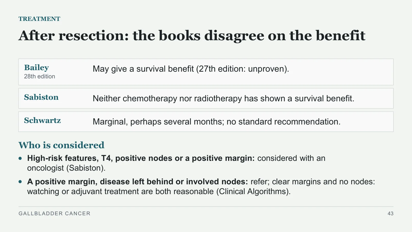
After resection: the books disagree on the benefit
TREATMENT
Bailey
28th edition
May give a survival benefit (27th edition: unproven).
Sabiston
Neither chemotherapy nor radiotherapy has shown a survival benefit.
Schwartz
Marginal, perhaps several months; no standard recommendation.
Who is considered
High-risk features, T4, positive nodes or a positive margin: considered with an oncologist (Sabiston).
A positive margin, disease left behind or involved nodes: refer; clear margins and no nodes: watching or adjuvant treatment are both reasonable (Clinical Algorithms).
GALLBLADDER CANCER
43
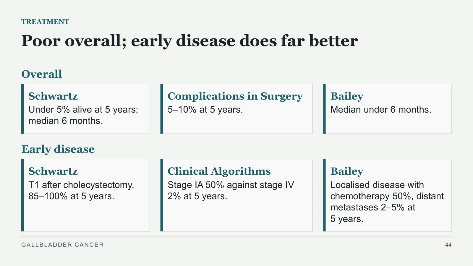
Poor overall; early disease does far better
TREATMENT
Overall
Schwartz
Under 5% alive at 5 years; median 6 months.
Complications in Surgery
5–10% at 5 years.
Bailey
Median under 6 months.
Early disease
Schwartz
T1 after cholecystectomy, 85–100% at 5 years.
Clinical Algorithms
Stage IA 50% against stage IV 2% at 5 years.
Bailey
Localised disease with chemotherapy 50%, distant metastases 2–5% at 5 years.
GALLBLADDER CANCER
44
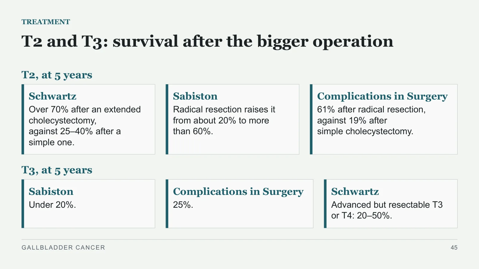
T2 and T3: survival after the bigger operation
TREATMENT
T2, at 5 years
Schwartz
Over 70% after an extended cholecystectomy, against 25–40% after a simple one.
Sabiston
Radical resection raises it from about 20% to more than 60%.
Complications in Surgery
61% after radical resection, against 19% after simple cholecystectomy.
T3, at 5 years
Sabiston
Under 20%.
Complications in Surgery
25%.
Schwartz
Advanced but resectable T3 or T4: 20–50%.
GALLBLADDER CANCER
45
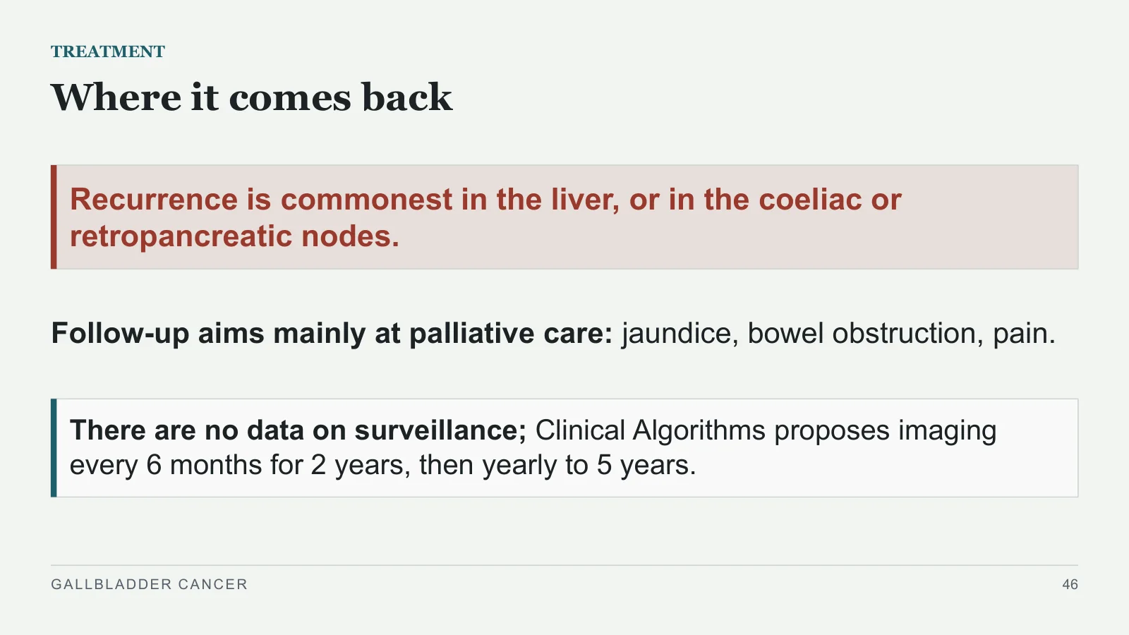
Where it comes back
TREATMENT
Recurrence is commonest in the liver, or in the coeliac or retropancreatic nodes.
Follow-up aims mainly at palliative care: jaundice, bowel obstruction, pain.
There are no data on surveillance; Clinical Algorithms proposes imaging every 6 months for 2 years, then yearly to 5 years.
GALLBLADDER CANCER
46
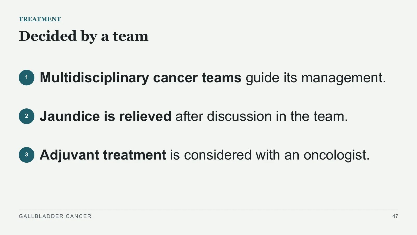
Decided by a team
TREATMENT
1
Multidisciplinary cancer teams guide its management.
2
Jaundice is relieved after discussion in the team.
3
Adjuvant treatment is considered with an oncologist.
GALLBLADDER CANCER
47
Palliation
Palliation
PART 7
GALLBLADDER CANCER
48
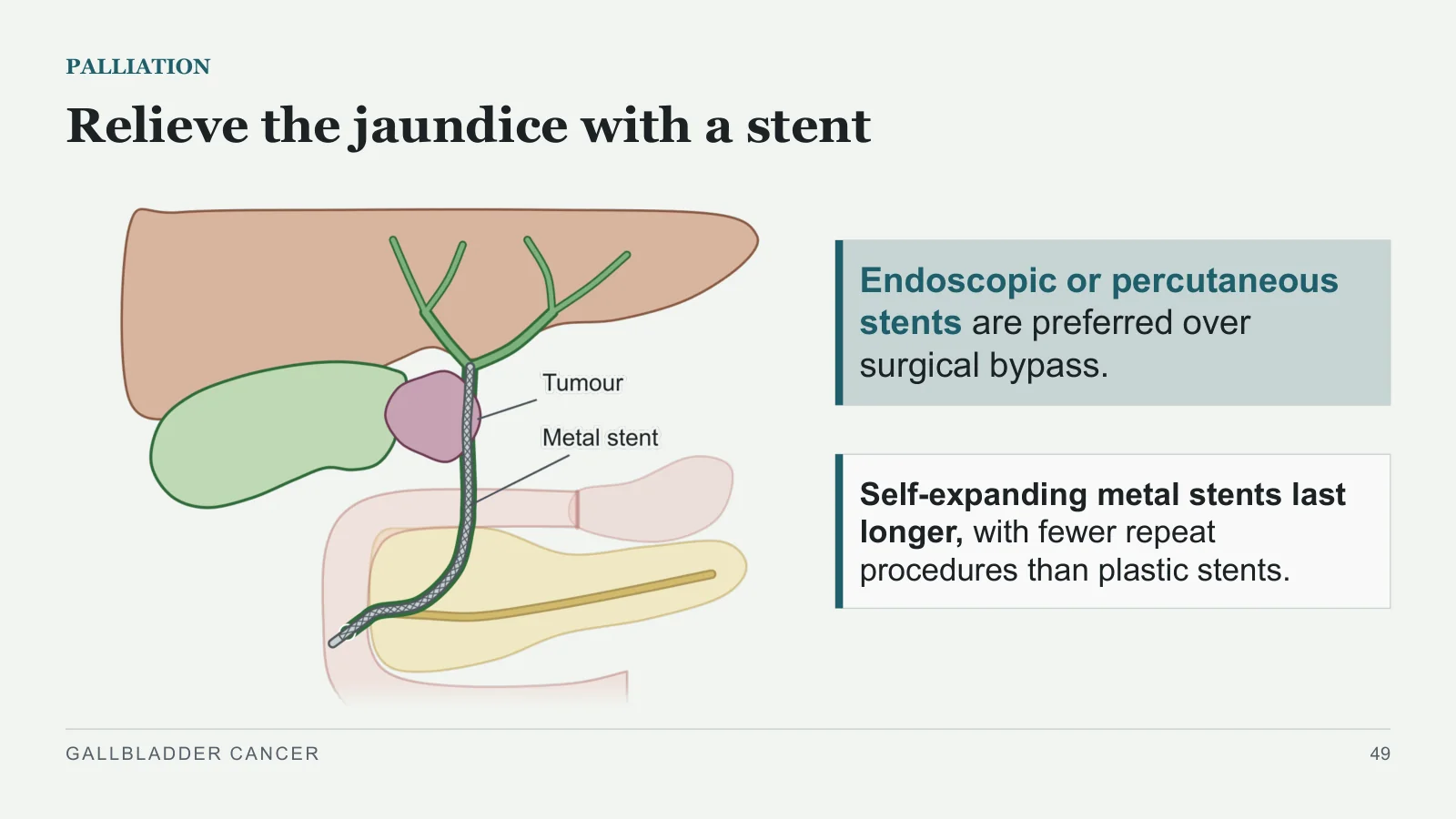
Relieve the jaundice with a stent
PALLIATION
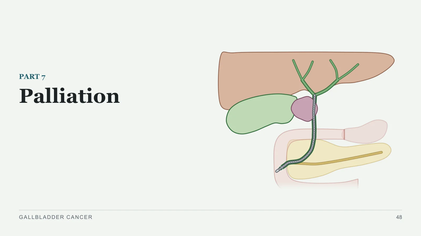
Endoscopic or percutaneous stents are preferred over surgical bypass.
Self-expanding metal stents last longer, with fewer repeat procedures than plastic stents.
GALLBLADDER CANCER
49
Palliation beyond the jaundice
PALLIATION
Metastatic disease is palliated without an operation.
A blocked duodenum from local spread: usually an endoscopic duodenal stent.
Pain: opioids by mouth, later by injection in hospice care; or a coeliac ganglion block through the skin.
Chemotherapy or radiotherapy can be offered, with no standard recommendation.
With distant metastases at presentation, median survival: the books disagree
Sabiston
13 months.
Schwartz
1 to 3 months.
GALLBLADDER CANCER
50
Early detection
Early detection
PART 8
GALLBLADDER CANCER
51
Taking the gallbladder out early
EARLY DETECTION
Recap: Lecture 3
Bailey: prophylactic cholecystectomy “may be performed”
Stones over 3 cm.
Polyps over 1 cm.
Anomalous drainage of the pancreatic duct.
Porcelain gallbladder.
Some groups and areas with a high rate of gallbladder cancer with stones, including parts of northern India.
Stone size: the books differ
Bailey
Over 3 cm.
Sabiston
Over 2.5 cm (“should consider cholecystectomy”, with porcelain gallbladder and a long common channel).
In populations at increased risk, it may be advisable (Schwartz).
GALLBLADDER CANCER
52
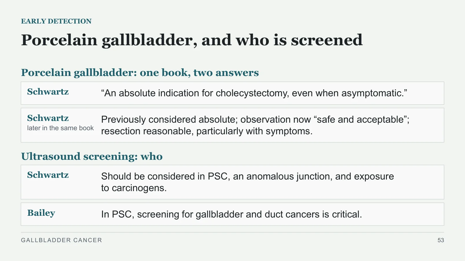
Porcelain gallbladder, and who is screened
EARLY DETECTION
Porcelain gallbladder: one book, two answers
Schwartz
“An absolute indication for cholecystectomy, even when asymptomatic.”
Schwartz
later in the same book
Previously considered absolute; observation now “safe and acceptable”; resection reasonable, particularly with symptoms.
Ultrasound screening: who
Schwartz
Should be considered in PSC, an anomalous junction, and exposure to carcinogens.
Bailey
In PSC, screening for gallbladder and duct cancers is critical.
GALLBLADDER CANCER
53
Case recap and take-home
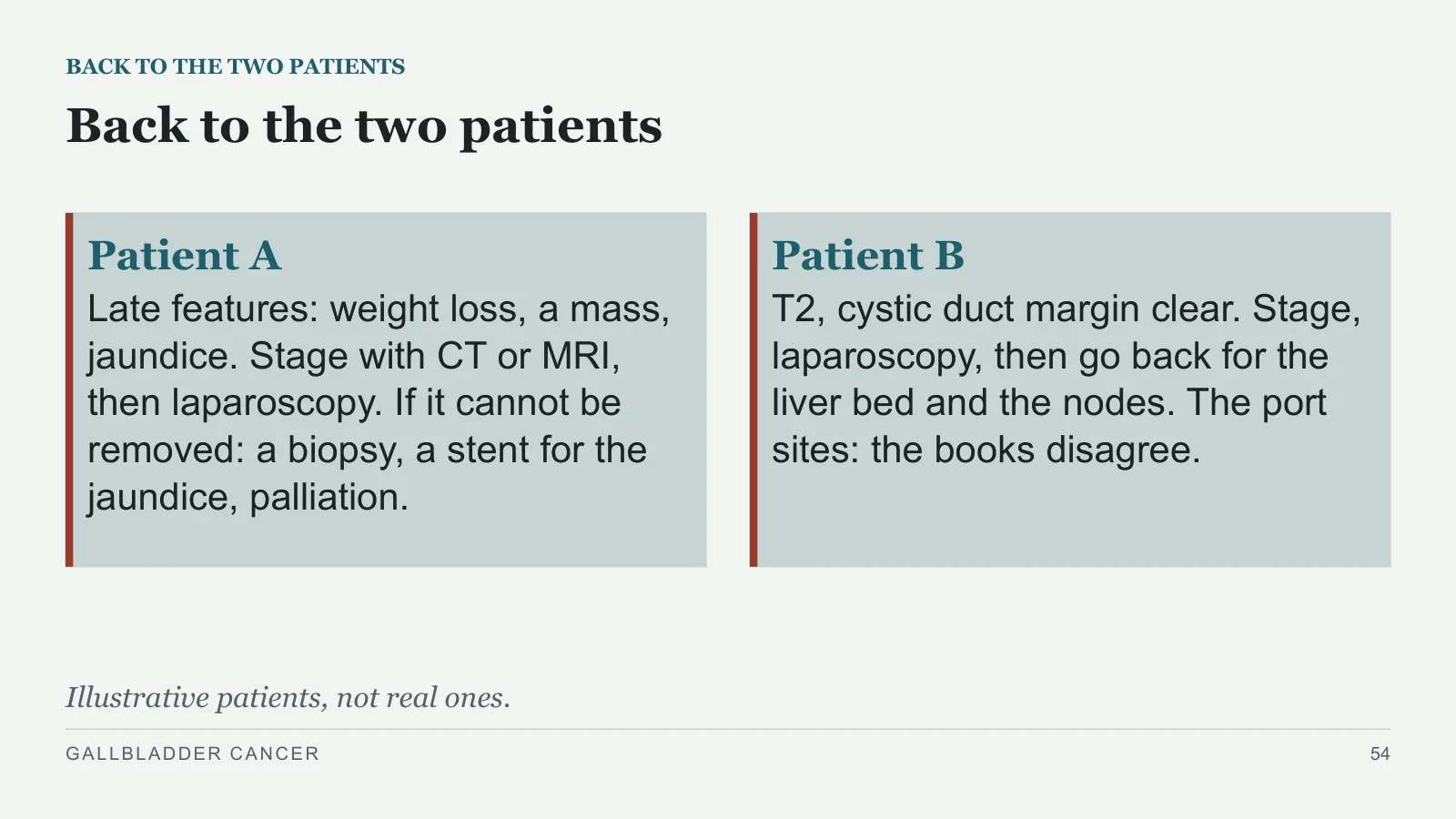
Back to the two patients
BACK TO THE TWO PATIENTS
Patient A
Late features: weight loss, a mass, jaundice. Stage with CT or MRI, then laparoscopy. If it cannot be removed: a biopsy, a stent for the jaundice, palliation.
Patient B
T2, cystic duct margin clear. Stage, laparoscopy, then go back for the liver bed and the nodes. The port sites: the books disagree.
Illustrative patients, not real ones.
GALLBLADDER CANCER
54
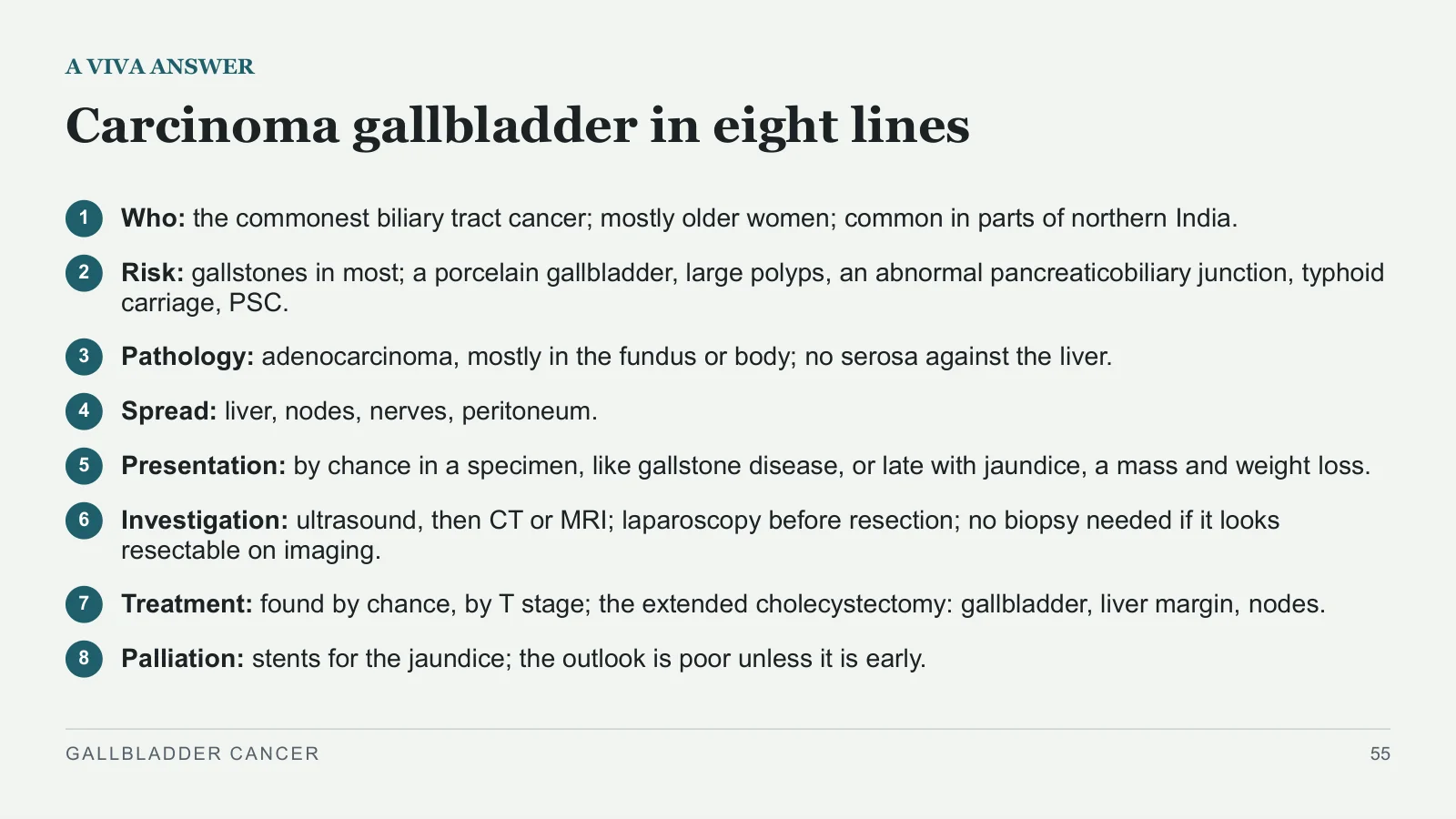
Carcinoma gallbladder in eight lines
A VIVA ANSWER
1
Who: the commonest biliary tract cancer; mostly older women; common in parts of northern India.
2
Risk: gallstones in most; a porcelain gallbladder, large polyps, an abnormal pancreaticobiliary junction, typhoid carriage, PSC.
3
Pathology: adenocarcinoma, mostly in the fundus or body; no serosa against the liver.
4
Spread: liver, nodes, nerves, peritoneum.
5
Presentation: by chance in a specimen, like gallstone disease, or late with jaundice, a mass and weight loss.
6
Investigation: ultrasound, then CT or MRI; laparoscopy before resection; no biopsy needed if it looks resectable on imaging.
7
Treatment: found by chance, by T stage; the extended cholecystectomy: gallbladder, liver margin, nodes.
8
Palliation: stents for the jaundice; the outlook is poor unless it is early.
GALLBLADDER CANCER
55
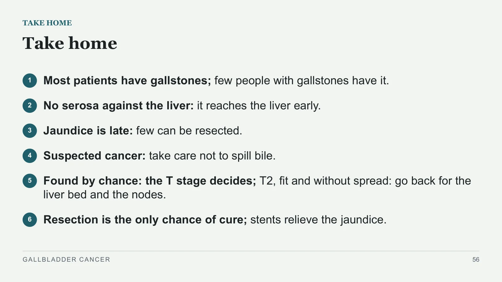
Take home
TAKE HOME
1
Most patients have gallstones; few people with gallstones have it.
2
No serosa against the liver: it reaches the liver early.
3
Jaundice is late: few can be resected.
4
Suspected cancer: take care not to spill bile.
5
Found by chance: the T stage decides; T2, fit and without spread: go back for the liver bed and the nodes.
6
Resection is the only chance of cure; stents relieve the jaundice.
GALLBLADDER CANCER
56
Sources and image credits
Books used
REFERENCES
Bailey & Love's Short Practice of Surgery, 28th edition. Chapter 71. 27th edition: pages 1210–1211.
Sabiston Textbook of Surgery, 21st edition. Chapter 55.
Schwartz's Principles of Surgery, 11th edition. Chapter 32.
Clinical Algorithms in General Surgery (Docimo and Pauli, editors). Pages 365–366.
Complications in Surgery, 2nd edition. Pages 429 and 444–445.
Fischer's Mastery of Surgery, 7th edition. Chapter 110 (Hunter and Fischer).
Clinical Scenarios in Surgery. Page 110.
GALLBLADDER CANCER
57
Image credits
REFERENCES
Diagrams: original drawings for this course (the recaps from Lecture 8 are labelled). Images: Wikimedia Commons, page commons.wikimedia.org/wiki/File: plus the name; licences at creativecommons.org/licenses.
GALLBLADDER CANCER
58
| Slide | File on Wikimedia Commons | Author, licence |
| 8 | Porzellangallenblase - 88j - CT axial WT und KF - 001.jpg | Hellerhoff, CC BY-SA 3.0 |
| 12 | Gallbladder adenocarcinoma (2) histopathology.jpg | KGH, CC BY-SA 3.0 |
| 23 | Adenokarzinom der Gallenblase mit Satellitenherden 66W - CT - 001.jpg | Hellerhoff, CC BY-SA 4.0 |
Sources and image credits
Sources and image credits are reproduced from the designated slides.
Images
- Slide 8 · Porzellangallenblase - 88j - CT axial WT und KF - 001.jpg · Hellerhoff, CC BY-SA 3.0 · CC BY-SA 3.0
- Slide 12 · Gallbladder adenocarcinoma (2) histopathology.jpg · KGH, CC BY-SA 3.0 · CC BY-SA 3.0
- Slide 23 · Adenokarzinom der Gallenblase mit Satellitenherden 66W - CT - 001.jpg · Hellerhoff, CC BY-SA 4.0 · CC BY-SA 4.0


Sources
- [1] REFERENCES
- [2] Books used
- [3] Bailey & Love's Short Practice of Surgery, 28th edition. Chapter 71. 27th edition: pages 1210–1211.
- [4] Sabiston Textbook of Surgery, 21st edition. Chapter 55.
- [5] Schwartz's Principles of Surgery, 11th edition. Chapter 32.
- [6] Clinical Algorithms in General Surgery (Docimo and Pauli, editors). Pages 365–366.
- [7] Complications in Surgery, 2nd edition. Pages 429 and 444–445.
- [8] Fischer's Mastery of Surgery, 7th edition. Chapter 110 (Hunter and Fischer).
- [9] Clinical Scenarios in Surgery. Page 110.