Hepatobiliary surgery · Lecture 4
Acute cholecystitis · 51 slides
The acutely inflamed gallbladder.
From a persistent pain to the next decision: recognise the inflamed gallbladder, assess severity and work through treatment and complications.
About the downloads
PowerPoint without speaker notes. SHA-256 checksums verify the downloaded files.
Contents · 10 parts
Opening
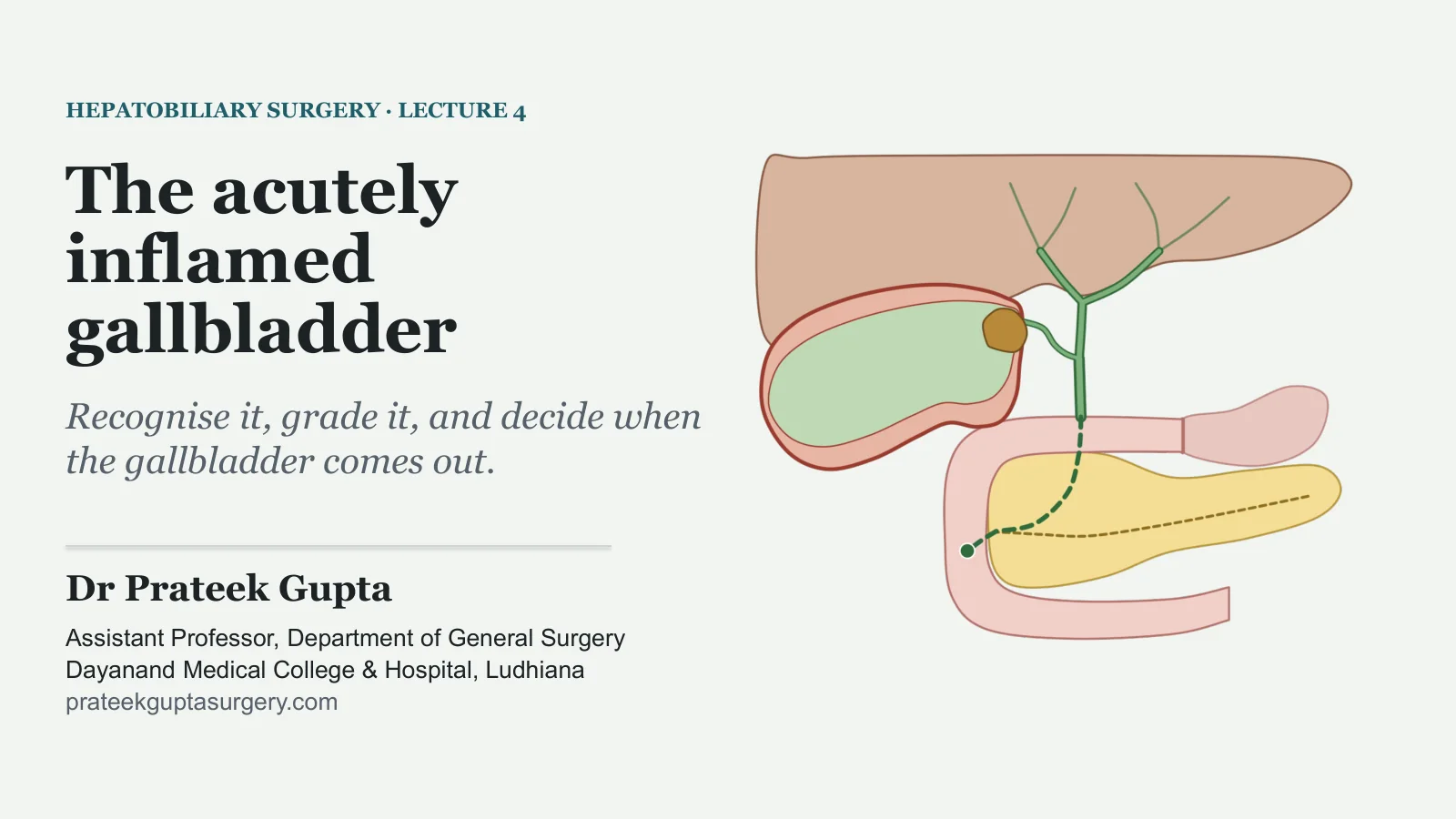
The acutely inflamed gallbladder
HEPATOBILIARY SURGERY · LECTURE 4
The acutely inflamed gallbladder
Recognise it, grade it, and decide when the gallbladder comes out.
Dr Prateek Gupta
Assistant Professor, Department of General Surgery Dayanand Medical College & Hospital, Ludhiana prateekguptasurgery.com
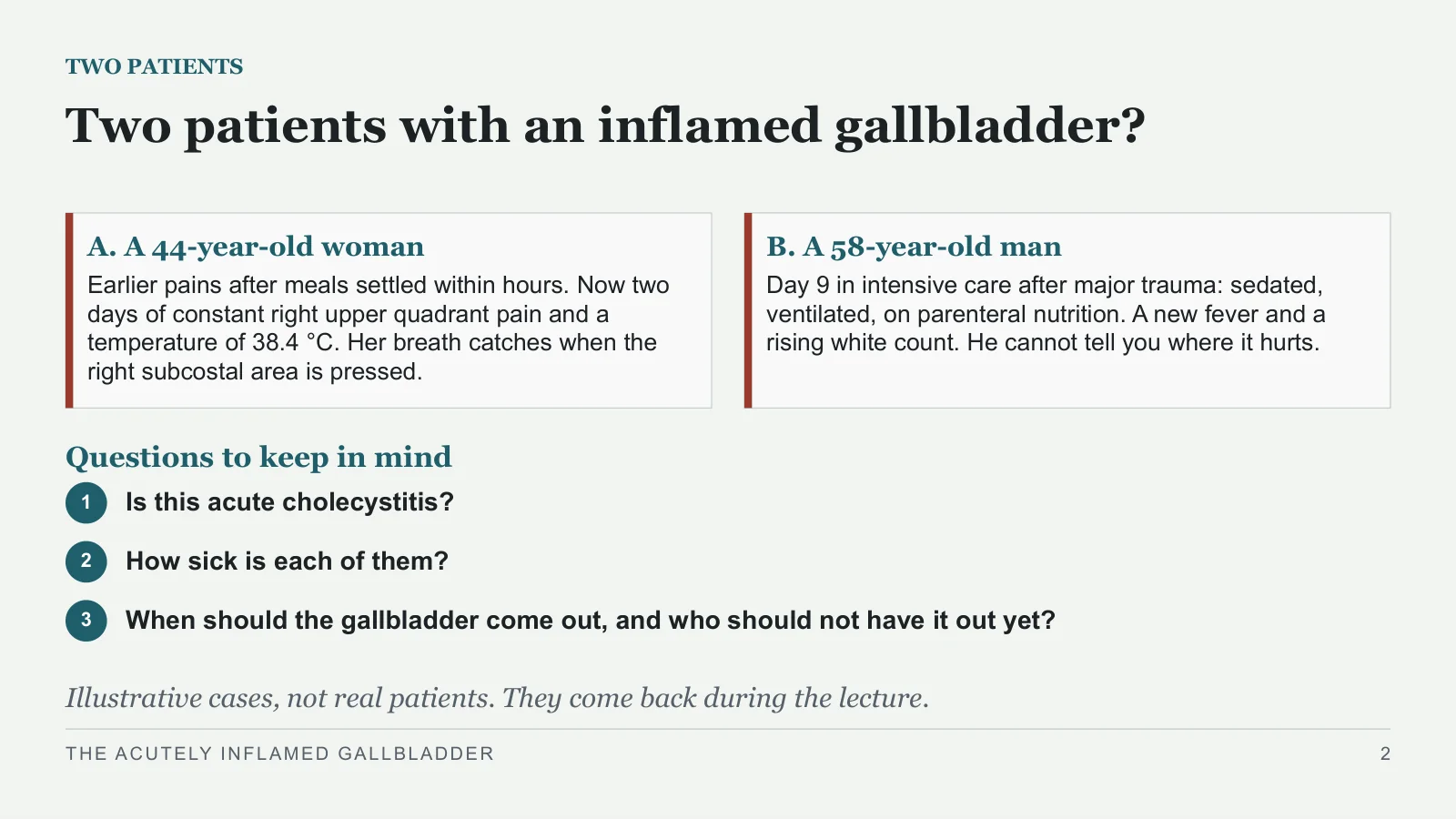
Two patients with an inflamed gallbladder?
TWO PATIENTS
A. A 44-year-old woman
Earlier pains after meals settled within hours. Now two days of constant right upper quadrant pain and a temperature of 38.4 °C. Her breath catches when the right subcostal area is pressed.
B. A 58-year-old man
Day 9 in intensive care after major trauma: sedated, ventilated, on parenteral nutrition. A new fever and a rising white count. He cannot tell you where it hurts.
Questions to keep in mind
1
Is this acute cholecystitis?
2
How sick is each of them?
3
When should the gallbladder come out, and who should not have it out yet?
Illustrative cases, not real patients. They come back during the lecture.
THE ACUTELY INFLAMED GALLBLADDER
2
From pain to inflammation
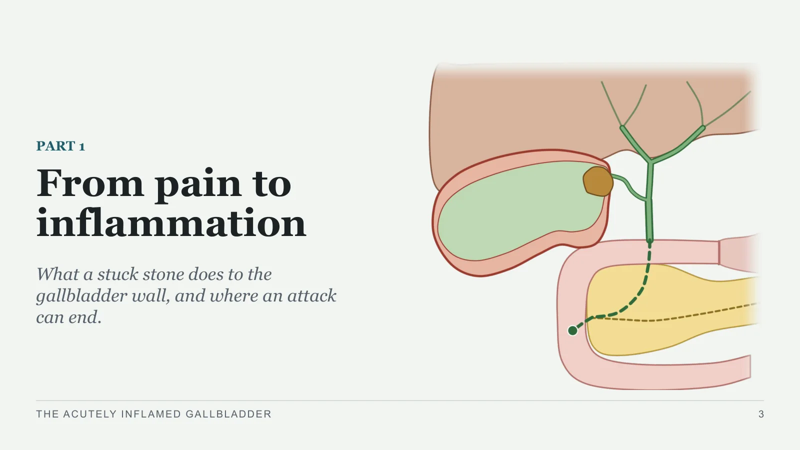
From pain to inflammation
PART 1
What a stuck stone does to the gallbladder wall, and where an attack can end.
THE ACUTELY INFLAMED GALLBLADDER
3
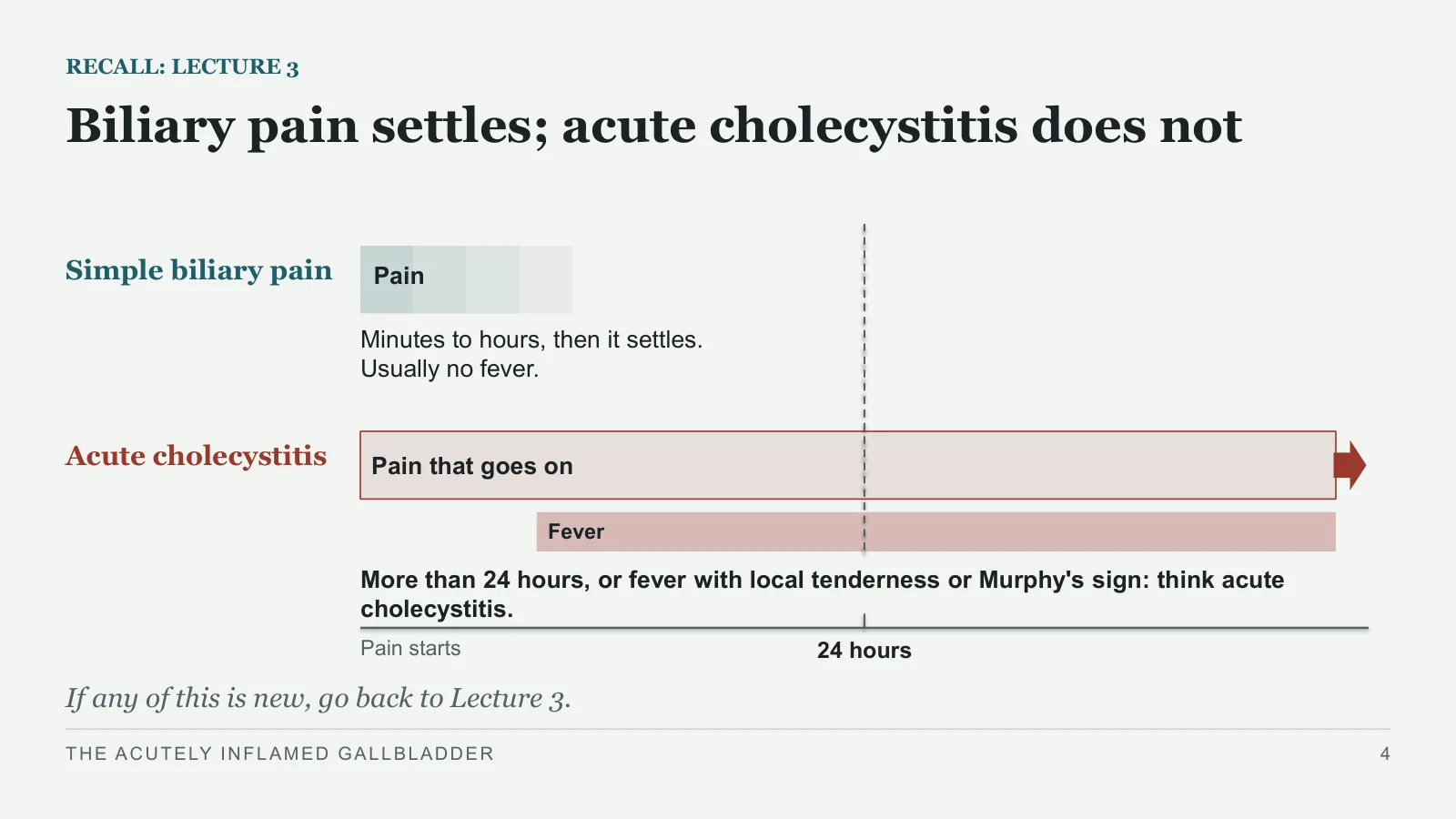
Biliary pain settles; acute cholecystitis does not
RECALL: LECTURE 3
Simple biliary pain
Acute cholecystitis
Pain
Minutes to hours, then it settles. Usually no fever.
Pain that goes on
Fever
More than 24 hours, or fever with local tenderness or Murphy's sign: think acute cholecystitis.
24 hours
Pain starts
If any of this is new, go back to Lecture 3.
THE ACUTELY INFLAMED GALLBLADDER
4
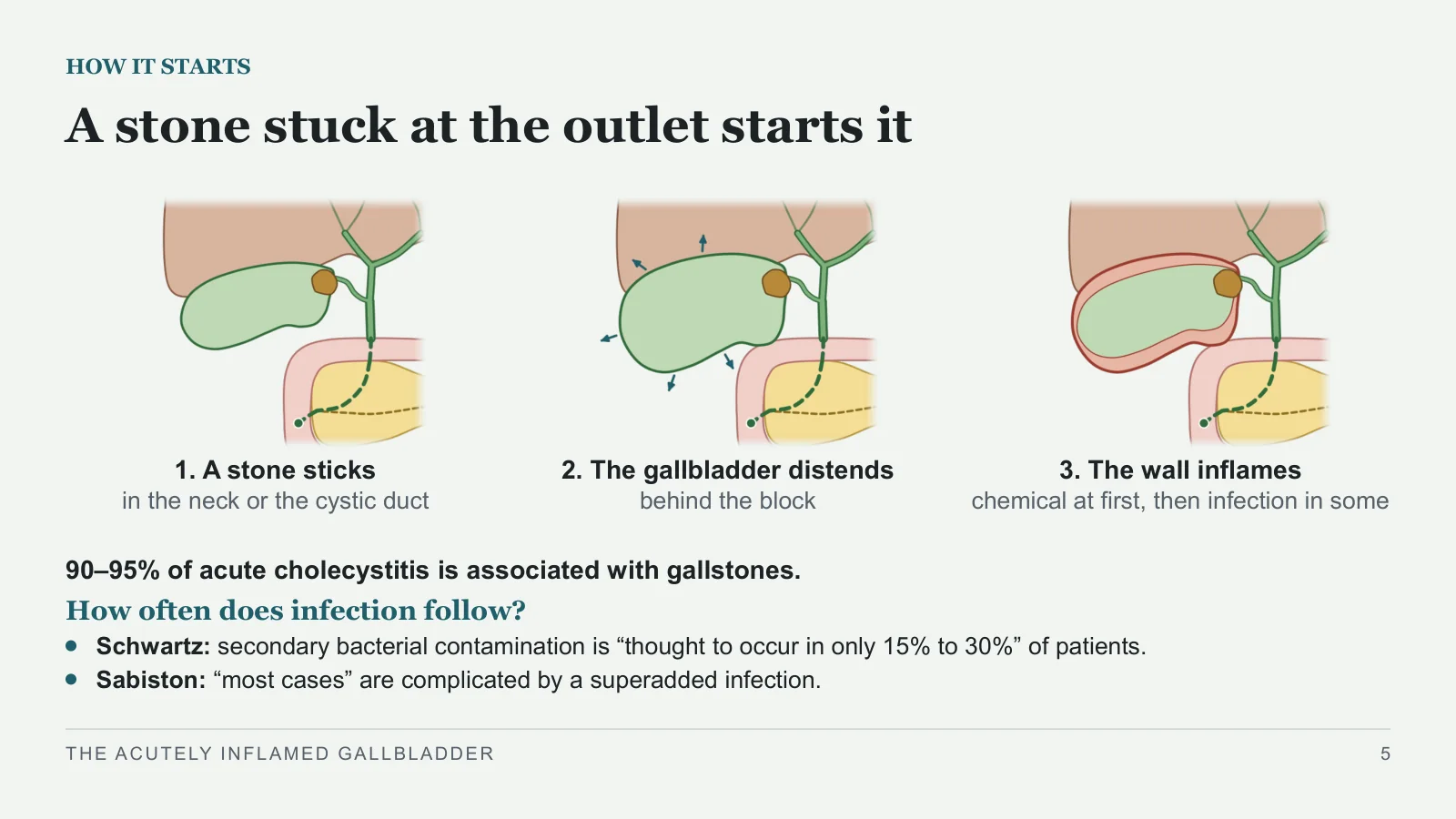
A stone stuck at the outlet starts it
HOW IT STARTS
1. A stone sticks
in the neck or the cystic duct
2. The gallbladder distends
behind the block
3. The wall inflames
chemical at first, then infection in some
90–95% of acute cholecystitis is associated with gallstones.
How often does infection follow?
Schwartz: secondary bacterial contamination is “thought to occur in only 15% to 30%” of patients.
Sabiston: “most cases” are complicated by a superadded infection.
THE ACUTELY INFLAMED GALLBLADDER
5
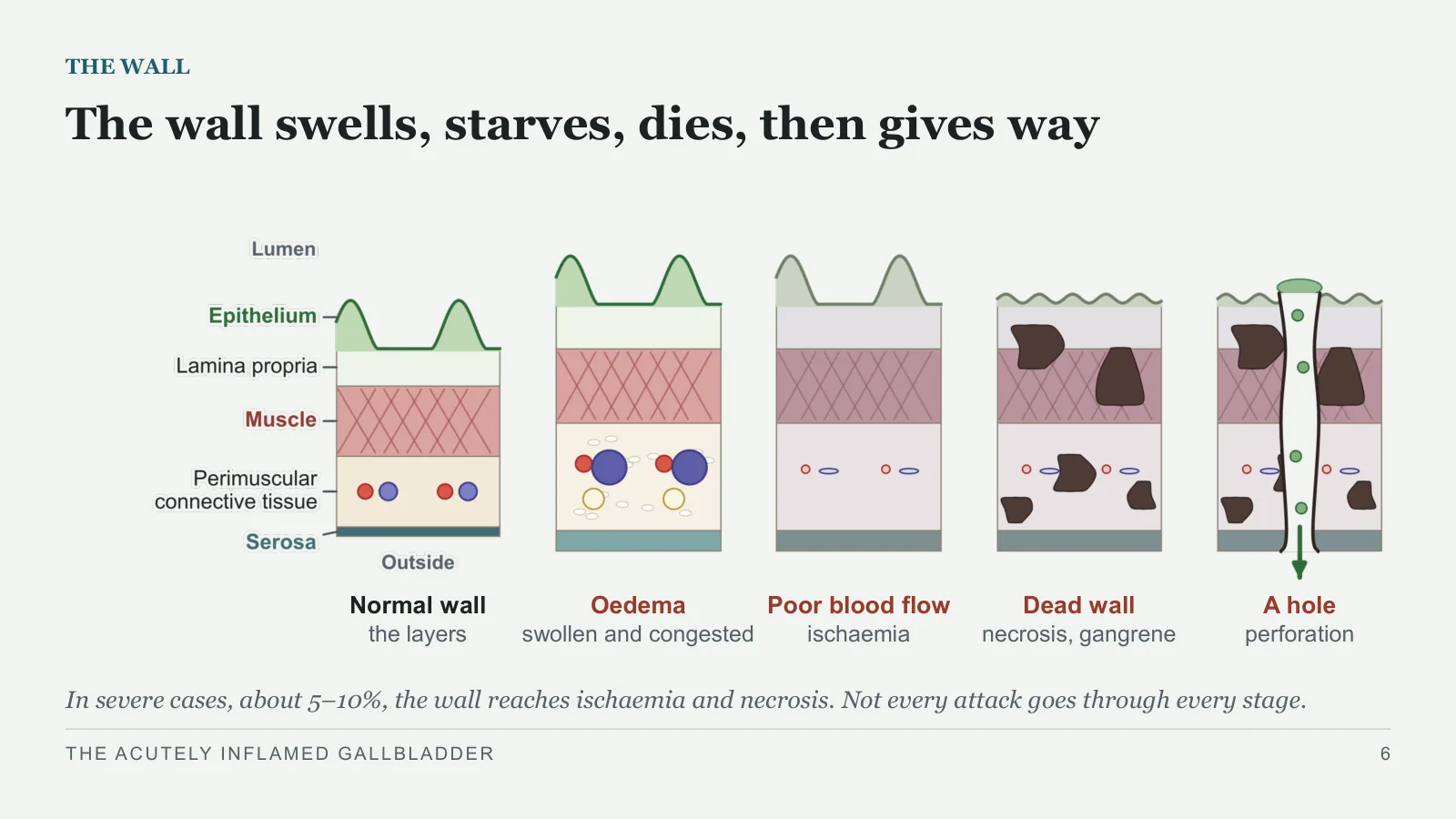
The wall swells, starves, dies, then gives way
THE WALL
Normal wall
the layers
Oedema
swollen and congested
Poor blood flow
ischaemia
Dead wall
necrosis, gangrene
A hole
perforation
In severe cases, about 5–10%, the wall reaches ischaemia and necrosis. Not every attack goes through every stage.
THE ACUTELY INFLAMED GALLBLADDER
6
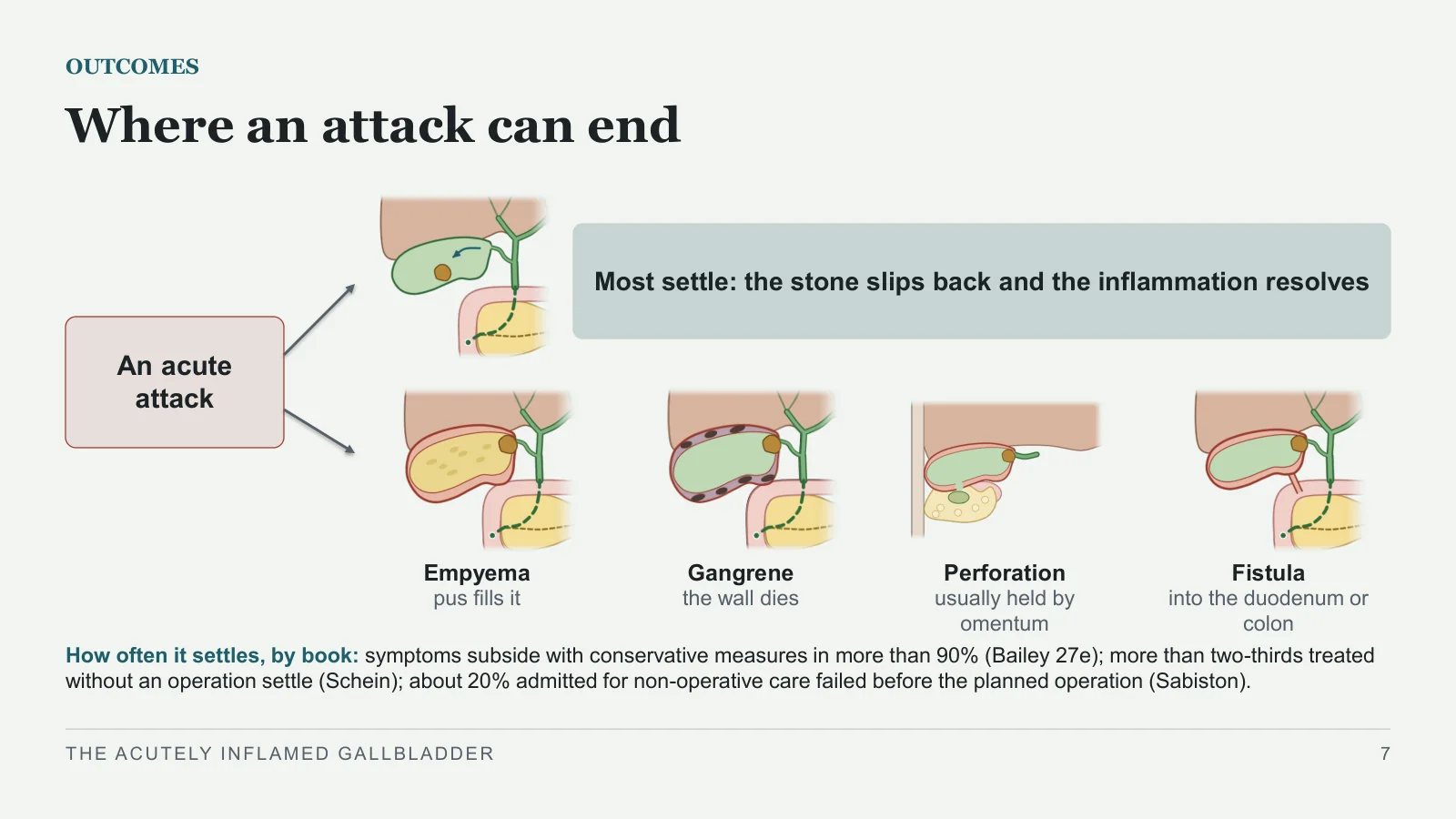
Where an attack can end
OUTCOMES
An acute attack
Most settle: the stone slips back and the inflammation resolves
Empyema
pus fills it
Gangrene
the wall dies
Perforation
usually held by omentum
Fistula
into the duodenum or colon
How often it settles, by book: symptoms subside with conservative measures in more than 90% (Bailey 27e); more than two-thirds treated without an operation settle (Schein); about 20% admitted for non-operative care failed before the planned operation (Sabiston).
THE ACUTELY INFLAMED GALLBLADDER
7
Recognising it
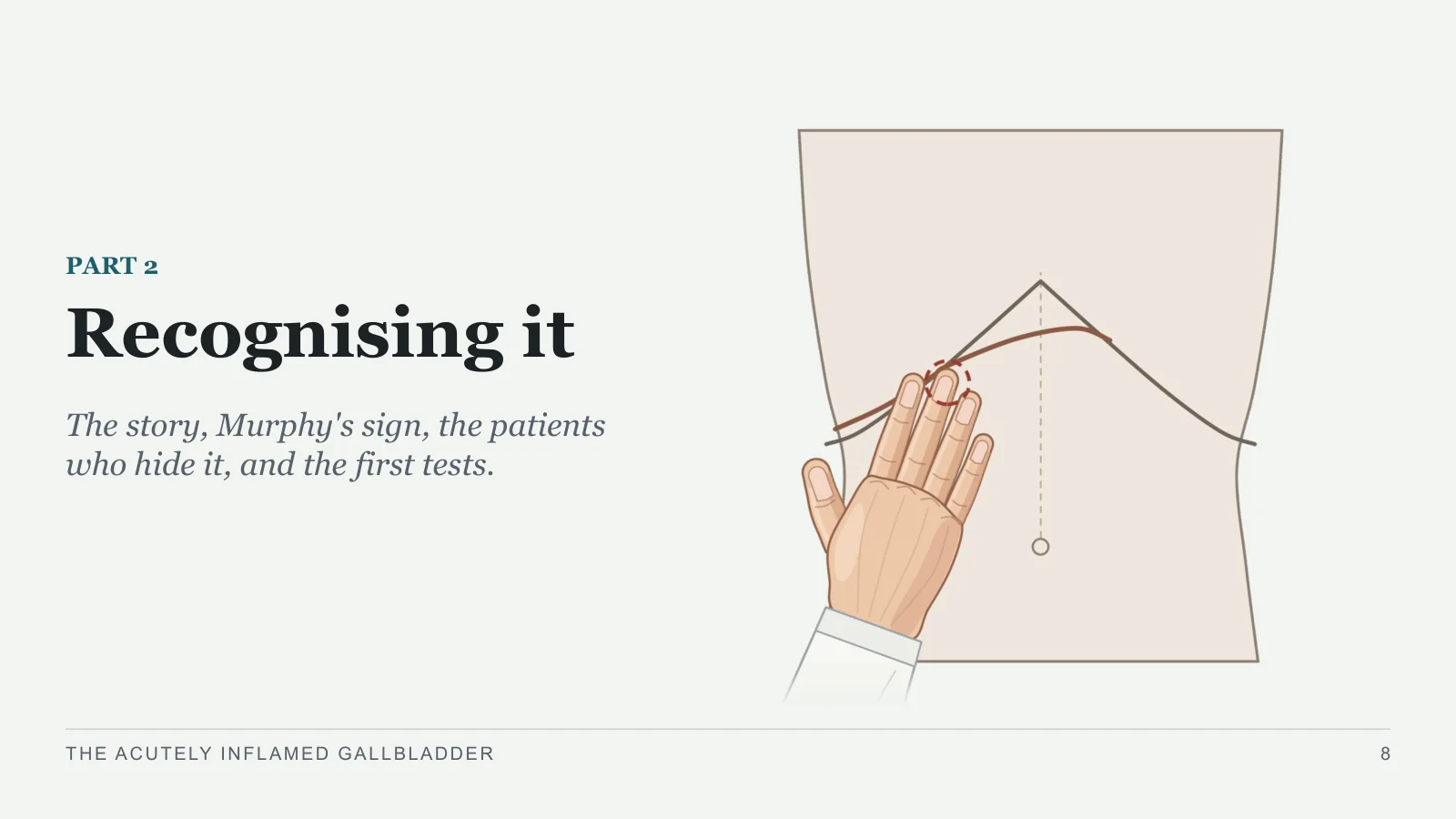
Recognising it
PART 2
The story, Murphy's sign, the patients who hide it, and the first tests.
THE ACUTELY INFLAMED GALLBLADDER
8
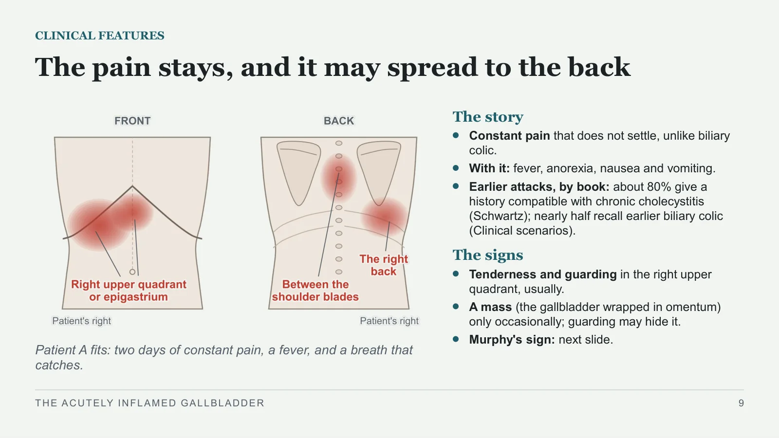
The pain stays, and it may spread to the back
CLINICAL FEATURES
Patient A fits: two days of constant pain, a fever, and a breath that catches.
The story
Constant pain that does not settle, unlike biliary colic.
With it: fever, anorexia, nausea and vomiting.
Earlier attacks, by book: about 80% give a history compatible with chronic cholecystitis (Schwartz); nearly half recall earlier biliary colic (Clinical scenarios).
The signs
Tenderness and guarding in the right upper quadrant, usually.
A mass (the gallbladder wrapped in omentum) only occasionally; guarding may hide it.
Murphy's sign: next slide.
THE ACUTELY INFLAMED GALLBLADDER
9
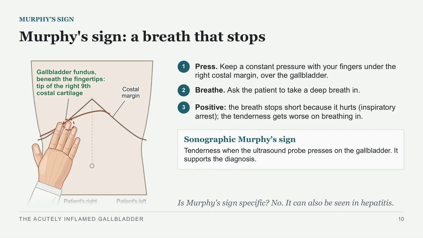
Murphy's sign: a breath that stops
MURPHY'S SIGN
1
Press. Keep a constant pressure with your fingers under the right costal margin, over the gallbladder.
2
Breathe. Ask the patient to take a deep breath in.
3
Positive: the breath stops short because it hurts (inspiratory arrest); the tenderness gets worse on breathing in.
Sonographic Murphy's sign
Tenderness when the ultrasound probe presses on the gallbladder. It supports the diagnosis.
Is Murphy's sign specific? No. It can also be seen in hepatitis.
THE ACUTELY INFLAMED GALLBLADDER
10
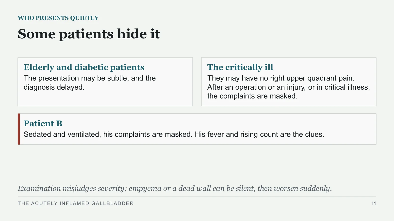
Some patients hide it
WHO PRESENTS QUIETLY
Elderly and diabetic patients
The presentation may be subtle, and the diagnosis delayed.
The critically ill
They may have no right upper quadrant pain. After an operation or an injury, or in critical illness, the complaints are masked.
Patient B
Sedated and ventilated, his complaints are masked. His fever and rising count are the clues.
Examination misjudges severity: empyema or a dead wall can be silent, then worsen suddenly.
THE ACUTELY INFLAMED GALLBLADDER
11
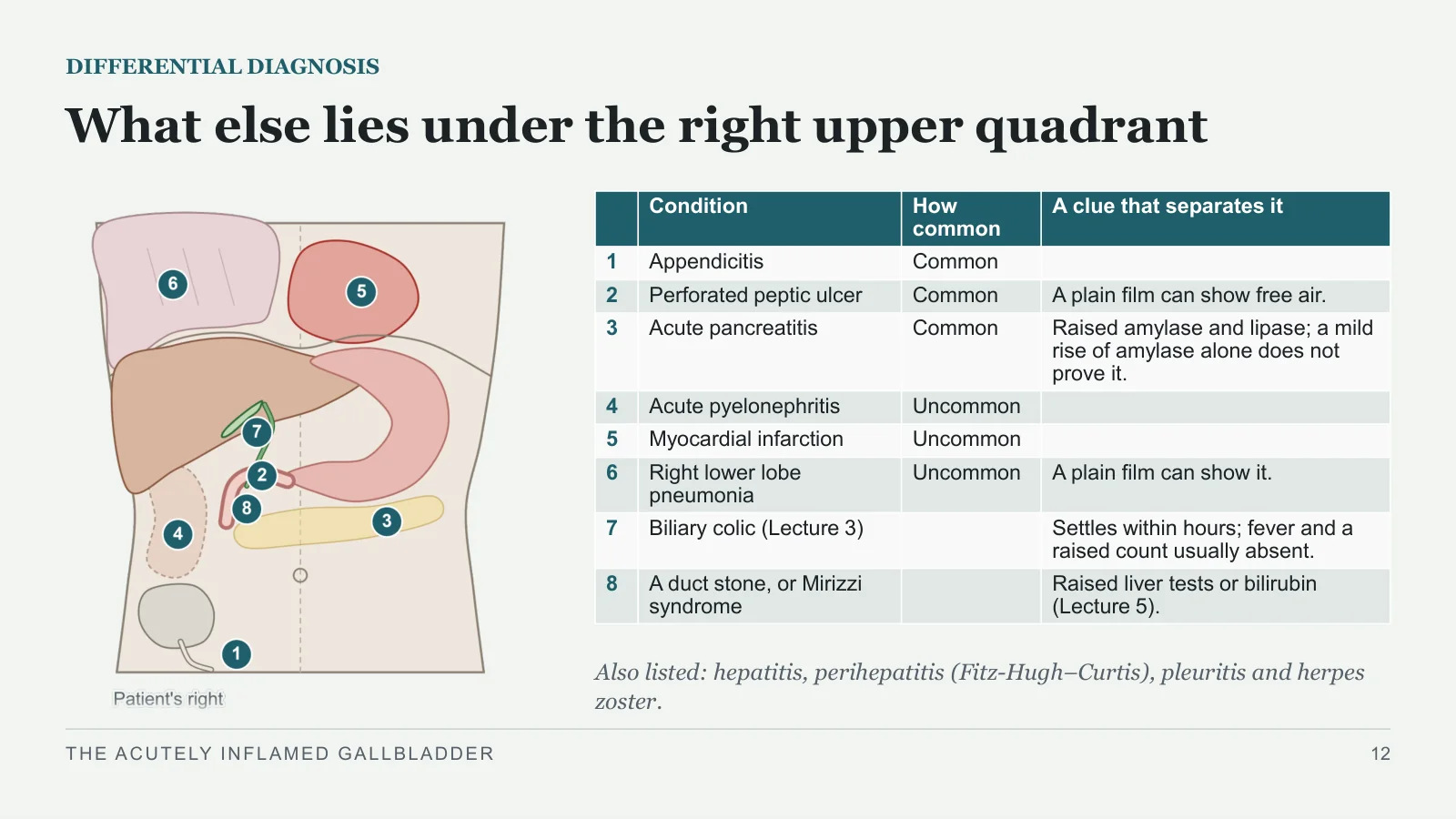
What else lies under the right upper quadrant
DIFFERENTIAL DIAGNOSIS
Also listed: hepatitis, perihepatitis (Fitz-Hugh–Curtis), pleuritis and herpes zoster.
THE ACUTELY INFLAMED GALLBLADDER
12
| Condition | How common | A clue that separates it | |
| 1 | Appendicitis | Common | |
| 2 | Perforated peptic ulcer | Common | A plain film can show free air. |
| 3 | Acute pancreatitis | Common | Raised amylase and lipase; a mild rise of amylase alone does not prove it. |
| 4 | Acute pyelonephritis | Uncommon | |
| 5 | Myocardial infarction | Uncommon | |
| 6 | Right lower lobe pneumonia | Uncommon | A plain film can show it. |
| 7 | Biliary colic (Lecture 3) | Settles within hours; fever and a raised count usually absent. | |
| 8 | A duct stone, or Mirizzi syndrome | Raised liver tests or bilirubin (Lecture 5). |
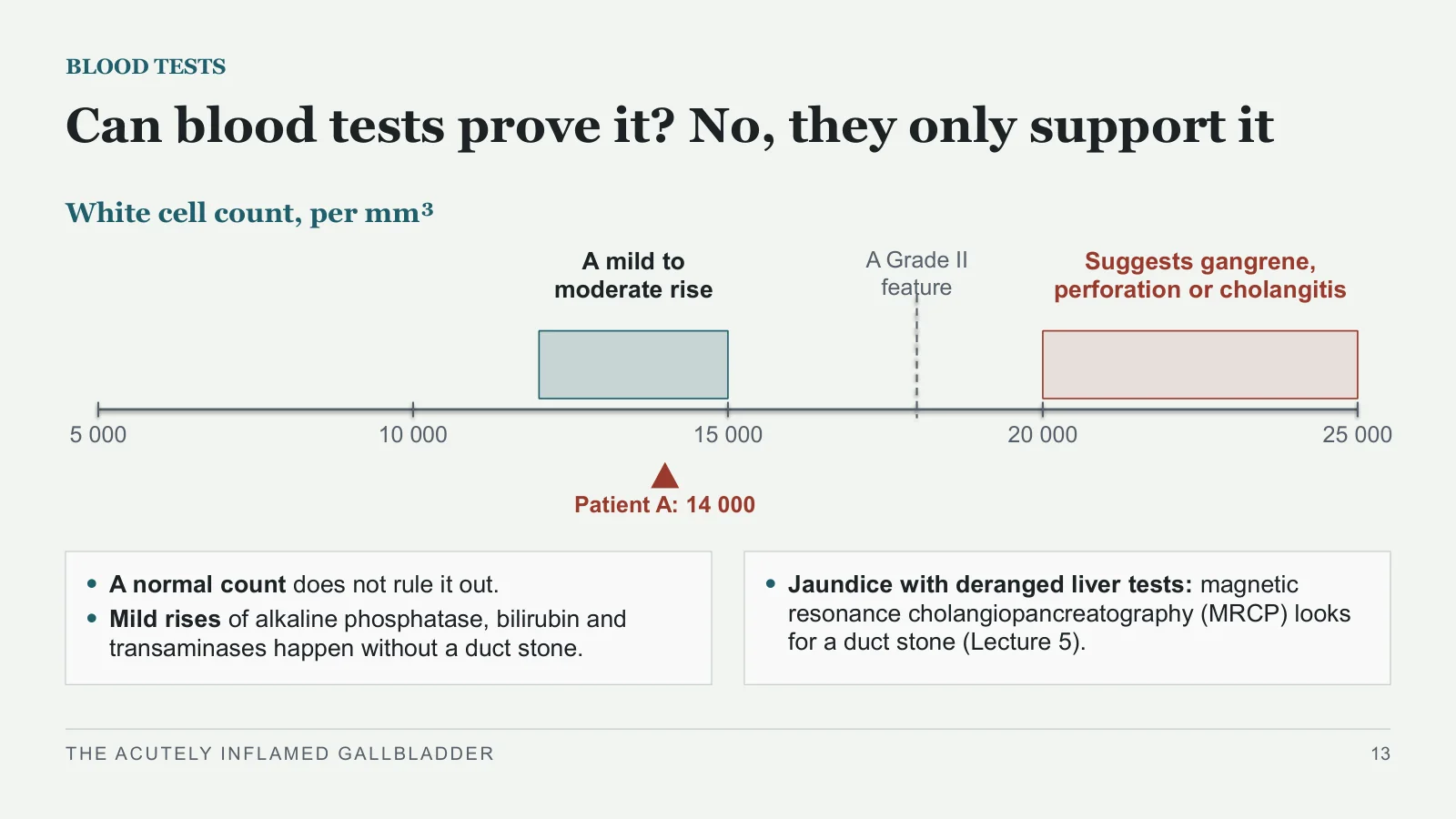
Can blood tests prove it? No, they only support it
BLOOD TESTS
White cell count, per mm³
A mild to moderate rise
Suggests gangrene, perforation or cholangitis
A Grade II feature
5 000
10 000
15 000
20 000
25 000
Patient A: 14 000
A normal count does not rule it out.
Mild rises of alkaline phosphatase, bilirubin and transaminases happen without a duct stone.
Jaundice with deranged liver tests: magnetic resonance cholangiopancreatography (MRCP) looks for a duct stone (Lecture 5).
THE ACUTELY INFLAMED GALLBLADDER
13
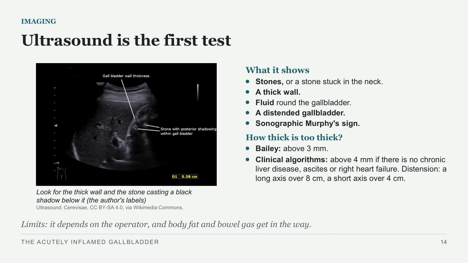
Ultrasound is the first test
IMAGING
Look for the thick wall and the stone casting a black shadow below it (the author's labels)
Ultrasound: Cerevisae, CC BY-SA 4.0, via Wikimedia Commons.
What it shows
Stones, or a stone stuck in the neck.
A thick wall.
Fluid round the gallbladder.
A distended gallbladder.
Sonographic Murphy's sign.
How thick is too thick?
Bailey: above 3 mm.
Clinical algorithms: above 4 mm if there is no chronic liver disease, ascites or right heart failure. Distension: a long axis over 8 cm, a short axis over 4 cm.
Limits: it depends on the operator, and body fat and bowel gas get in the way.
THE ACUTELY INFLAMED GALLBLADDER
14
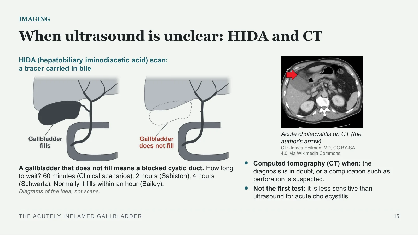
When ultrasound is unclear: HIDA and CT
IMAGING
HIDA (hepatobiliary iminodiacetic acid) scan: a tracer carried in bile
A gallbladder that does not fill means a blocked cystic duct. How long to wait? 60 minutes (Clinical scenarios), 2 hours (Sabiston), 4 hours (Schwartz). Normally it fills within an hour (Bailey).
Diagrams of the idea, not scans.
Acute cholecystitis on CT (the author's arrow)
CT: James Heilman, MD, CC BY-SA 4.0, via Wikimedia Commons.
Computed tomography (CT) when: the diagnosis is in doubt, or a complication such as perforation is suspected.
Not the first test: it is less sensitive than ultrasound for acute cholecystitis.
THE ACUTELY INFLAMED GALLBLADDER
15
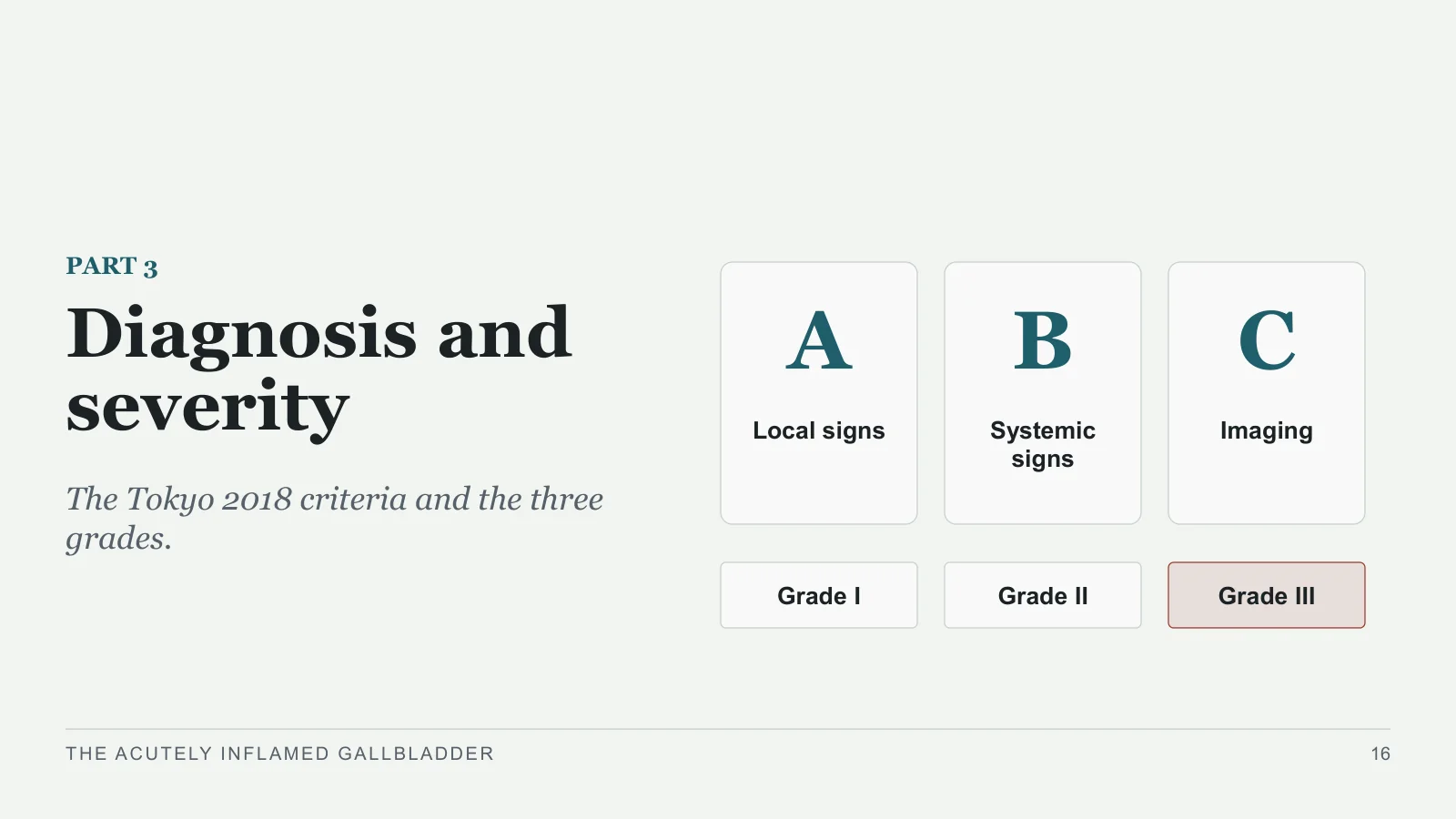
Diagnosis and severity
A
PART 3
Diagnosis and severity
The Tokyo 2018 criteria and the three grades.
Local signs
B
Systemic signs
C
Imaging
Grade I
Grade II
Grade III
THE ACUTELY INFLAMED GALLBLADDER
16
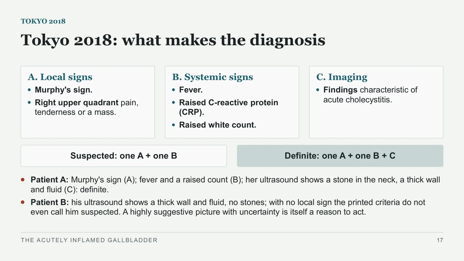
Tokyo 2018: what makes the diagnosis
TOKYO 2018
A. Local signs
Murphy's sign.
Right upper quadrant pain, tenderness or a mass.
B. Systemic signs
Fever.
Raised C-reactive protein (CRP).
Raised white count.
C. Imaging
Findings characteristic of acute cholecystitis.
Suspected: one A + one B
Definite: one A + one B + C
Patient A: Murphy's sign (A); fever and a raised count (B); her ultrasound shows a stone in the neck, a thick wall and fluid (C): definite.
Patient B: his ultrasound shows a thick wall and fluid, no stones; with no local sign the printed criteria do not even call him suspected. A highly suggestive picture with uncertainty is itself a reason to act.
THE ACUTELY INFLAMED GALLBLADDER
17
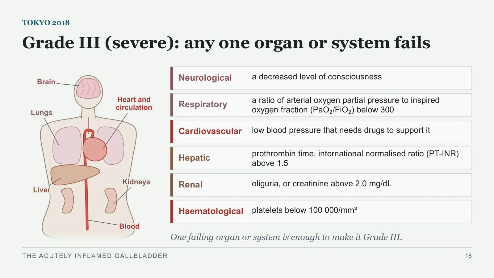
Grade III (severe): any one organ or system fails
TOKYO 2018
Neurological
a decreased level of consciousness
Respiratory
a ratio of arterial oxygen partial pressure to inspired oxygen fraction (PaO₂/FiO₂) below 300
Cardiovascular
low blood pressure that needs drugs to support it
Hepatic
prothrombin time, international normalised ratio (PT-INR) above 1.5
Renal
oliguria, or creatinine above 2.0 mg/dL
Haematological
platelets below 100 000/mm³
One failing organ or system is enough to make it Grade III.
THE ACUTELY INFLAMED GALLBLADDER
18
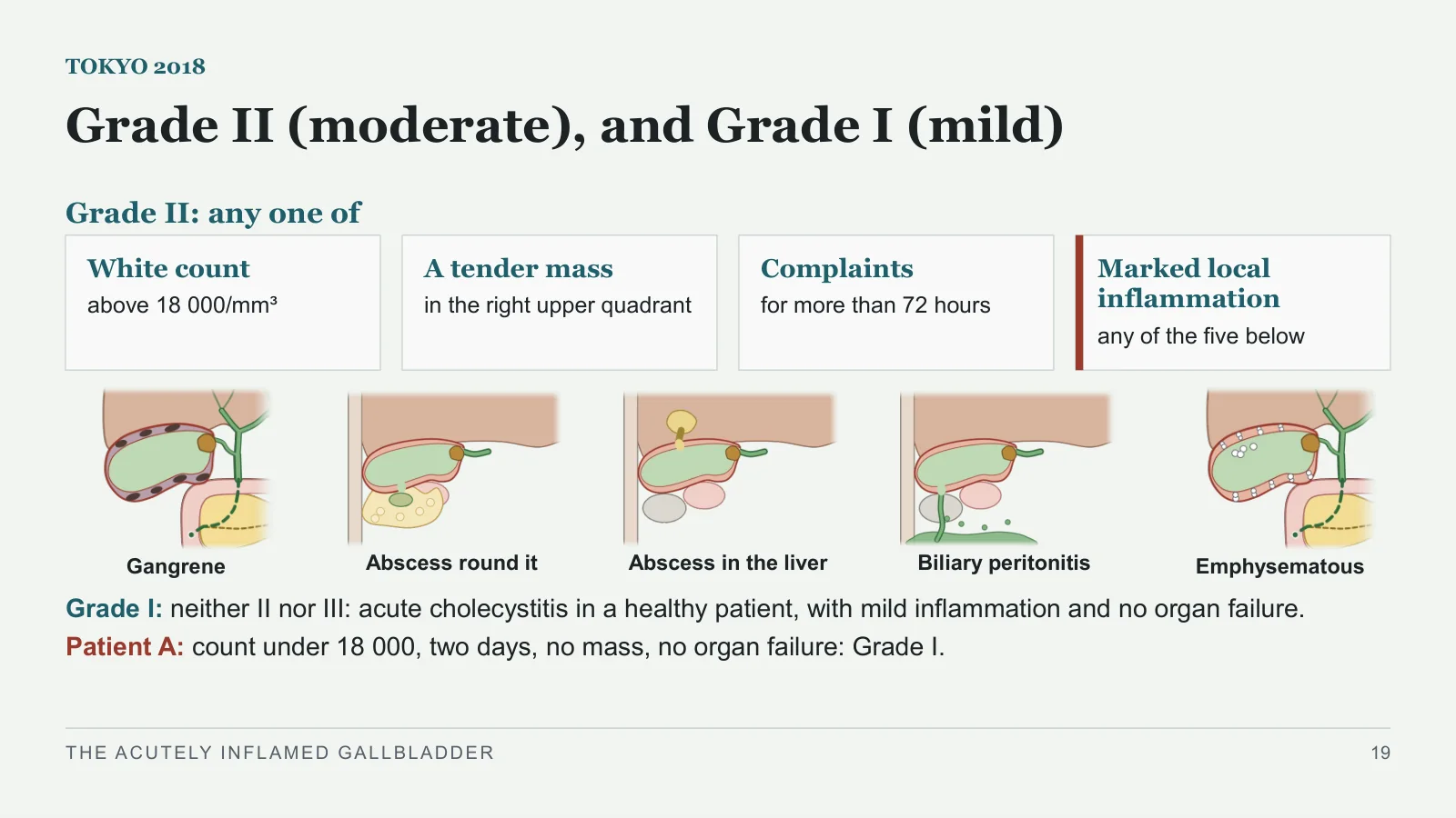
Grade II (moderate), and Grade I (mild)
TOKYO 2018
Grade II: any one of
White count
above 18 000/mm³
A tender mass
in the right upper quadrant
Complaints
for more than 72 hours
Marked local inflammation
any of the five below
Gangrene
Abscess round it
Abscess in the liver
Biliary peritonitis
Emphysematous
Grade I: neither II nor III: acute cholecystitis in a healthy patient, with mild inflammation and no organ failure.
Patient A: count under 18 000, two days, no mass, no organ failure: Grade I.
THE ACUTELY INFLAMED GALLBLADDER
19
First treatment
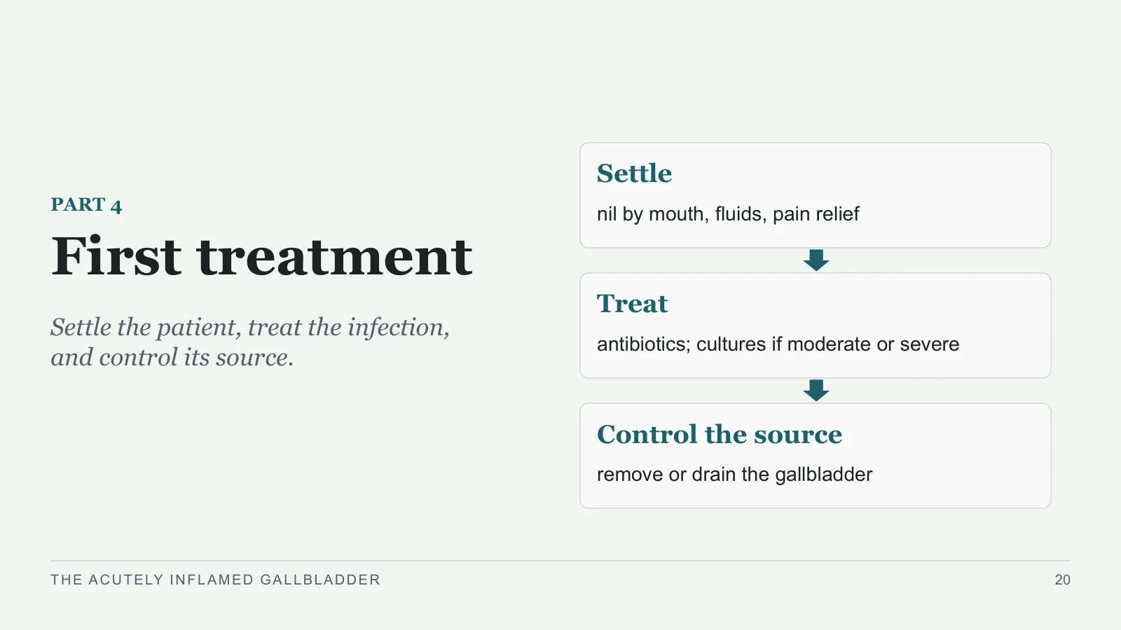
First treatment
PART 4
Settle the patient, treat the infection, and control its source.
Settle
nil by mouth, fluids, pain relief
Treat
antibiotics; cultures if moderate or severe
Control the source
remove or drain the gallbladder
THE ACUTELY INFLAMED GALLBLADDER
20
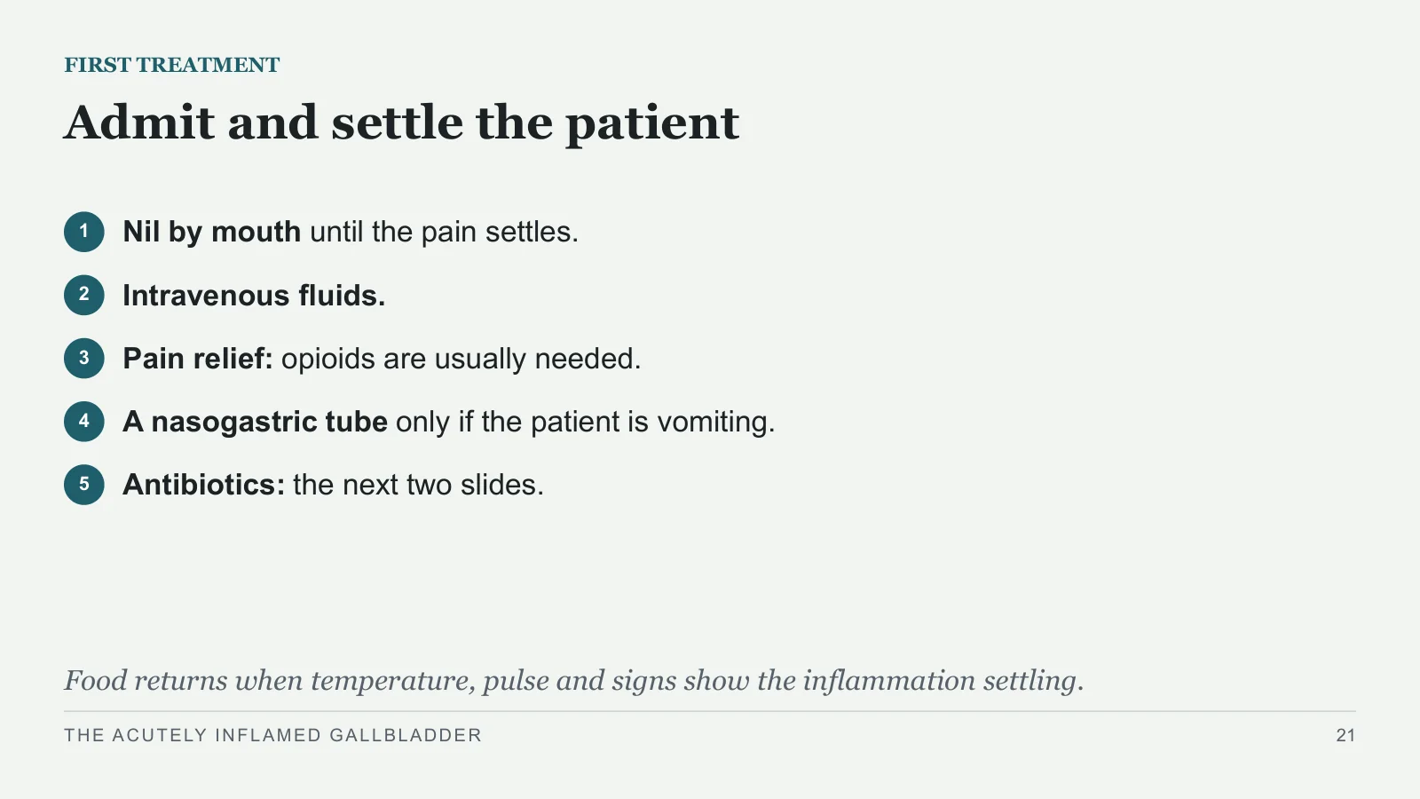
Admit and settle the patient
FIRST TREATMENT
1
Nil by mouth until the pain settles.
2
Intravenous fluids.
3
Pain relief: opioids are usually needed.
4
A nasogastric tube only if the patient is vomiting.
5
Antibiotics: the next two slides.
Food returns when temperature, pulse and signs show the inflammation settling.
THE ACUTELY INFLAMED GALLBLADDER
21
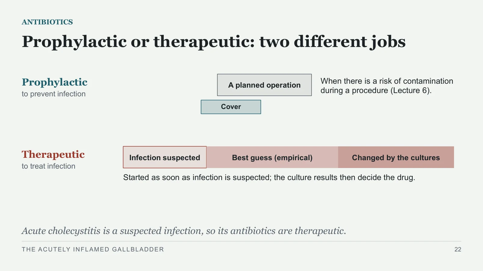
Prophylactic or therapeutic: two different jobs
ANTIBIOTICS
Prophylactic to prevent infection
Therapeutic to treat infection
A planned operation
Cover
When there is a risk of contamination during a procedure (Lecture 6).
Infection suspected
Best guess (empirical)
Changed by the cultures
Started as soon as infection is suspected; the culture results then decide the drug.
Acute cholecystitis is a suspected infection, so its antibiotics are therapeutic.
THE ACUTELY INFLAMED GALLBLADDER
22
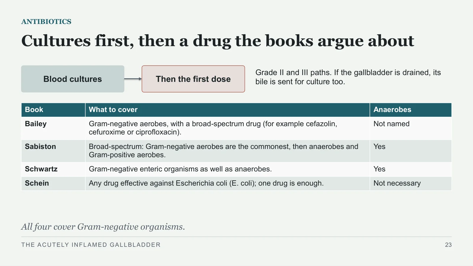
Cultures first, then a drug the books argue about
ANTIBIOTICS
Blood cultures
Then the first dose
Grade II and III paths. If the gallbladder is drained, its bile is sent for culture too.
All four cover Gram-negative organisms.
THE ACUTELY INFLAMED GALLBLADDER
23
| Book | What to cover | Anaerobes |
| Bailey | Gram-negative aerobes, with a broad-spectrum drug (for example cefazolin, cefuroxime or ciprofloxacin). | Not named |
| Sabiston | Broad-spectrum: Gram-negative aerobes are the commonest, then anaerobes and Gram-positive aerobes. | Yes |
| Schwartz | Gram-negative enteric organisms as well as anaerobes. | Yes |
| Schein | Any drug effective against Escherichia coli (E. coli); one drug is enough. | Not necessary |
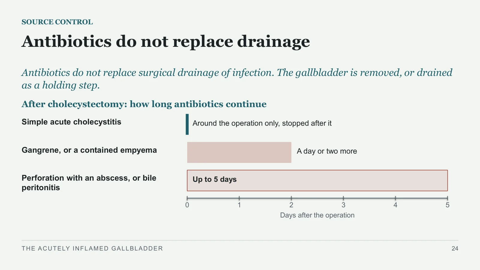
Antibiotics do not replace drainage
SOURCE CONTROL
Antibiotics do not replace surgical drainage of infection. The gallbladder is removed, or drained as a holding step.
After cholecystectomy: how long antibiotics continue
Simple acute cholecystitis
Around the operation only, stopped after it
Gangrene, or a contained empyema
A day or two more
Perforation with an abscess, or bile peritonitis
Up to 5 days
0
1
2
3
4
5
Days after the operation
THE ACUTELY INFLAMED GALLBLADDER
24
When the gallbladder comes out
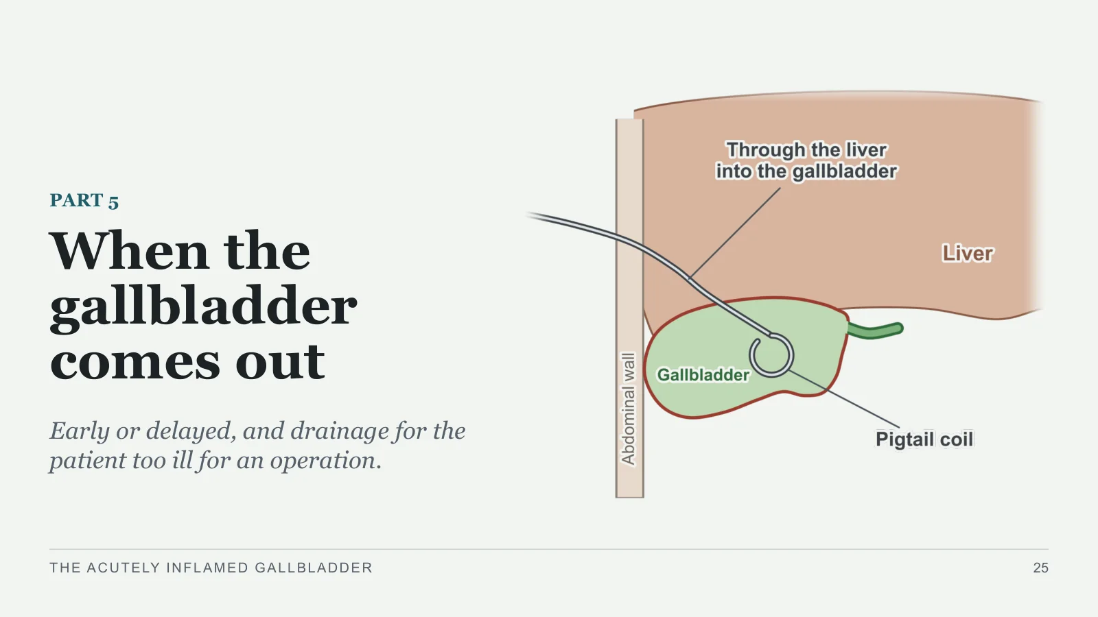
When the gallbladder comes out
PART 5
Early or delayed, and drainage for the patient too ill for an operation.
THE ACUTELY INFLAMED GALLBLADDER
25
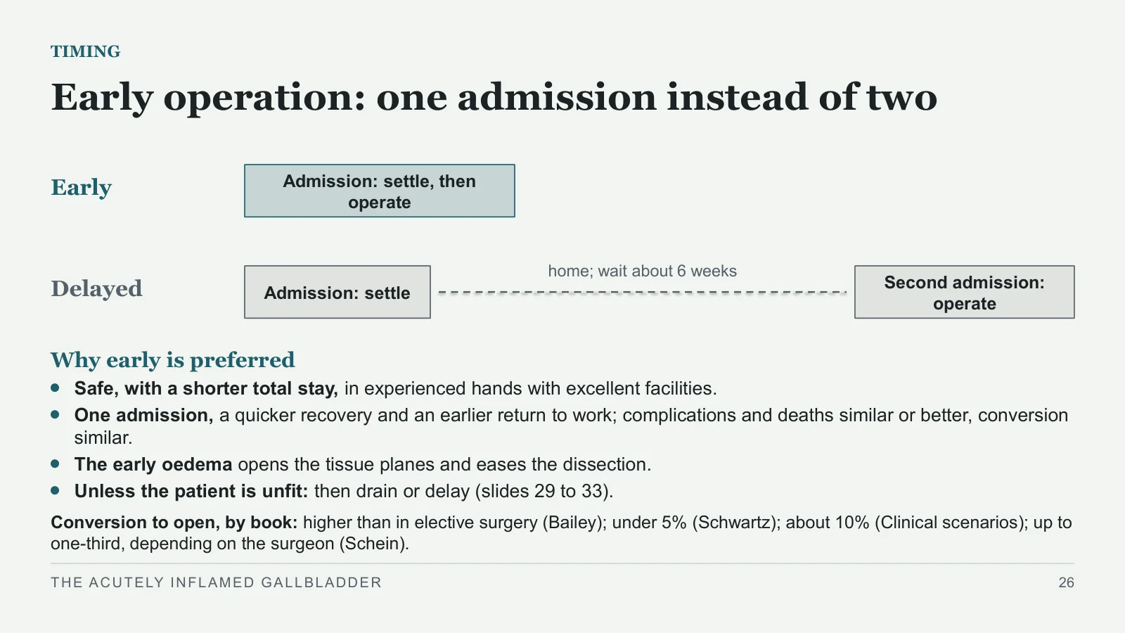
Early operation: one admission instead of two
TIMING
Early
Delayed
Admission: settle, then operate
Admission: settle
home; wait about 6 weeks
Second admission: operate
Why early is preferred
Safe, with a shorter total stay, in experienced hands with excellent facilities.
One admission, a quicker recovery and an earlier return to work; complications and deaths similar or better, conversion similar.
The early oedema opens the tissue planes and eases the dissection.
Unless the patient is unfit: then drain or delay (slides 29 to 33).
Conversion to open, by book: higher than in elective surgery (Bailey); under 5% (Schwartz); about 10% (Clinical scenarios); up to one-third, depending on the surgeon (Schein).
THE ACUTELY INFLAMED GALLBLADDER
26
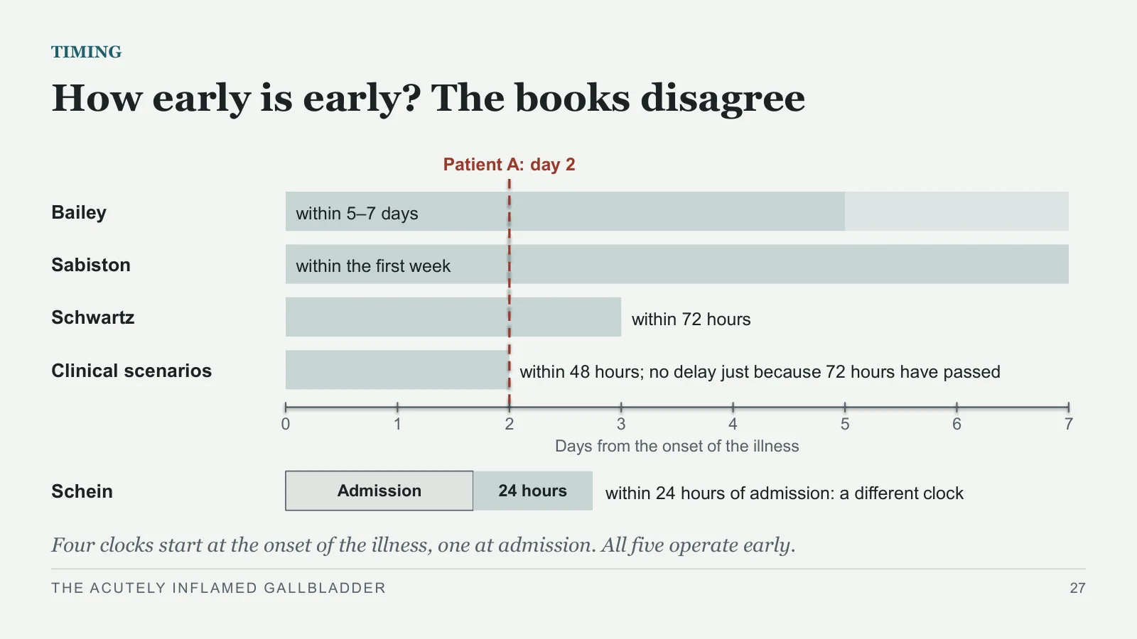
How early is early? The books disagree
TIMING
Bailey
within 5–7 days
Sabiston
within the first week
Schwartz
within 72 hours
Clinical scenarios
within 48 hours; no delay just because 72 hours have passed
0
1
2
3
4
5
6
7
Days from the onset of the illness
Patient A: day 2
Schein
Admission
24 hours
within 24 hours of admission: a different clock
Four clocks start at the onset of the illness, one at admission. All five operate early.
THE ACUTELY INFLAMED GALLBLADDER
27
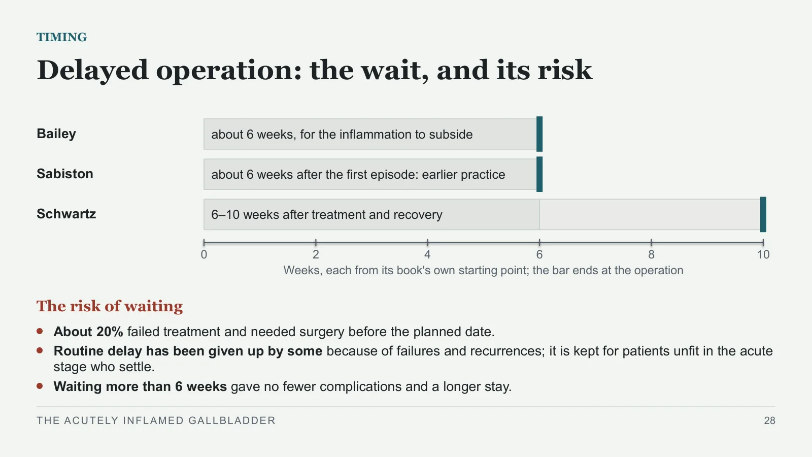
Delayed operation: the wait, and its risk
TIMING
Bailey
about 6 weeks, for the inflammation to subside
Sabiston
about 6 weeks after the first episode: earlier practice
Schwartz
6–10 weeks after treatment and recovery
0
2
4
6
8
10
Weeks, each from its book's own starting point; the bar ends at the operation
The risk of waiting
About 20% failed treatment and needed surgery before the planned date.
Routine delay has been given up by some because of failures and recurrences; it is kept for patients unfit in the acute stage who settle.
Waiting more than 6 weeks gave no fewer complications and a longer stay.
THE ACUTELY INFLAMED GALLBLADDER
28
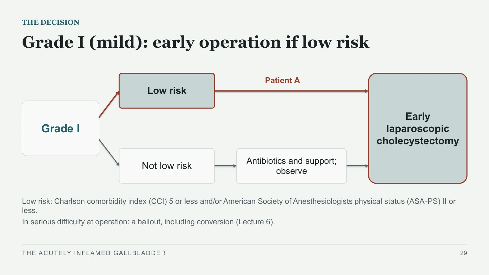
Grade I (mild): early operation if low risk
THE DECISION
Grade I
Low risk
Not low risk
Antibiotics and support; observe
Early laparoscopic cholecystectomy
Patient A
Low risk: Charlson comorbidity index (CCI) 5 or less and/or American Society of Anesthesiologists physical status (ASA-PS) II or less.
In serious difficulty at operation: a bailout, including conversion (Lecture 6).
THE ACUTELY INFLAMED GALLBLADDER
29
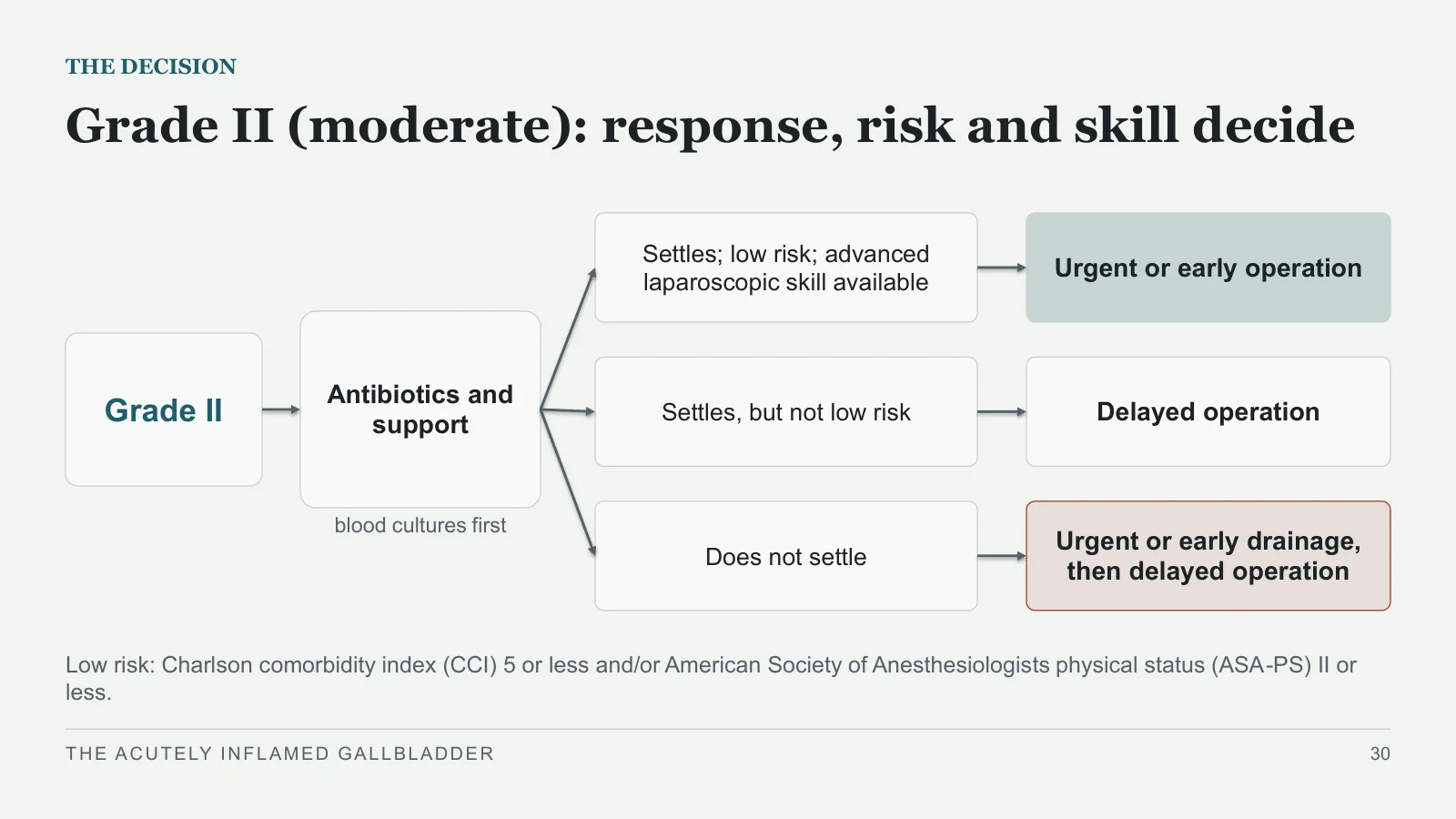
Grade II (moderate): response, risk and skill decide
THE DECISION
Grade II
Antibiotics and support
blood cultures first
Settles; low risk; advanced laparoscopic skill available
Urgent or early operation
Settles, but not low risk
Delayed operation
Does not settle
Urgent or early drainage, then delayed operation
Low risk: Charlson comorbidity index (CCI) 5 or less and/or American Society of Anesthesiologists physical status (ASA-PS) II or less.
THE ACUTELY INFLAMED GALLBLADDER
30
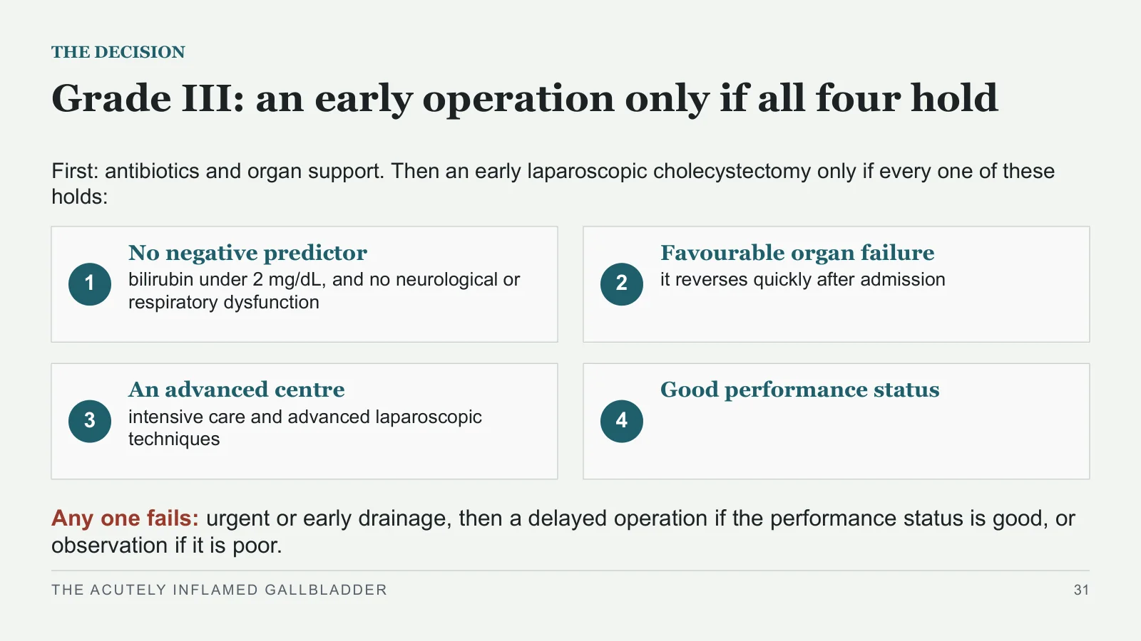
Grade III: an early operation only if all four hold
THE DECISION
First: antibiotics and organ support. Then an early laparoscopic cholecystectomy only if every one of these holds:
1
No negative predictor
bilirubin under 2 mg/dL, and no neurological or respiratory dysfunction
2
Favourable organ failure
it reverses quickly after admission
3
An advanced centre
intensive care and advanced laparoscopic techniques
4
Good performance status
Any one fails: urgent or early drainage, then a delayed operation if the performance status is good, or observation if it is poor.
THE ACUTELY INFLAMED GALLBLADDER
31
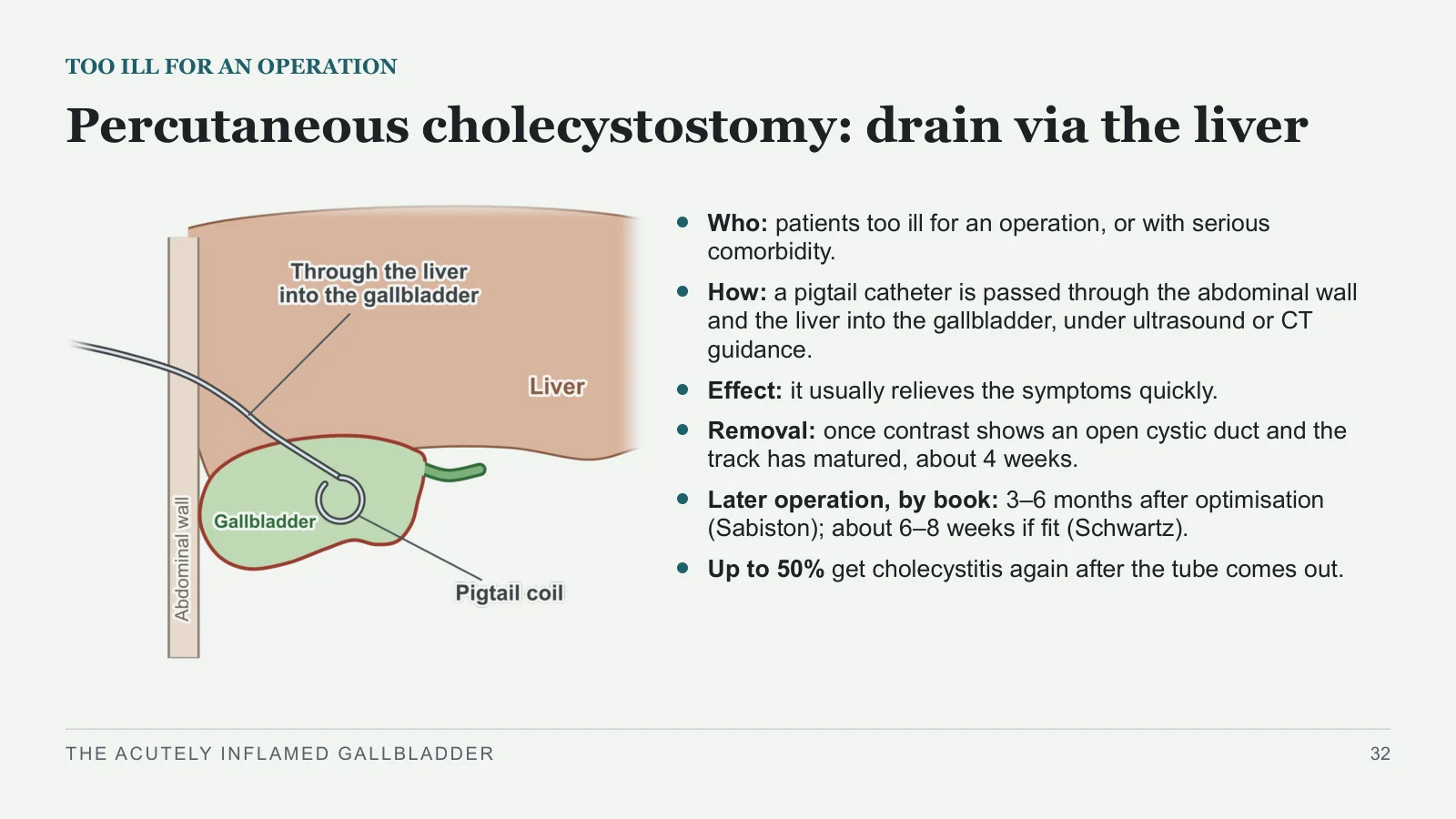
Percutaneous cholecystostomy: drain via the liver
TOO ILL FOR AN OPERATION
Who: patients too ill for an operation, or with serious comorbidity.
How: a pigtail catheter is passed through the abdominal wall and the liver into the gallbladder, under ultrasound or CT guidance.
Effect: it usually relieves the symptoms quickly.
Removal: once contrast shows an open cystic duct and the track has matured, about 4 weeks.
Later operation, by book: 3–6 months after optimisation (Sabiston); about 6–8 weeks if fit (Schwartz).
Up to 50% get cholecystitis again after the tube comes out.
THE ACUTELY INFLAMED GALLBLADDER
32
Not better in a day or two: look again
AFTER DRAINAGE
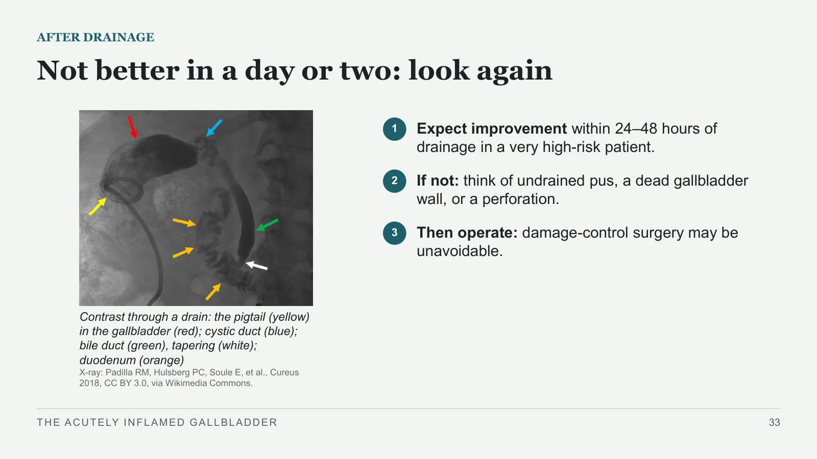
Contrast through a drain: the pigtail (yellow) in the gallbladder (red); cystic duct (blue); bile duct (green), tapering (white); duodenum (orange)
X-ray: Padilla RM, Hulsberg PC, Soule E, et al., Cureus 2018, CC BY 3.0, via Wikimedia Commons.
1
Expect improvement within 24–48 hours of drainage in a very high-risk patient.
2
If not: think of undrained pus, a dead gallbladder wall, or a perforation.
3
Then operate: damage-control surgery may be unavoidable.
THE ACUTELY INFLAMED GALLBLADDER
33
When it gets worse
When it gets worse
PART 6
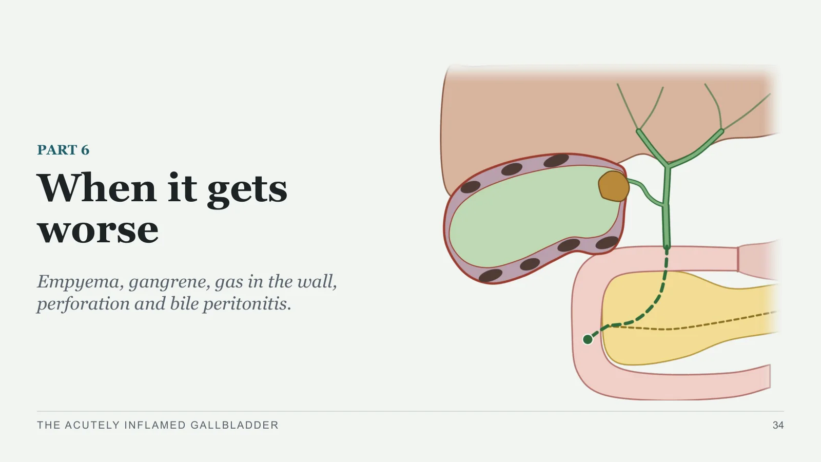
Empyema, gangrene, gas in the wall, perforation and bile peritonitis.
THE ACUTELY INFLAMED GALLBLADDER
34
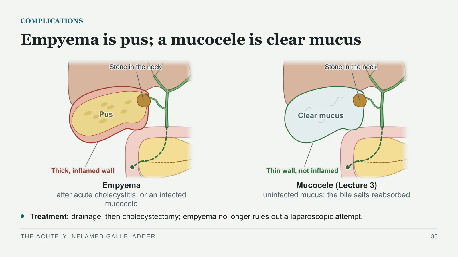
Empyema is pus; a mucocele is clear mucus
COMPLICATIONS
Empyema
after acute cholecystitis, or an infected mucocele
Mucocele (Lecture 3)
uninfected mucus; the bile salts reabsorbed
Treatment: drainage, then cholecystectomy; empyema no longer rules out a laparoscopic attempt.
THE ACUTELY INFLAMED GALLBLADDER
35
Gangrene: a wall that has died
COMPLICATIONS
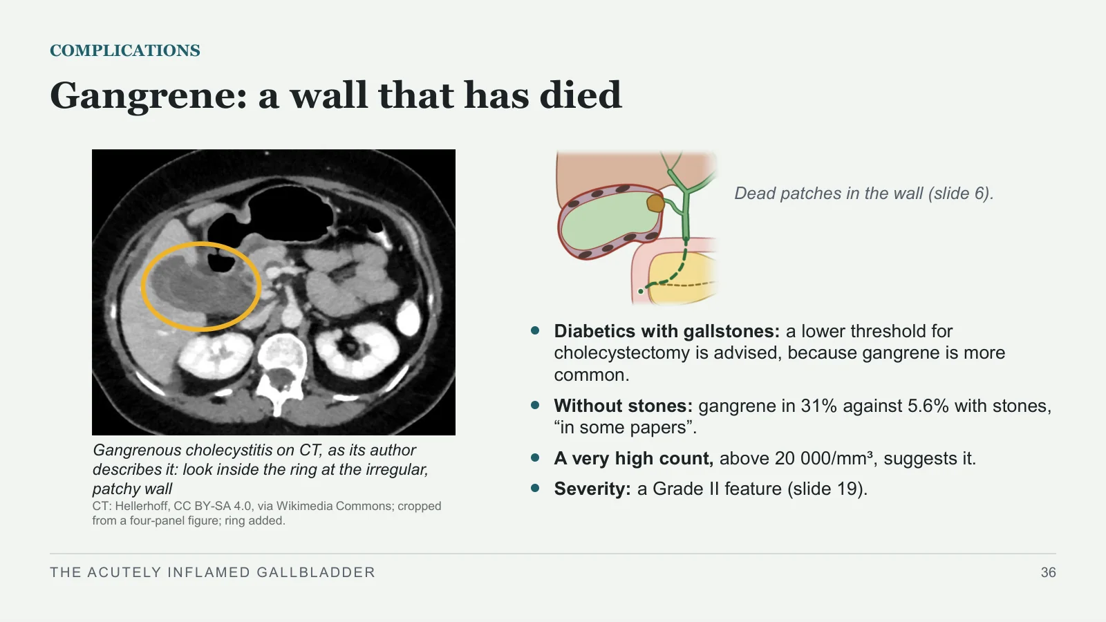
Gangrenous cholecystitis on CT, as its author describes it: look inside the ring at the irregular, patchy wall
CT: Hellerhoff, CC BY-SA 4.0, via Wikimedia Commons; cropped from a four-panel figure; ring added.
Dead patches in the wall (slide 6).
Diabetics with gallstones: a lower threshold for cholecystectomy is advised, because gangrene is more common.
Without stones: gangrene in 31% against 5.6% with stones, “in some papers”.
A very high count, above 20 000/mm³, suggests it.
Severity: a Grade II feature (slide 19).
THE ACUTELY INFLAMED GALLBLADDER
36
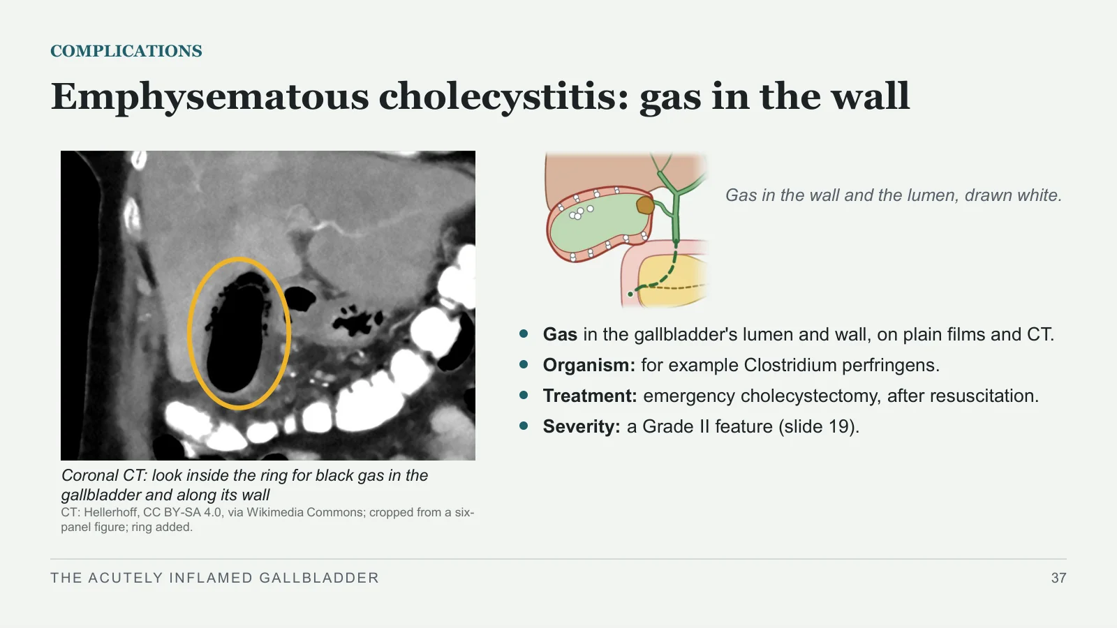
Emphysematous cholecystitis: gas in the wall
COMPLICATIONS
Coronal CT: look inside the ring for black gas in the gallbladder and along its wall
CT: Hellerhoff, CC BY-SA 4.0, via Wikimedia Commons; cropped from a six-panel figure; ring added.
Gas in the wall and the lumen, drawn white.
Gas in the gallbladder's lumen and wall, on plain films and CT.
Organism: for example Clostridium perfringens.
Treatment: emergency cholecystectomy, after resuscitation.
Severity: a Grade II feature (slide 19).
THE ACUTELY INFLAMED GALLBLADDER
37
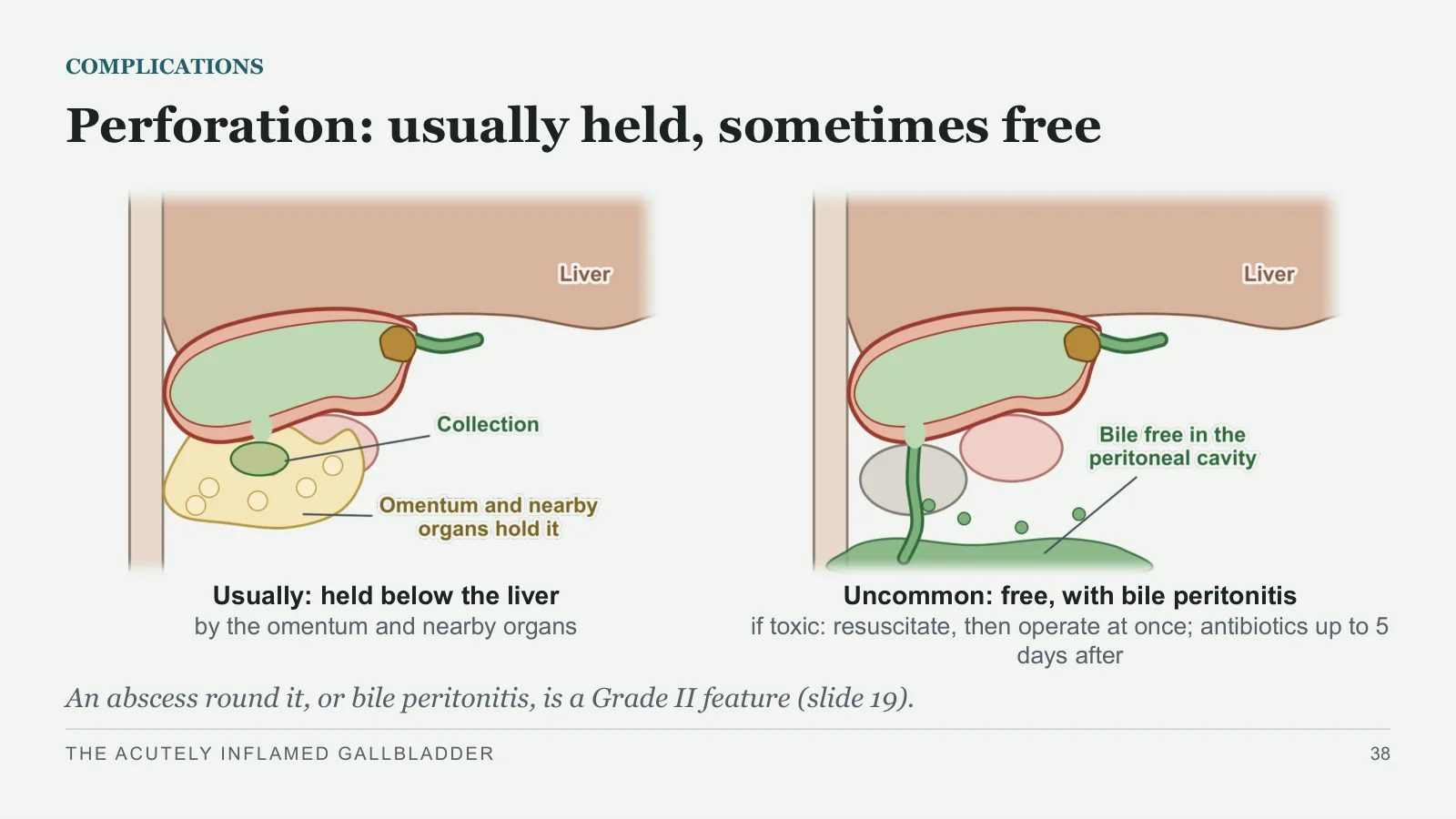
Perforation: usually held, sometimes free
COMPLICATIONS
Usually: held below the liver
by the omentum and nearby organs
Uncommon: free, with bile peritonitis
if toxic: resuscitate, then operate at once; antibiotics up to 5 days after
An abscess round it, or bile peritonitis, is a Grade II feature (slide 19).
THE ACUTELY INFLAMED GALLBLADDER
38
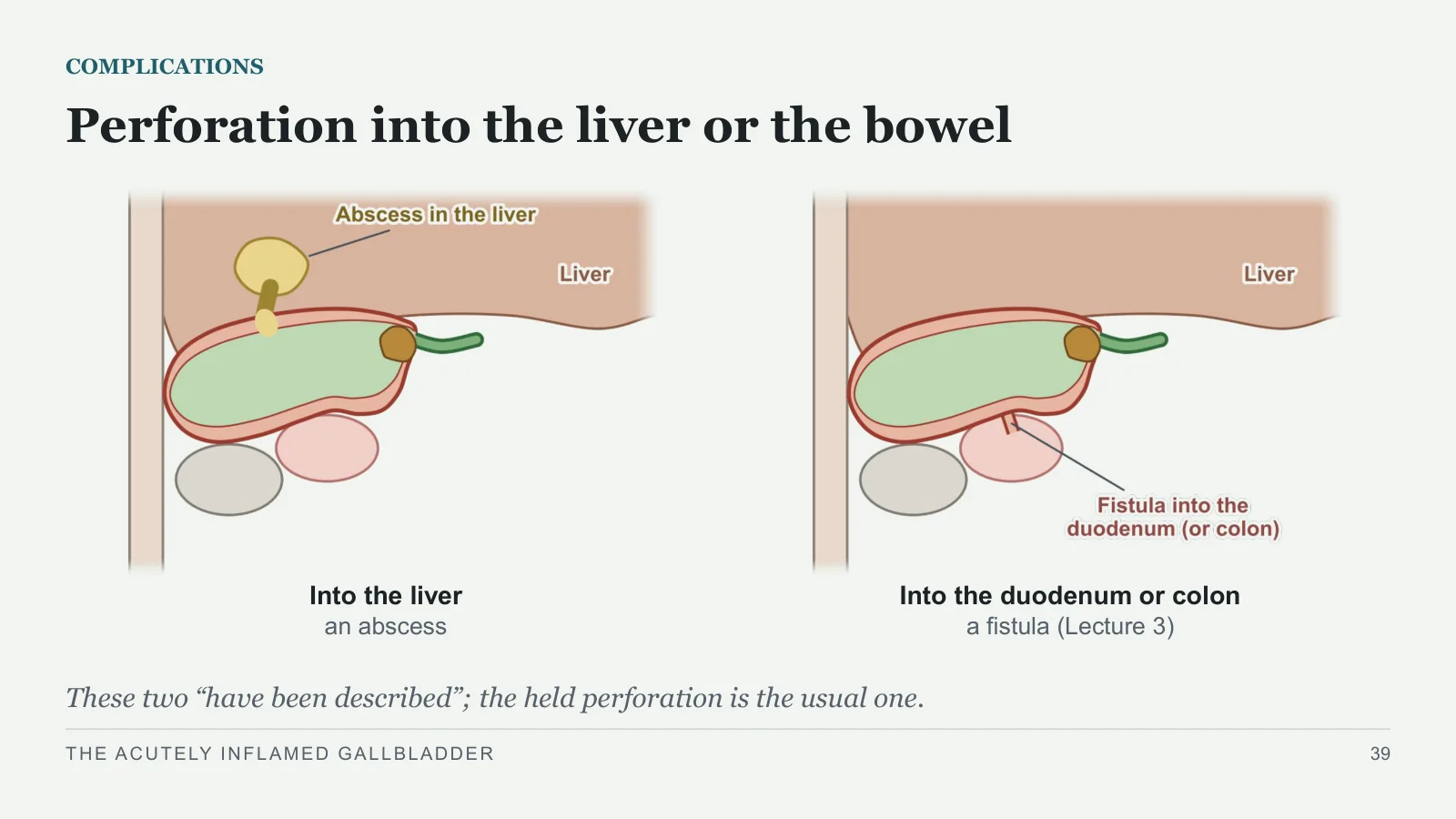
Perforation into the liver or the bowel
COMPLICATIONS
Into the liver
an abscess
Into the duodenum or colon
a fistula (Lecture 3)
These two “have been described”; the held perforation is the usual one.
THE ACUTELY INFLAMED GALLBLADDER
39
Without stones
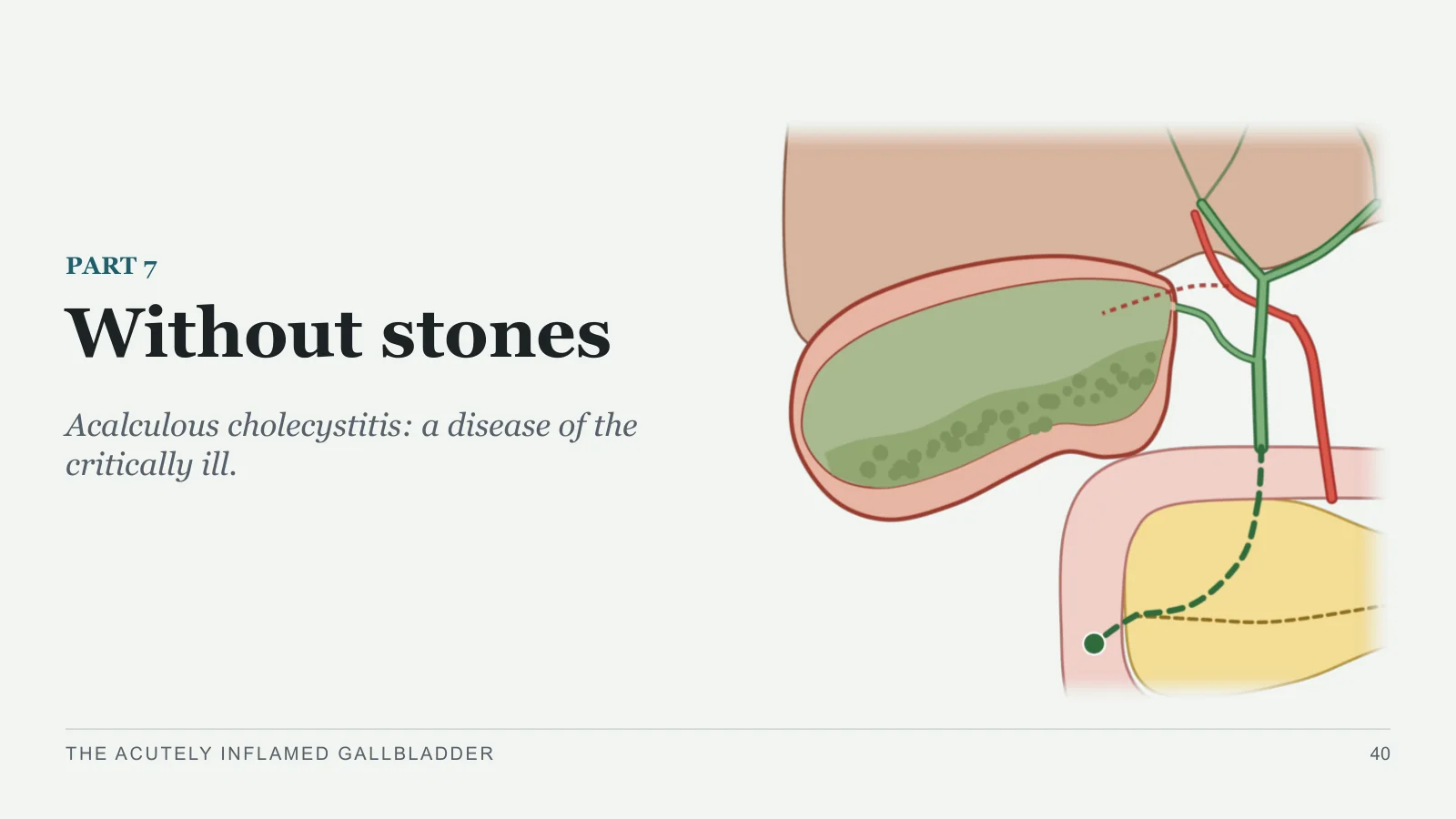
Without stones
PART 7
Acalculous cholecystitis: a disease of the critically ill.
THE ACUTELY INFLAMED GALLBLADDER
40
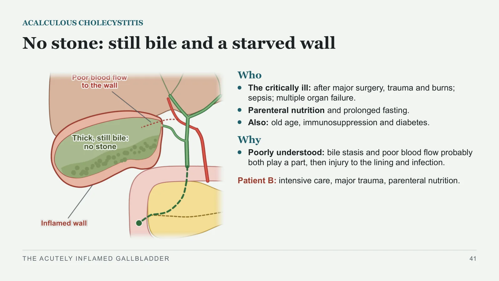
No stone: still bile and a starved wall
ACALCULOUS CHOLECYSTITIS
Who
The critically ill: after major surgery, trauma and burns; sepsis; multiple organ failure.
Parenteral nutrition and prolonged fasting.
Also: old age, immunosuppression and diabetes.
Why
Poorly understood: bile stasis and poor blood flow probably both play a part, then injury to the lining and infection.
Patient B: intensive care, major trauma, parenteral nutrition.
THE ACUTELY INFLAMED GALLBLADDER
41
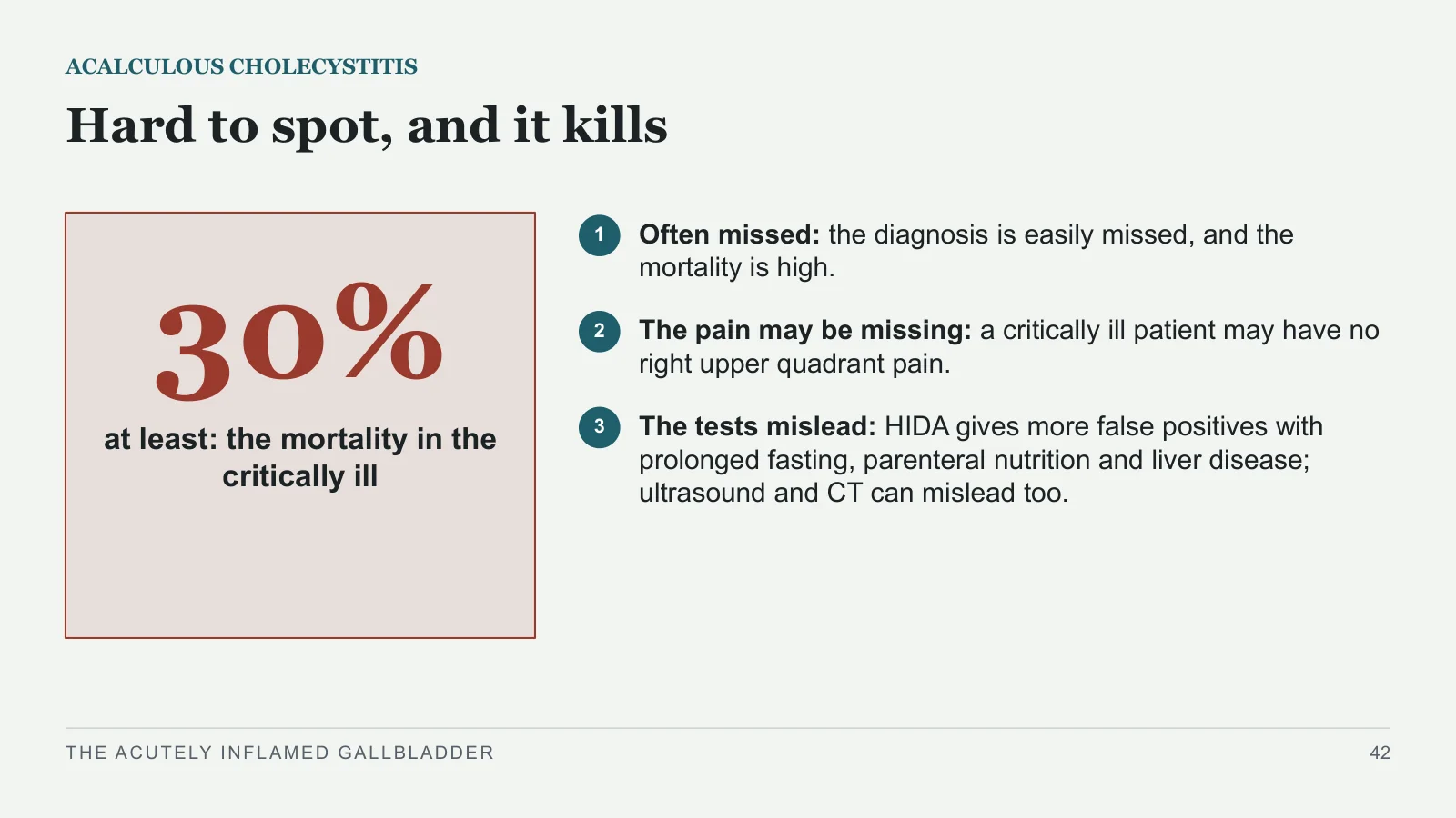
30%
ACALCULOUS CHOLECYSTITIS
Hard to spot, and it kills
at least: the mortality in the critically ill
1
Often missed: the diagnosis is easily missed, and the mortality is high.
2
The pain may be missing: a critically ill patient may have no right upper quadrant pain.
3
The tests mislead: HIDA gives more false positives with prolonged fasting, parenteral nutrition and liver disease; ultrasound and CT can mislead too.
THE ACUTELY INFLAMED GALLBLADDER
42
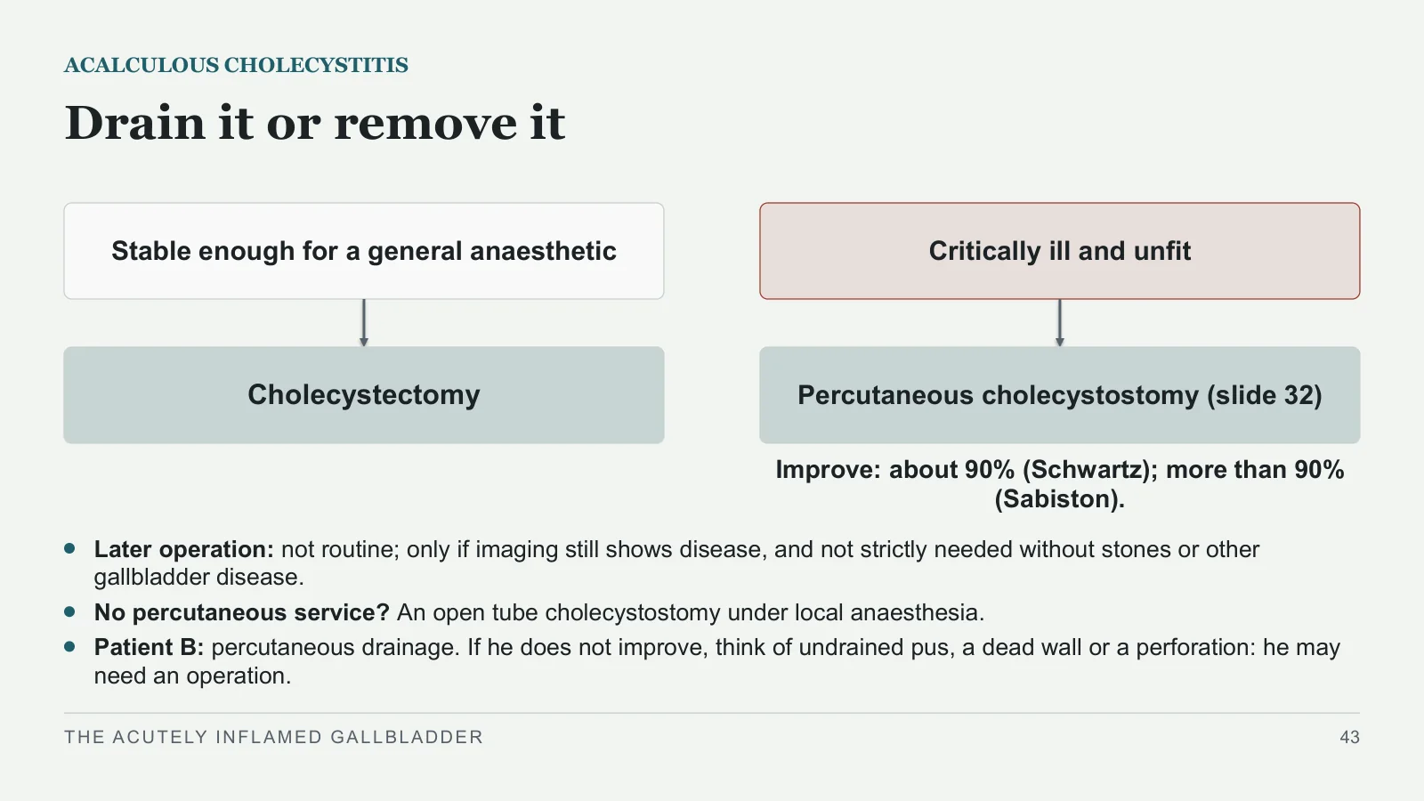
Drain it or remove it
ACALCULOUS CHOLECYSTITIS
Stable enough for a general anaesthetic
Cholecystectomy
Critically ill and unfit
Percutaneous cholecystostomy (slide 32)
Improve: about 90% (Schwartz); more than 90% (Sabiston).
Later operation: not routine; only if imaging still shows disease, and not strictly needed without stones or other gallbladder disease.
No percutaneous service? An open tube cholecystostomy under local anaesthesia.
Patient B: percutaneous drainage. If he does not improve, think of undrained pus, a dead wall or a perforation: he may need an operation.
THE ACUTELY INFLAMED GALLBLADDER
43
Two special cases
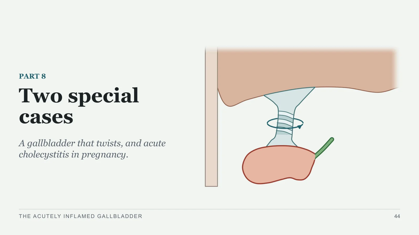
Two special cases
PART 8
A gallbladder that twists, and acute cholecystitis in pregnancy.
THE ACUTELY INFLAMED GALLBLADDER
44
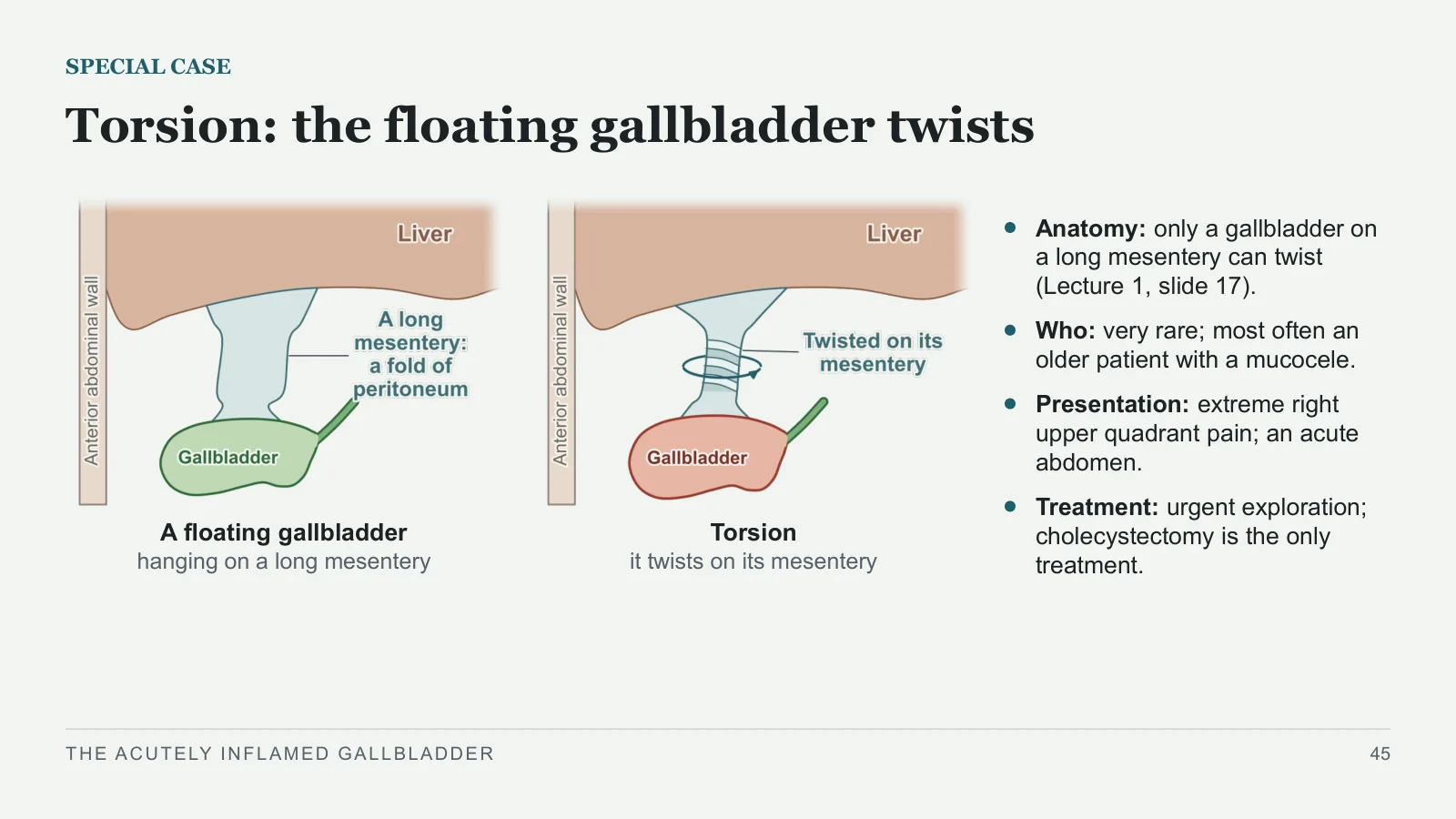
Torsion: the floating gallbladder twists
SPECIAL CASE
A floating gallbladder
hanging on a long mesentery
Torsion
it twists on its mesentery
Anatomy: only a gallbladder on a long mesentery can twist (Lecture 1, slide 17).
Who: very rare; most often an older patient with a mucocele.
Presentation: extreme right upper quadrant pain; an acute abdomen.
Treatment: urgent exploration; cholecystectomy is the only treatment.
THE ACUTELY INFLAMED GALLBLADDER
45
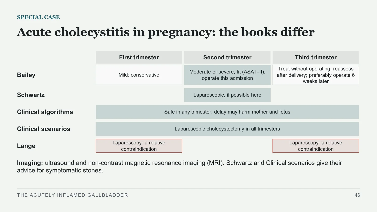
Acute cholecystitis in pregnancy: the books differ
SPECIAL CASE
First trimester
Second trimester
Third trimester
Bailey
Mild: conservative
Moderate or severe, fit (ASA I–II): operate this admission
Treat without operating; reassess after delivery; preferably operate 6 weeks later
Schwartz
Laparoscopic, if possible here
Clinical algorithms
Safe in any trimester; delay may harm mother and fetus
Clinical scenarios
Laparoscopic cholecystectomy in all trimesters
Lange
Laparoscopy: a relative contraindication
Laparoscopy: a relative contraindication
Imaging: ultrasound and non-contrast magnetic resonance imaging (MRI). Schwartz and Clinical scenarios give their advice for symptomatic stones.
THE ACUTELY INFLAMED GALLBLADDER
46
Recap and references
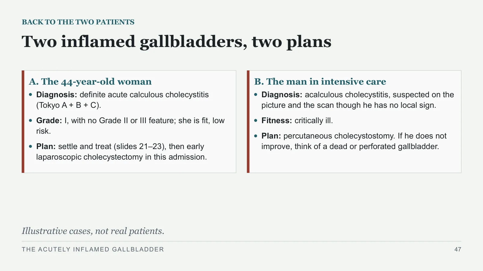
Two inflamed gallbladders, two plans
BACK TO THE TWO PATIENTS
A. The 44-year-old woman
Diagnosis: definite acute calculous cholecystitis (Tokyo A + B + C).
Grade: I, with no Grade II or III feature; she is fit, low risk.
Plan: settle and treat (slides 21–23), then early laparoscopic cholecystectomy in this admission.
B. The man in intensive care
Diagnosis: acalculous cholecystitis, suspected on the picture and the scan though he has no local sign.
Fitness: critically ill.
Plan: percutaneous cholecystostomy. If he does not improve, think of a dead or perforated gallbladder.
Illustrative cases, not real patients.
THE ACUTELY INFLAMED GALLBLADDER
47
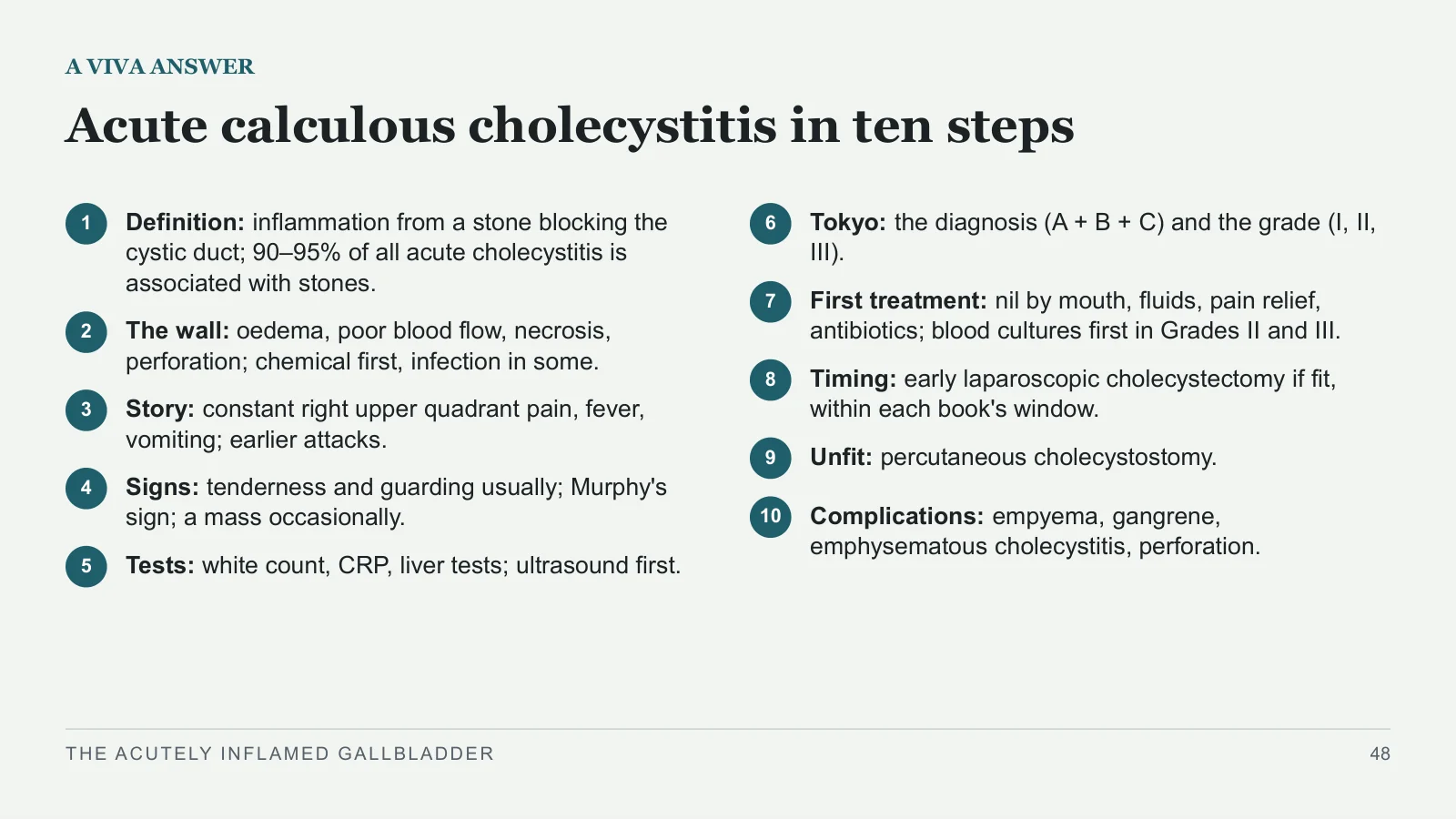
Acute calculous cholecystitis in ten steps
A VIVA ANSWER
1
Definition: inflammation from a stone blocking the cystic duct; 90–95% of all acute cholecystitis is associated with stones.
2
The wall: oedema, poor blood flow, necrosis, perforation; chemical first, infection in some.
3
Story: constant right upper quadrant pain, fever, vomiting; earlier attacks.
4
Signs: tenderness and guarding usually; Murphy's sign; a mass occasionally.
5
Tests: white count, CRP, liver tests; ultrasound first.
6
Tokyo: the diagnosis (A + B + C) and the grade (I, II, III).
7
First treatment: nil by mouth, fluids, pain relief, antibiotics; blood cultures first in Grades II and III.
8
Timing: early laparoscopic cholecystectomy if fit, within each book's window.
9
Unfit: percutaneous cholecystostomy.
10
Complications: empyema, gangrene, emphysematous cholecystitis, perforation.
THE ACUTELY INFLAMED GALLBLADDER
48
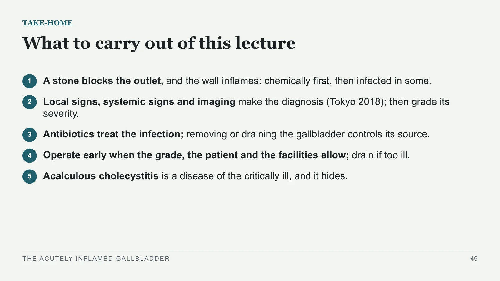
What to carry out of this lecture
TAKE-HOME
1
A stone blocks the outlet, and the wall inflames: chemically first, then infected in some.
2
Local signs, systemic signs and imaging make the diagnosis (Tokyo 2018); then grade its severity.
3
Antibiotics treat the infection; removing or draining the gallbladder controls its source.
4
Operate early when the grade, the patient and the facilities allow; drain if too ill.
5
Acalculous cholecystitis is a disease of the critically ill, and it hides.
THE ACUTELY INFLAMED GALLBLADDER
49
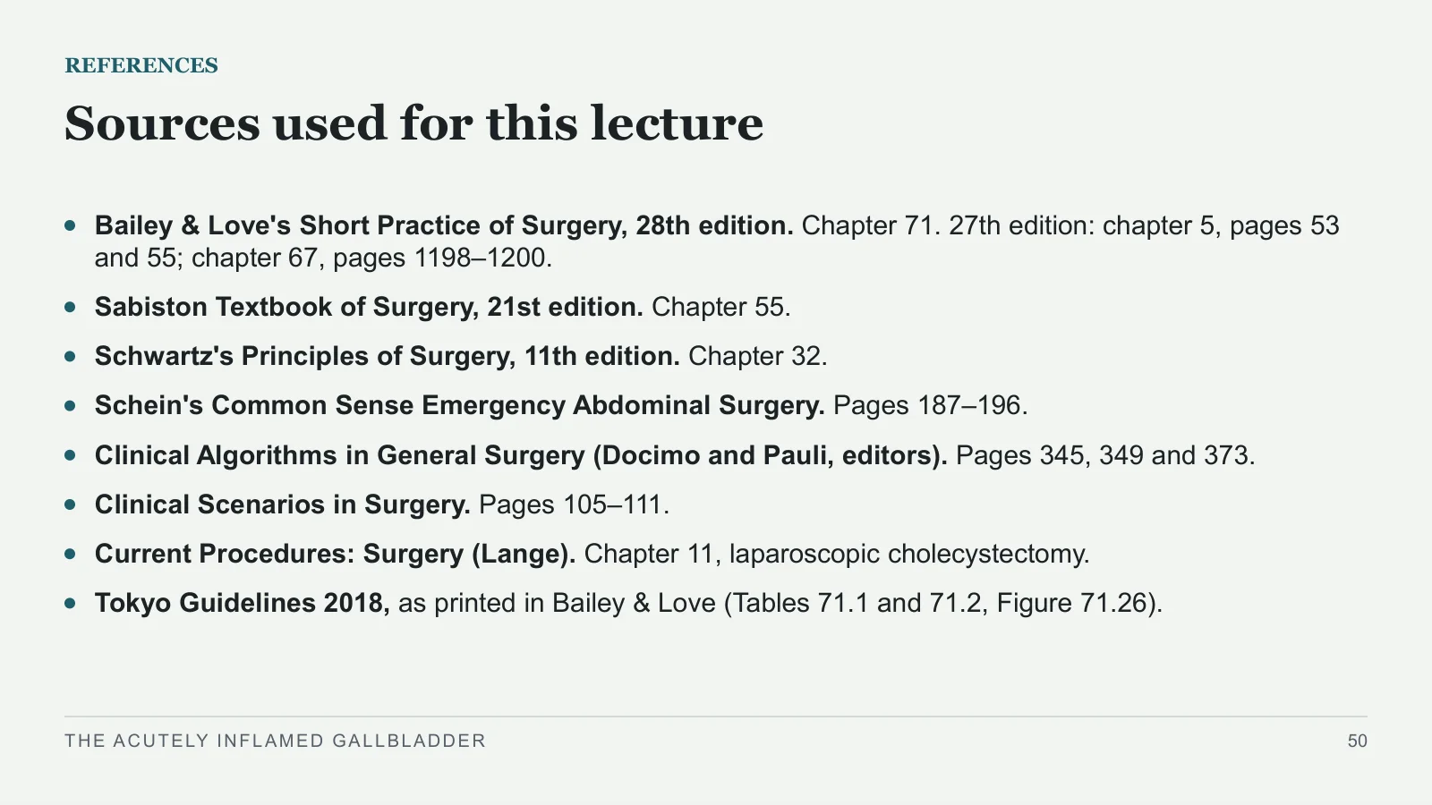
Sources used for this lecture
REFERENCES
Bailey & Love's Short Practice of Surgery, 28th edition. Chapter 71. 27th edition: chapter 5, pages 53 and 55; chapter 67, pages 1198–1200.
Sabiston Textbook of Surgery, 21st edition. Chapter 55.
Schwartz's Principles of Surgery, 11th edition. Chapter 32.
Schein's Common Sense Emergency Abdominal Surgery. Pages 187–196.
Clinical Algorithms in General Surgery (Docimo and Pauli, editors). Pages 345, 349 and 373.
Clinical Scenarios in Surgery. Pages 105–111.
Current Procedures: Surgery (Lange). Chapter 11, laparoscopic cholecystectomy.
Tokyo Guidelines 2018, as printed in Bailey & Love (Tables 71.1 and 71.2, Figure 71.26).
THE ACUTELY INFLAMED GALLBLADDER
50
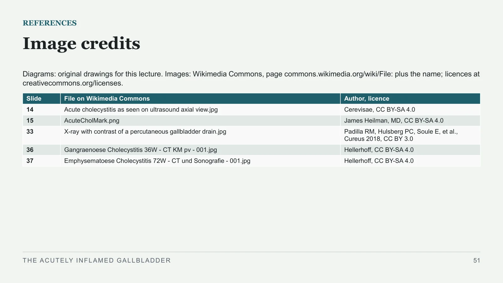
Image credits
REFERENCES
Diagrams: original drawings for this lecture. Images: Wikimedia Commons, page commons.wikimedia.org/wiki/File: plus the name; licences at creativecommons.org/licenses.
THE ACUTELY INFLAMED GALLBLADDER
51
| Slide | File on Wikimedia Commons | Author, licence |
| 14 | Acute cholecystitis as seen on ultrasound axial view.jpg | Cerevisae, CC BY-SA 4.0 |
| 15 | AcuteCholMark.png | James Heilman, MD, CC BY-SA 4.0 |
| 33 | X-ray with contrast of a percutaneous gallbladder drain.jpg | Padilla RM, Hulsberg PC, Soule E, et al., Cureus 2018, CC BY 3.0 |
| 36 | Gangraenoese Cholecystitis 36W - CT KM pv - 001.jpg | Hellerhoff, CC BY-SA 4.0 |
| 37 | Emphysematoese Cholecystitis 72W - CT und Sonografie - 001.jpg | Hellerhoff, CC BY-SA 4.0 |
Sources and image credits
Original lecture diagrams and source/image credits are preserved from the PowerPoint. Each photograph or scan below links to its source and applicable licence. Changes are described in the slide captions; adapted ShareAlike images retain that licence.
Images
- Slide 14: Acute cholecystitis as seen on ultrasound axial view.jpg · Cerevisae, CC BY-SA 4.0. Slide caption: Ultrasound: Cerevisae, CC BY-SA 4.0, via Wikimedia Commons. Adaptations of this image retain the same Creative Commons ShareAlike licence.
- Licence — Acute cholecystitis as seen on ultrasound axial view.jpg · CC BY-SA 4.0
- Slide 15: AcuteCholMark.png · James Heilman, MD, CC BY-SA 4.0. Slide caption: CT: James Heilman, MD, CC BY-SA 4.0, via Wikimedia Commons. Adaptations of this image retain the same Creative Commons ShareAlike licence.
- Licence — AcuteCholMark.png · CC BY-SA 4.0
- Slide 33: X-ray with contrast of a percutaneous gallbladder drain.jpg · Padilla RM, Hulsberg PC, Soule E, et al., Cureus 2018, CC BY 3.0. Slide caption: X-ray: Padilla RM, Hulsberg PC, Soule E, et al., Cureus 2018, CC BY 3.0, via Wikimedia Commons.
- Licence — X-ray with contrast of a percutaneous gallbladder drain.jpg · CC BY 3.0
- Original article — drain image, Figure 7 · Padilla RM, Hulsberg PC, Soule E, et al. Cureus 2018;10(11):e3577. DOI 10.7759/cureus.3577. CC BY 3.0.
- Slide 36: Gangraenoese Cholecystitis 36W - CT KM pv - 001.jpg · Hellerhoff, CC BY-SA 4.0. Slide caption: CT: Hellerhoff, CC BY-SA 4.0, via Wikimedia Commons; cropped from a four-panel figure; ring added. Adaptations of this image retain the same Creative Commons ShareAlike licence.
- Licence — Gangraenoese Cholecystitis 36W - CT KM pv - 001.jpg · CC BY-SA 4.0
- Slide 37: Emphysematoese Cholecystitis 72W - CT und Sonografie - 001.jpg · Hellerhoff, CC BY-SA 4.0. Slide caption: CT: Hellerhoff, CC BY-SA 4.0, via Wikimedia Commons; cropped from a six-panel figure; ring added. Adaptations of this image retain the same Creative Commons ShareAlike licence.
- Licence — Emphysematoese Cholecystitis 72W - CT und Sonografie - 001.jpg · CC BY-SA 4.0


Sources
- [1] REFERENCES
- [2] Sources used for this lecture
- [3] Bailey & Love's Short Practice of Surgery, 28th edition. Chapter 71. 27th edition: chapter 5, pages 53 and 55; chapter 67, pages 1198–1200.
- [4] Sabiston Textbook of Surgery, 21st edition. Chapter 55.
- [5] Schwartz's Principles of Surgery, 11th edition. Chapter 32.
- [6] Schein's Common Sense Emergency Abdominal Surgery. Pages 187–196.
- [7] Clinical Algorithms in General Surgery (Docimo and Pauli, editors). Pages 345, 349 and 373.
- [8] Clinical Scenarios in Surgery. Pages 105–111.
- [9] Current Procedures: Surgery (Lange). Chapter 11, laparoscopic cholecystectomy.
- [10] Tokyo Guidelines 2018, as printed in Bailey & Love (Tables 71.1 and 71.2, Figure 71.26).