Hepatobiliary surgery · Lecture 5
Duct stones · Acute cholangitis · 52 slides
Stones in the duct and acute cholangitis.
When a stone leaves the gallbladder: follow obstruction and infection through the first hour, drainage, investigation and duct clearance.
About the downloads
PowerPoint without speaker notes. SHA-256 checksums verify the downloaded files.
Contents · 8 parts
Opening
Stones in the duct and acute cholangitis
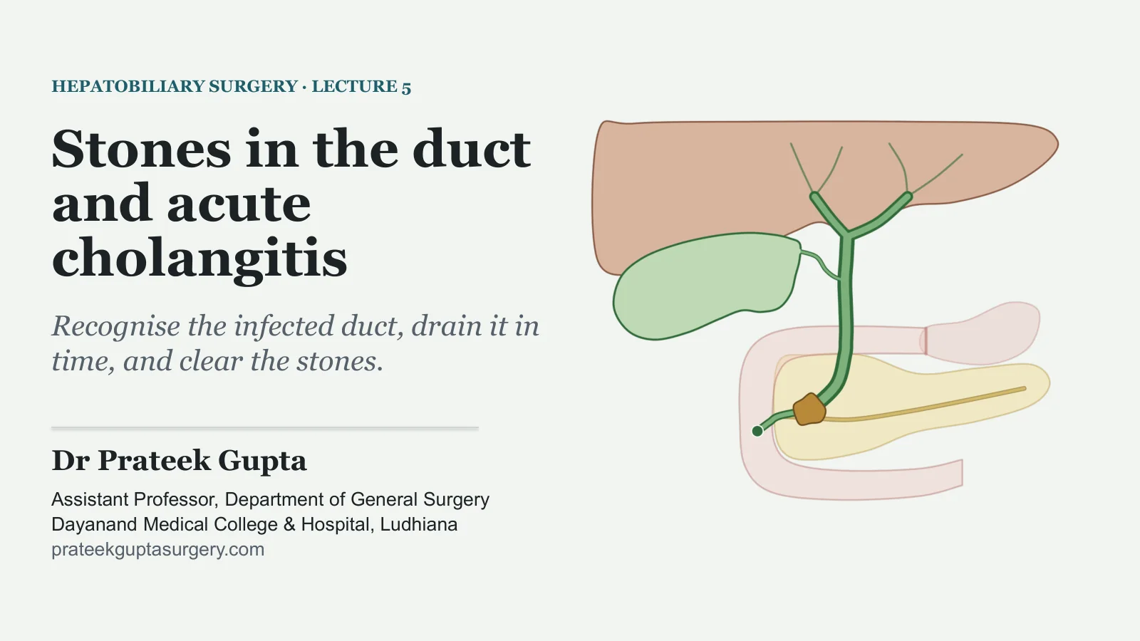
HEPATOBILIARY SURGERY · LECTURE 5
Stones in the duct and acute cholangitis
Recognise the infected duct, drain it in time, and clear the stones.
Dr Prateek Gupta
Assistant Professor, Department of General Surgery Dayanand Medical College & Hospital, Ludhiana prateekguptasurgery.com
Two patients with stones in the duct?
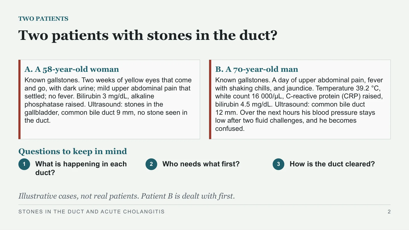
TWO PATIENTS
A. A 58-year-old woman
Known gallstones. Two weeks of yellow eyes that come and go, with dark urine; mild upper abdominal pain that settled; no fever. Bilirubin 3 mg/dL, alkaline phosphatase raised. Ultrasound: stones in the gallbladder, common bile duct 9 mm, no stone seen in the duct.
B. A 70-year-old man
Known gallstones. A day of upper abdominal pain, fever with shaking chills, and jaundice. Temperature 39.2 °C, white count 16 000/μL, C-reactive protein (CRP) raised, bilirubin 4.5 mg/dL. Ultrasound: common bile duct 12 mm. Over the next hours his blood pressure stays low after two fluid challenges, and he becomes confused.
Questions to keep in mind
1
What is happening in each duct?
2
Who needs what first?
3
How is the duct cleared?
Illustrative cases, not real patients. Patient B is dealt with first.
STONES IN THE DUCT AND ACUTE CHOLANGITIS
2
Stones in the duct
Stones in the duct
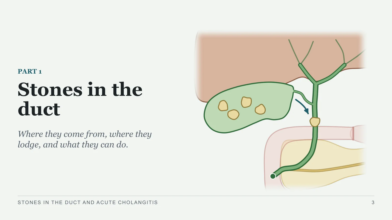
PART 1
Where they come from, where they lodge, and what they can do.
STONES IN THE DUCT AND ACUTE CHOLANGITIS
3
Secondary or primary
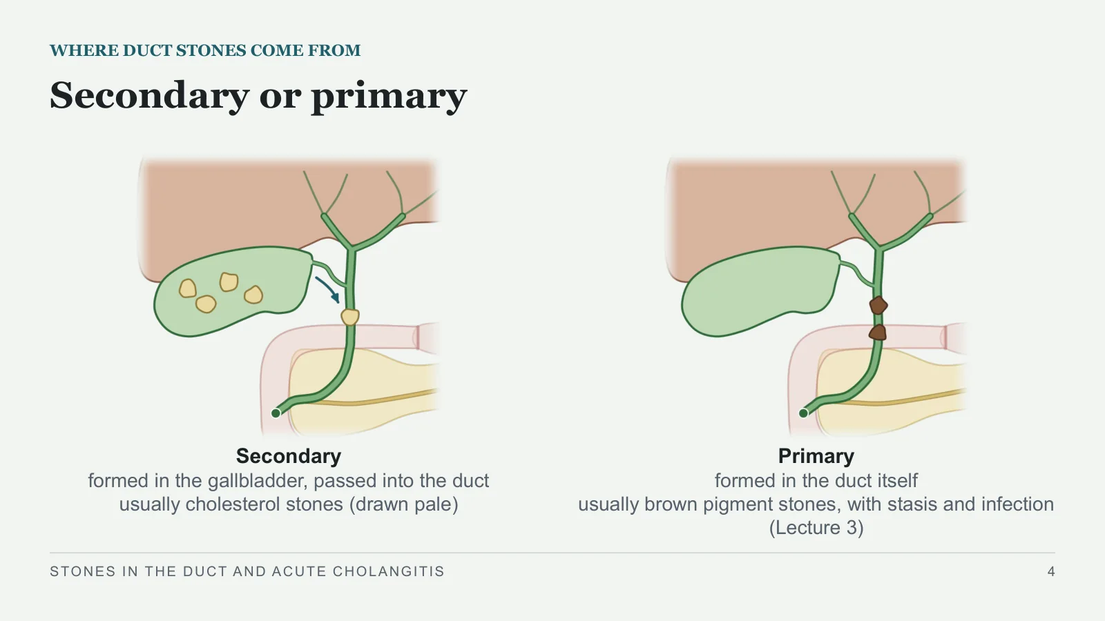
WHERE DUCT STONES COME FROM
Secondary
formed in the gallbladder, passed into the duct
usually cholesterol stones (drawn pale)
Primary
formed in the duct itself
usually brown pigment stones, with stasis and infection (Lecture 3)
STONES IN THE DUCT AND ACUTE CHOLANGITIS
4
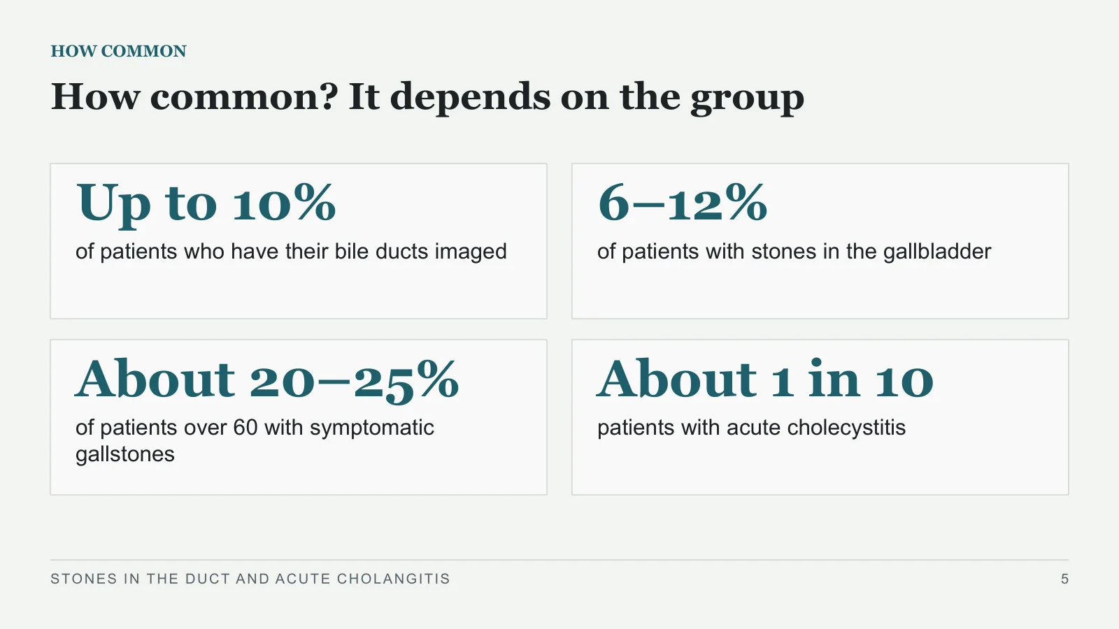
Up to 10%
HOW COMMON
How common? It depends on the group
of patients who have their bile ducts imaged
6–12%
of patients with stones in the gallbladder
About 20–25%
of patients over 60 with symptomatic gallstones
About 1 in 10
patients with acute cholecystitis
STONES IN THE DUCT AND ACUTE CHOLANGITIS
5
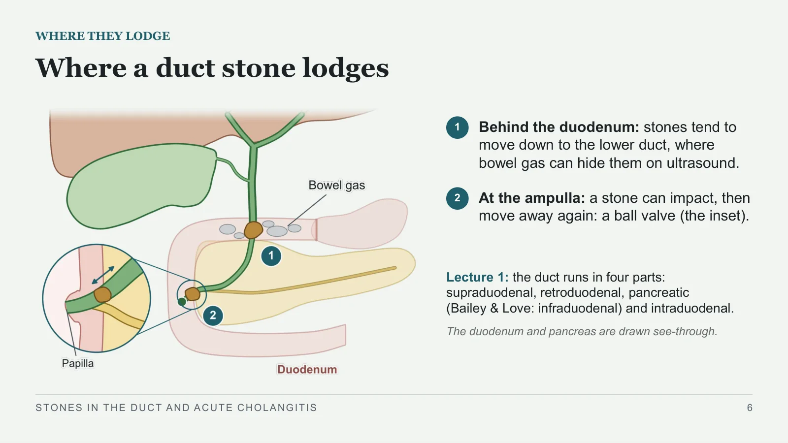
Where a duct stone lodges
WHERE THEY LODGE
1
Behind the duodenum: stones tend to move down to the lower duct, where bowel gas can hide them on ultrasound.
2
At the ampulla: a stone can impact, then move away again: a ball valve (the inset).
Lecture 1: the duct runs in four parts: supraduodenal, retroduodenal, pancreatic (Bailey & Love: infraduodenal) and intraduodenal.
The duodenum and pancreas are drawn see-through.
STONES IN THE DUCT AND ACUTE CHOLANGITIS
6
What a duct stone can do
WHAT A STONE CAN DO
1
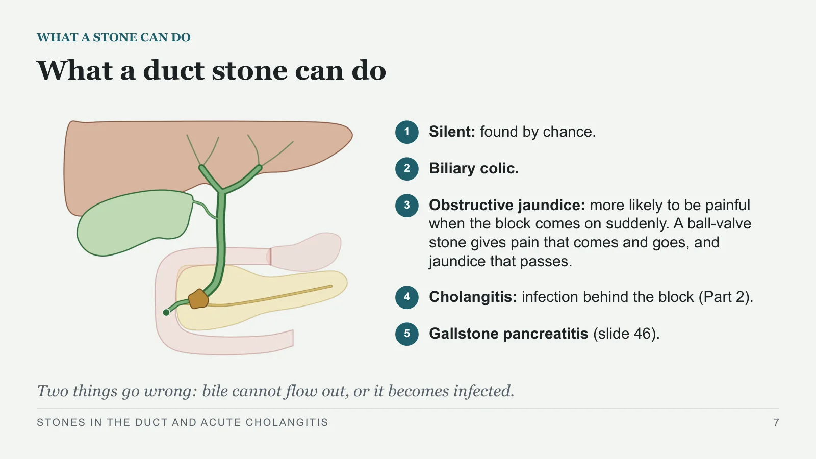
Silent: found by chance.
2
Biliary colic.
3
Obstructive jaundice: more likely to be painful when the block comes on suddenly. A ball-valve stone gives pain that comes and goes, and jaundice that passes.
4
Cholangitis: infection behind the block (Part 2).
5
Gallstone pancreatitis (slide 46).
Two things go wrong: bile cannot flow out, or it becomes infected.
STONES IN THE DUCT AND ACUTE CHOLANGITIS
7
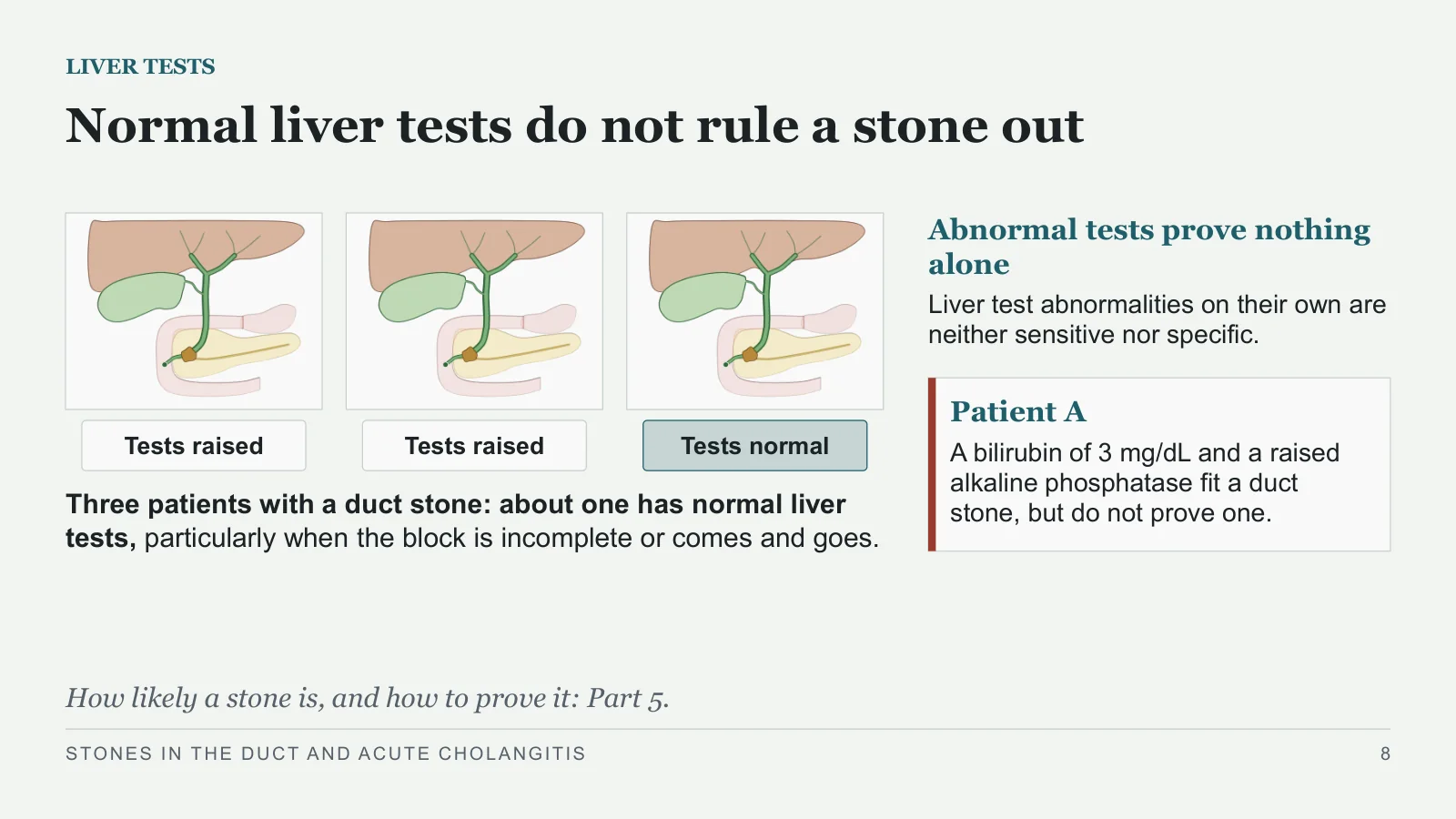
Normal liver tests do not rule a stone out
LIVER TESTS
Tests raised
Tests raised
Tests normal
Three patients with a duct stone: about one has normal liver tests, particularly when the block is incomplete or comes and goes.
Abnormal tests prove nothing alone
Liver test abnormalities on their own are neither sensitive nor specific.
Patient A
A bilirubin of 3 mg/dL and a raised alkaline phosphatase fit a duct stone, but do not prove one.
How likely a stone is, and how to prove it: Part 5.
STONES IN THE DUCT AND ACUTE CHOLANGITIS
8
Acute cholangitis
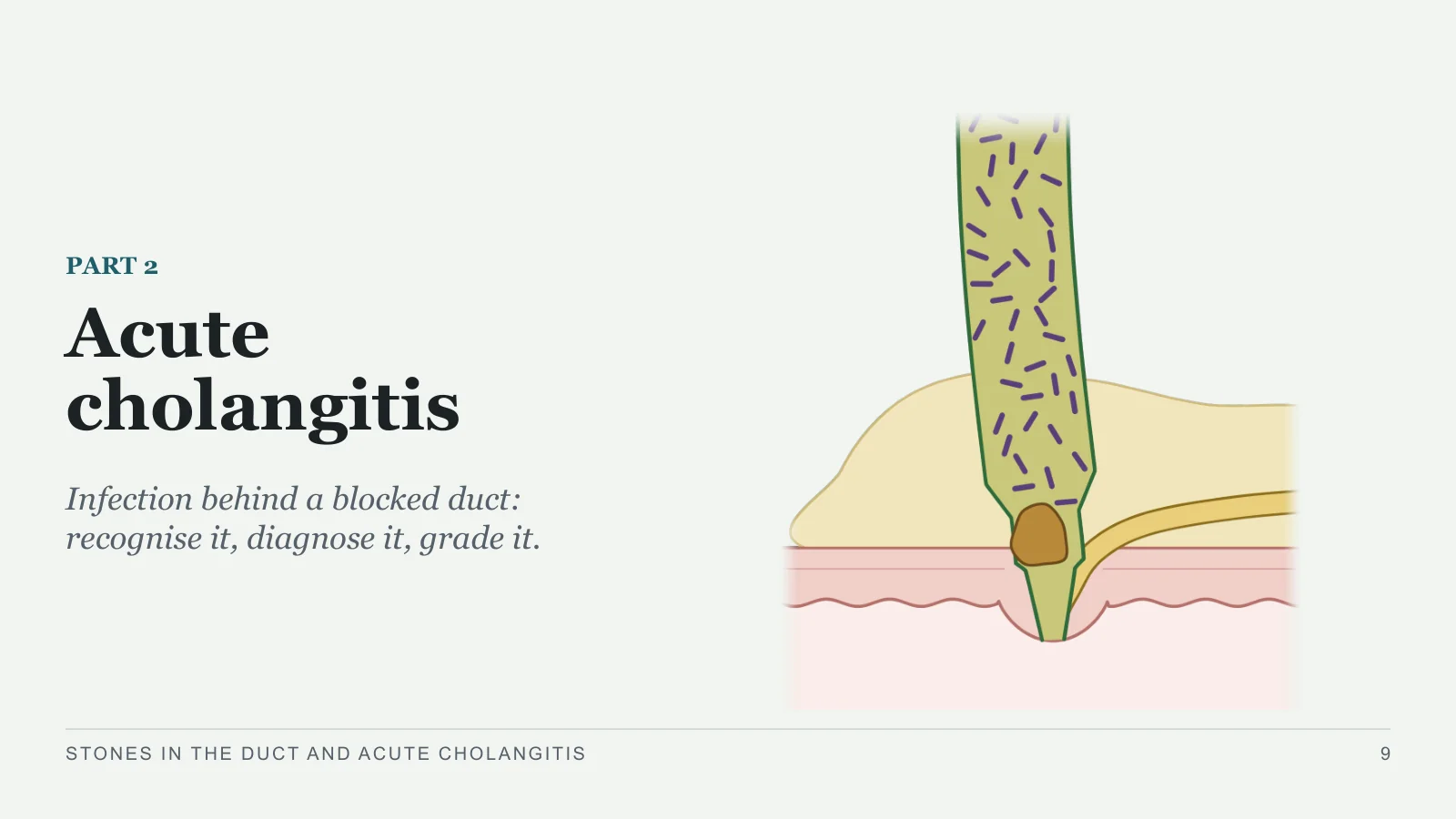
Acute cholangitis
PART 2
Infection behind a blocked duct: recognise it, diagnose it, grade it.
STONES IN THE DUCT AND ACUTE CHOLANGITIS
9
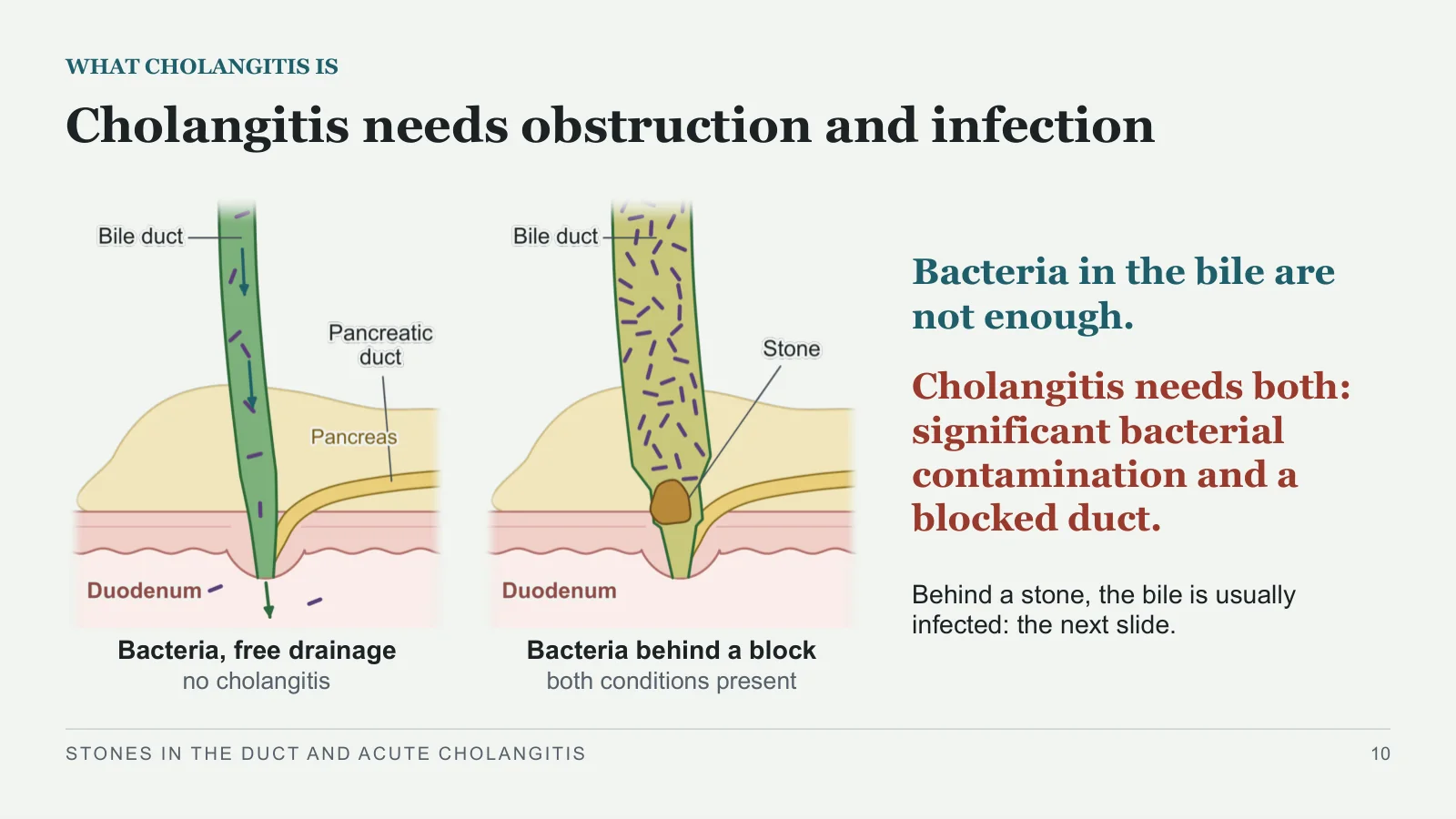
Cholangitis needs obstruction and infection
WHAT CHOLANGITIS IS
Bacteria, free drainage
no cholangitis
Bacteria behind a block
both conditions present
Bacteria in the bile are not enough.
Cholangitis needs both: significant bacterial contamination and a blocked duct.
Behind a stone, the bile is usually infected: the next slide.
STONES IN THE DUCT AND ACUTE CHOLANGITIS
10
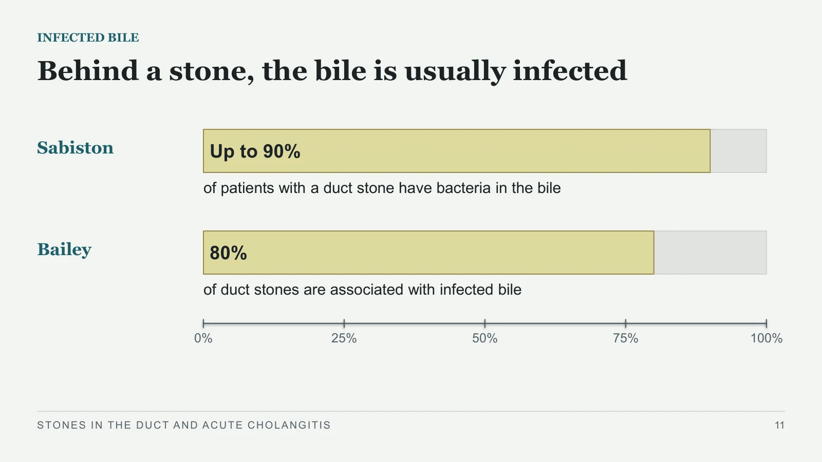
Behind a stone, the bile is usually infected
INFECTED BILE
Sabiston
Up to 90%
of patients with a duct stone have bacteria in the bile
Bailey
80%
of duct stones are associated with infected bile
0%
25%
50%
75%
100%
STONES IN THE DUCT AND ACUTE CHOLANGITIS
11
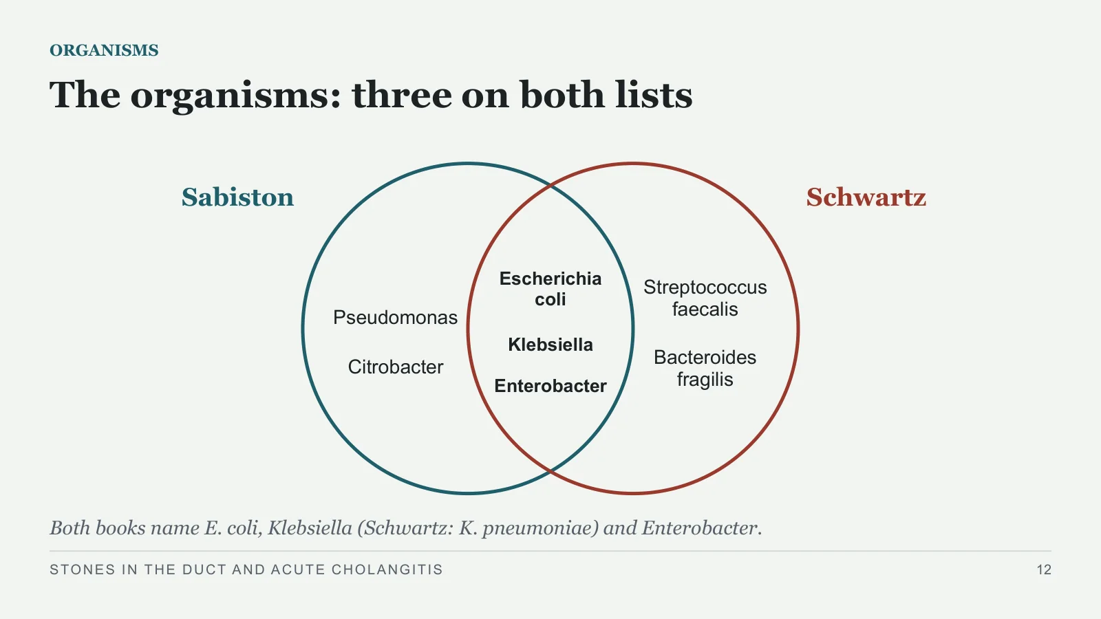
The organisms: three on both lists
ORGANISMS
Sabiston
Schwartz
Escherichia coli
Klebsiella
Enterobacter
Pseudomonas
Citrobacter
Streptococcus faecalis
Bacteroides fragilis
Both books name E. coli, Klebsiella (Schwartz: K. pneumoniae) and Enterobacter.
STONES IN THE DUCT AND ACUTE CHOLANGITIS
12
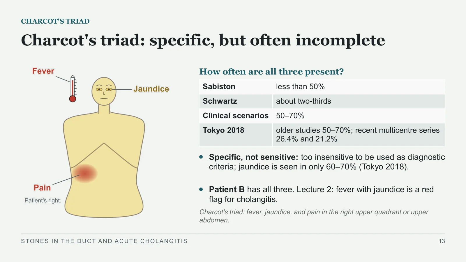
Charcot's triad: specific, but often incomplete
CHARCOT'S TRIAD
How often are all three present?
Specific, not sensitive: too insensitive to be used as diagnostic criteria; jaundice is seen in only 60–70% (Tokyo 2018).
Patient B has all three. Lecture 2: fever with jaundice is a red flag for cholangitis.
Charcot's triad: fever, jaundice, and pain in the right upper quadrant or upper abdomen.
STONES IN THE DUCT AND ACUTE CHOLANGITIS
13
| Sabiston | less than 50% |
| Schwartz | about two-thirds |
| Clinical scenarios | 50–70% |
| Tokyo 2018 | older studies 50–70%; recent multicentre series 26.4% and 21.2% |
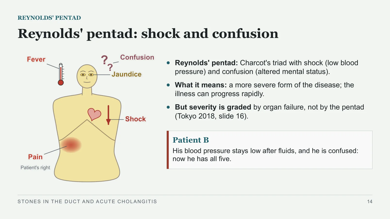
Reynolds' pentad: shock and confusion
REYNOLDS' PENTAD
Reynolds' pentad: Charcot's triad with shock (low blood pressure) and confusion (altered mental status).
What it means: a more severe form of the disease; the illness can progress rapidly.
But severity is graded by organ failure, not by the pentad (Tokyo 2018, slide 16).
Patient B
His blood pressure stays low after fluids, and he is confused: now he has all five.
STONES IN THE DUCT AND ACUTE CHOLANGITIS
14
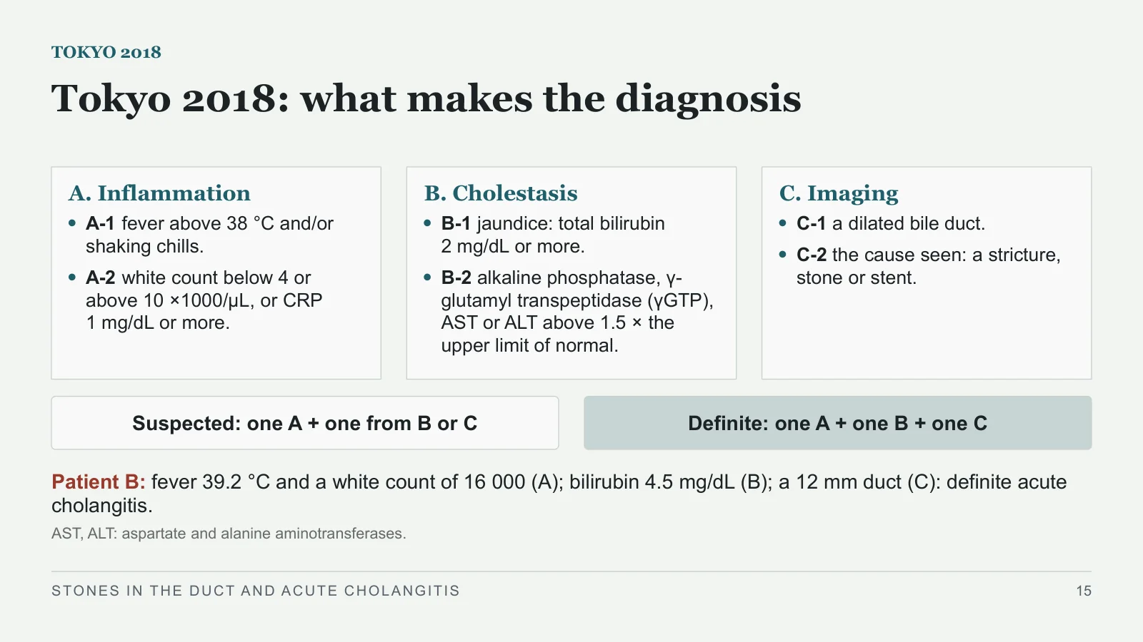
Tokyo 2018: what makes the diagnosis
TOKYO 2018
A. Inflammation
A-1 fever above 38 °C and/or shaking chills.
A-2 white count below 4 or above 10 ×1000/μL, or CRP 1 mg/dL or more.
B. Cholestasis
B-1 jaundice: total bilirubin 2 mg/dL or more.
B-2 alkaline phosphatase, γ-glutamyl transpeptidase (γGTP), AST or ALT above 1.5 × the upper limit of normal.
C. Imaging
C-1 a dilated bile duct.
C-2 the cause seen: a stricture, stone or stent.
Suspected: one A + one from B or C
Definite: one A + one B + one C
Patient B: fever 39.2 °C and a white count of 16 000 (A); bilirubin 4.5 mg/dL (B); a 12 mm duct (C): definite acute cholangitis.
AST, ALT: aspartate and alanine aminotransferases.
STONES IN THE DUCT AND ACUTE CHOLANGITIS
15
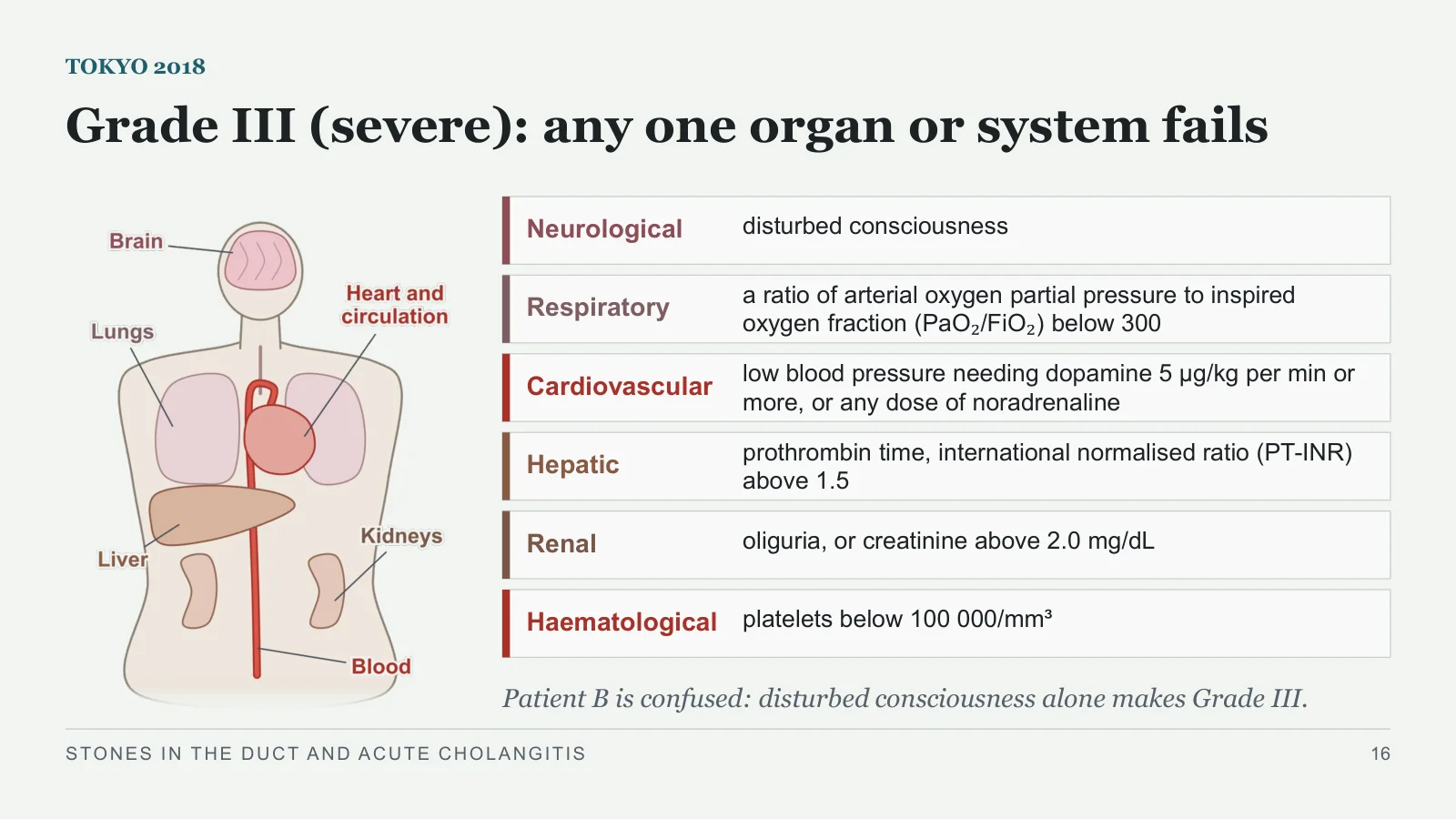
Grade III (severe): any one organ or system fails
TOKYO 2018
Neurological
disturbed consciousness
Respiratory
a ratio of arterial oxygen partial pressure to inspired oxygen fraction (PaO₂/FiO₂) below 300
Cardiovascular
low blood pressure needing dopamine 5 μg/kg per min or more, or any dose of noradrenaline
Hepatic
prothrombin time, international normalised ratio (PT-INR) above 1.5
Renal
oliguria, or creatinine above 2.0 mg/dL
Haematological
platelets below 100 000/mm³
Patient B is confused: disturbed consciousness alone makes Grade III.
STONES IN THE DUCT AND ACUTE CHOLANGITIS
16
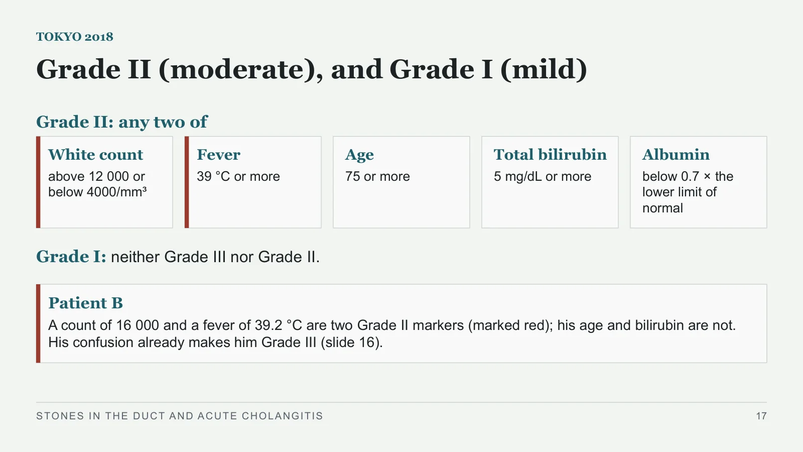
Grade II (moderate), and Grade I (mild)
TOKYO 2018
Grade II: any two of
White count
above 12 000 or below 4000/mm³
Fever
39 °C or more
Age
75 or more
Total bilirubin
5 mg/dL or more
Albumin
below 0.7 × the lower limit of normal
Grade I: neither Grade III nor Grade II.
Patient B
A count of 16 000 and a fever of 39.2 °C are two Grade II markers (marked red); his age and bilirubin are not. His confusion already makes him Grade III (slide 16).
STONES IN THE DUCT AND ACUTE CHOLANGITIS
17
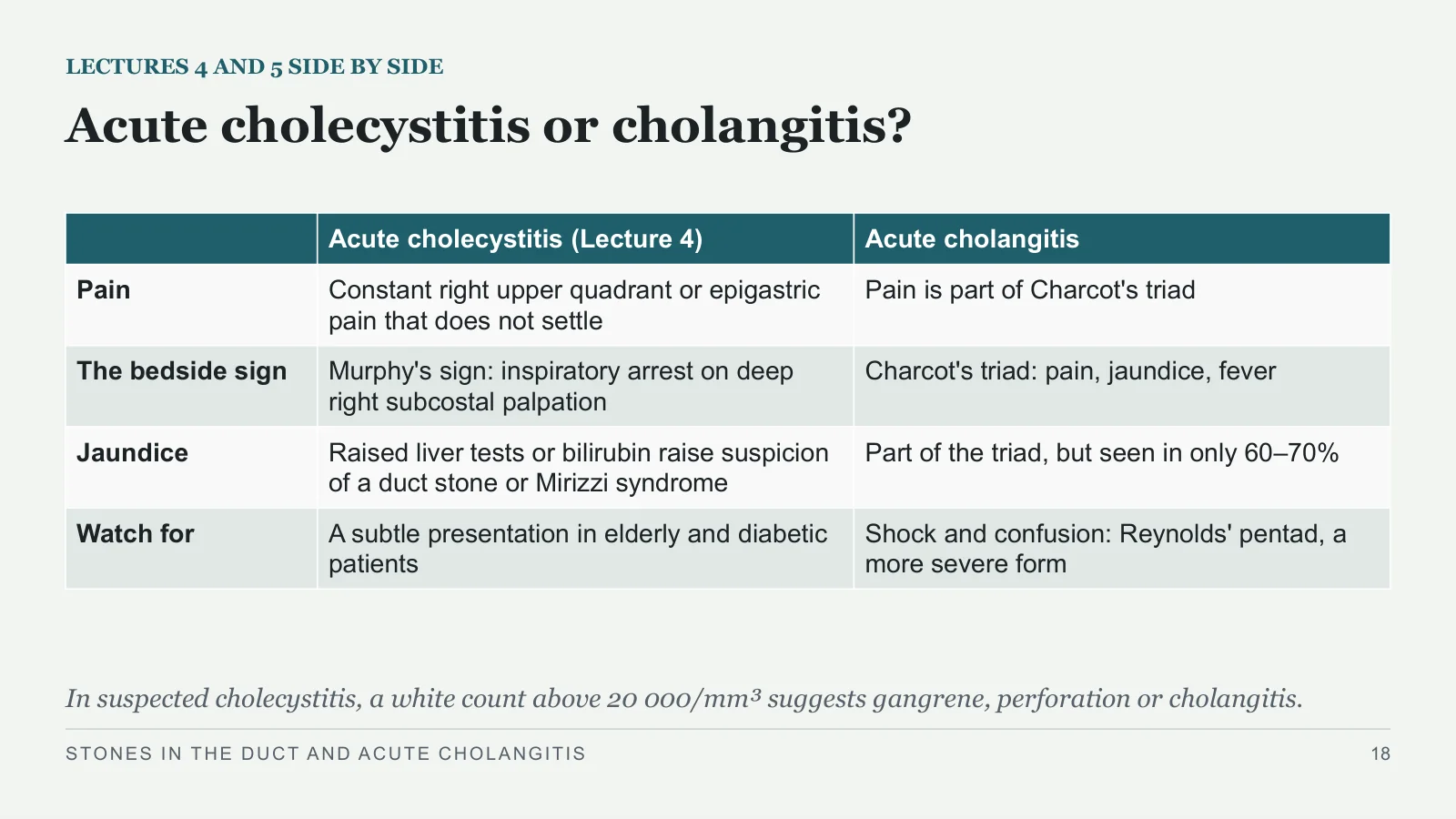
Acute cholecystitis or cholangitis?
LECTURES 4 AND 5 SIDE BY SIDE
In suspected cholecystitis, a white count above 20 000/mm³ suggests gangrene, perforation or cholangitis.
STONES IN THE DUCT AND ACUTE CHOLANGITIS
18
| Acute cholecystitis (Lecture 4) | Acute cholangitis | |
| Pain | Constant right upper quadrant or epigastric pain that does not settle | Pain is part of Charcot's triad |
| The bedside sign | Murphy's sign: inspiratory arrest on deep right subcostal palpation | Charcot's triad: pain, jaundice, fever |
| Jaundice | Raised liver tests or bilirubin raise suspicion of a duct stone or Mirizzi syndrome | Part of the triad, but seen in only 60–70% |
| Watch for | A subtle presentation in elderly and diabetic patients | Shock and confusion: Reynolds' pentad, a more severe form |
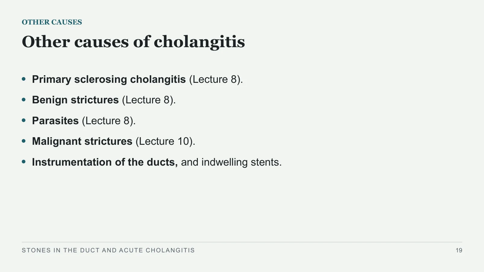
Other causes of cholangitis
OTHER CAUSES
Primary sclerosing cholangitis (Lecture 8).
Benign strictures (Lecture 8).
Parasites (Lecture 8).
Malignant strictures (Lecture 10).
Instrumentation of the ducts, and indwelling stents.
STONES IN THE DUCT AND ACUTE CHOLANGITIS
19
The first hour
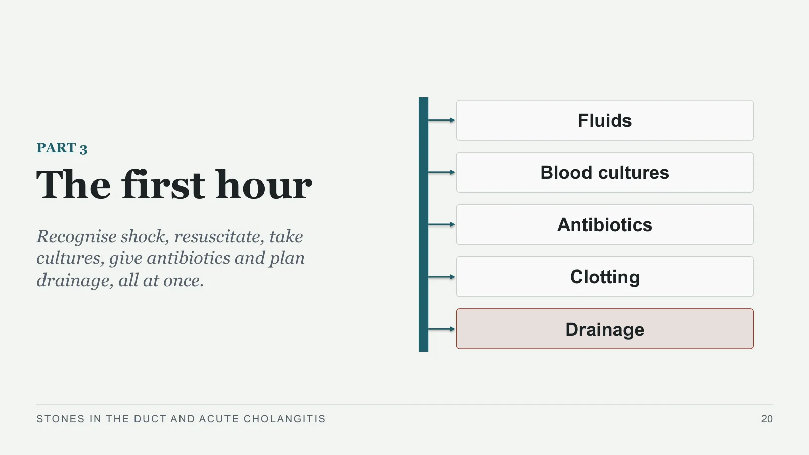
The first hour
PART 3
Recognise shock, resuscitate, take cultures, give antibiotics and plan drainage, all at once.
Fluids
Blood cultures
Antibiotics
Clotting
Drainage
STONES IN THE DUCT AND ACUTE CHOLANGITIS
20
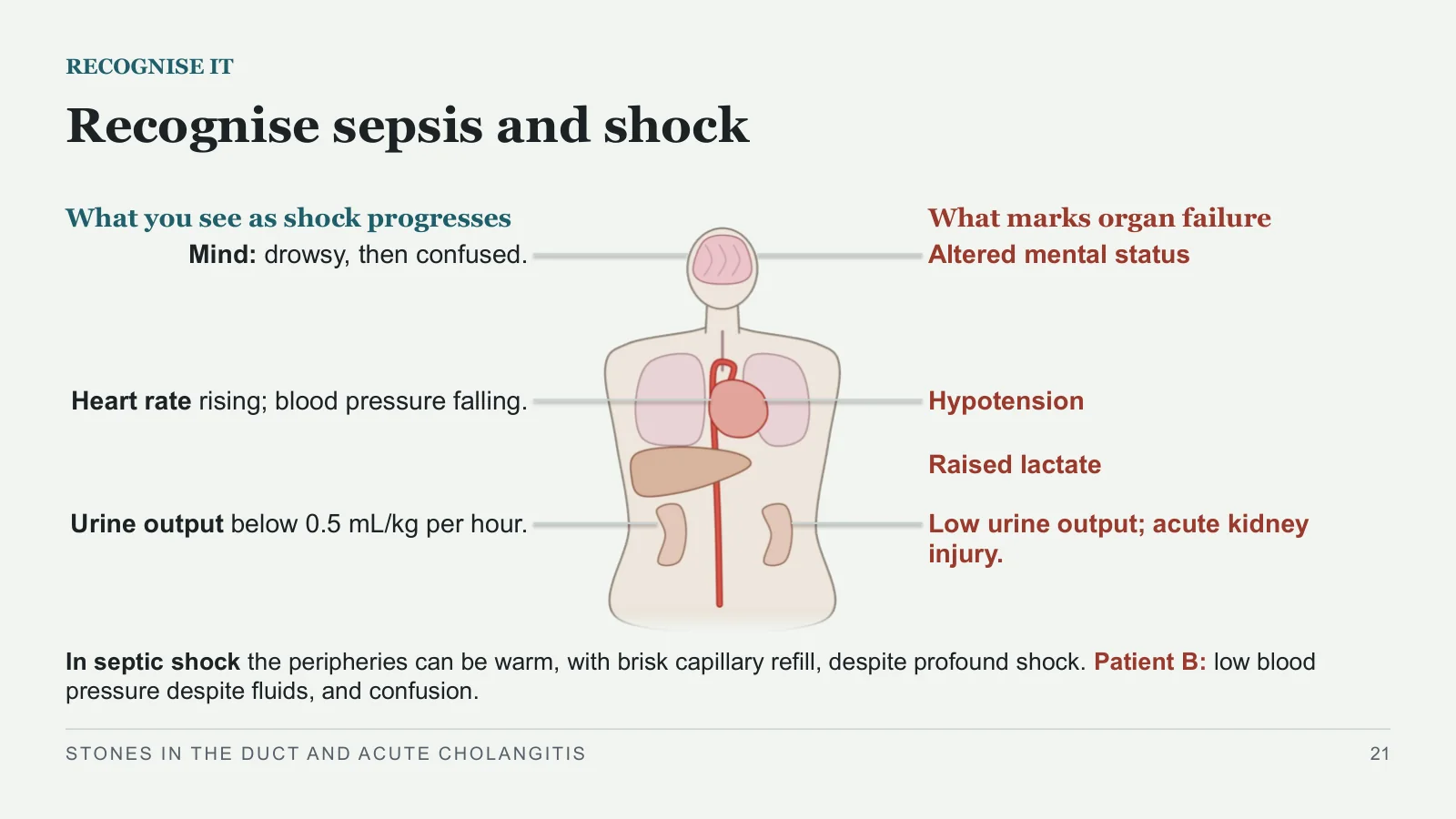
Recognise sepsis and shock
RECOGNISE IT
What you see as shock progresses
What marks organ failure
Mind: drowsy, then confused.
Heart rate rising; blood pressure falling.
Urine output below 0.5 mL/kg per hour.
Altered mental status
Hypotension
Raised lactate
Low urine output; acute kidney injury.
In septic shock the peripheries can be warm, with brisk capillary refill, despite profound shock. Patient B: low blood pressure despite fluids, and confusion.
STONES IN THE DUCT AND ACUTE CHOLANGITIS
21
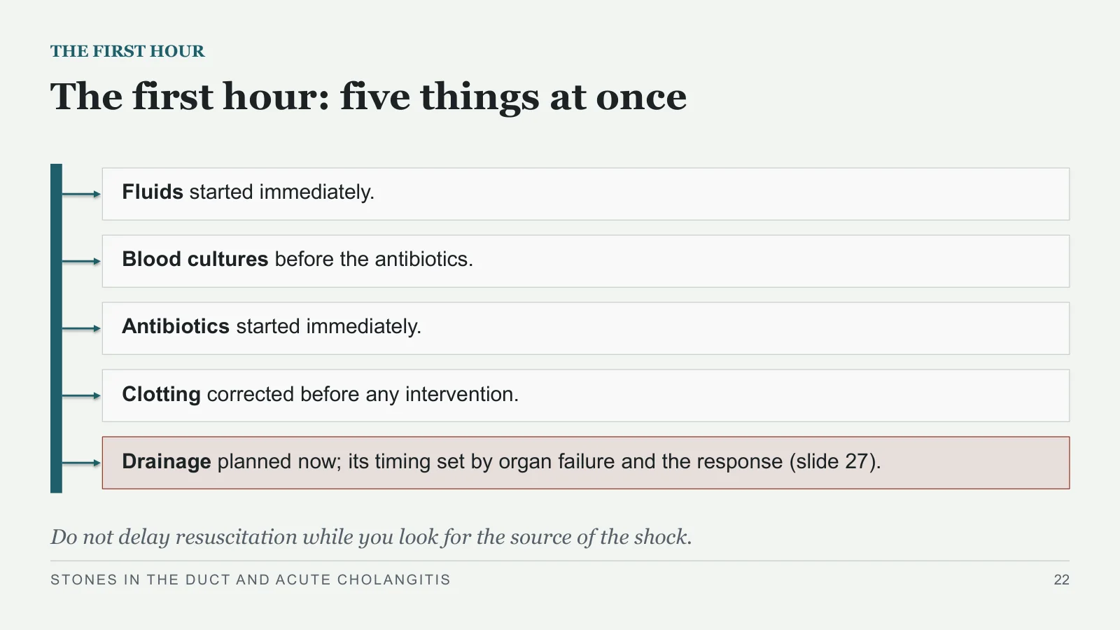
The first hour: five things at once
THE FIRST HOUR
Fluids started immediately.
Blood cultures before the antibiotics.
Antibiotics started immediately.
Clotting corrected before any intervention.
Drainage planned now; its timing set by organ failure and the response (slide 27).
Do not delay resuscitation while you look for the source of the shock.
STONES IN THE DUCT AND ACUTE CHOLANGITIS
22
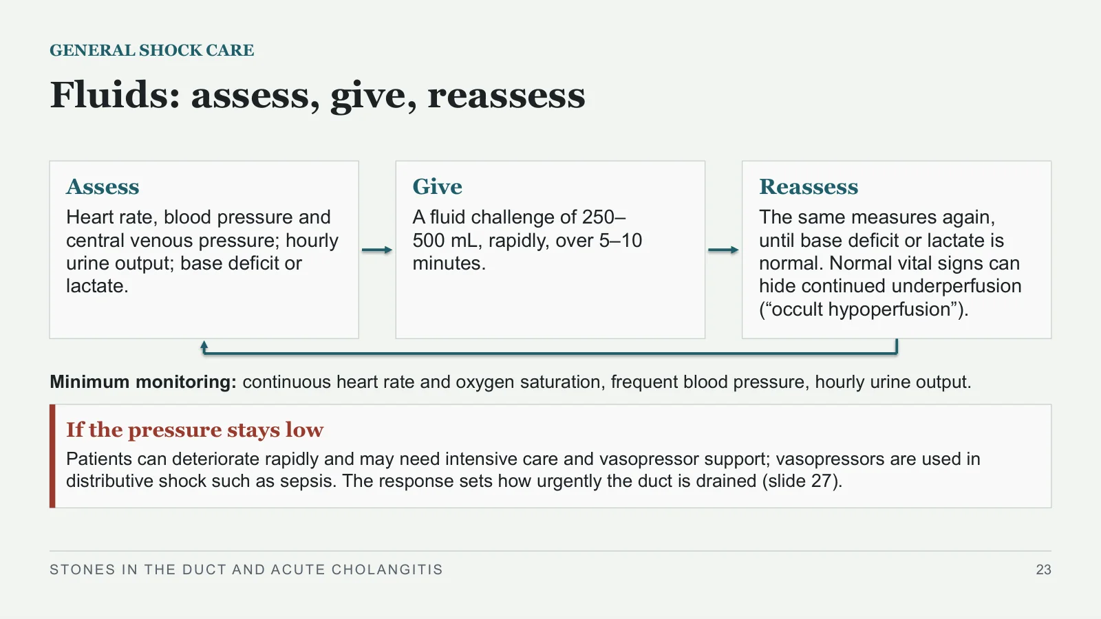
Fluids: assess, give, reassess
GENERAL SHOCK CARE
Assess
Heart rate, blood pressure and central venous pressure; hourly urine output; base deficit or lactate.
Give
A fluid challenge of 250–500 mL, rapidly, over 5–10 minutes.
Reassess
The same measures again, until base deficit or lactate is normal. Normal vital signs can hide continued underperfusion (“occult hypoperfusion”).
Minimum monitoring: continuous heart rate and oxygen saturation, frequent blood pressure, hourly urine output.
If the pressure stays low
Patients can deteriorate rapidly and may need intensive care and vasopressor support; vasopressors are used in distributive shock such as sepsis. The response sets how urgently the duct is drained (slide 27).
STONES IN THE DUCT AND ACUTE CHOLANGITIS
23
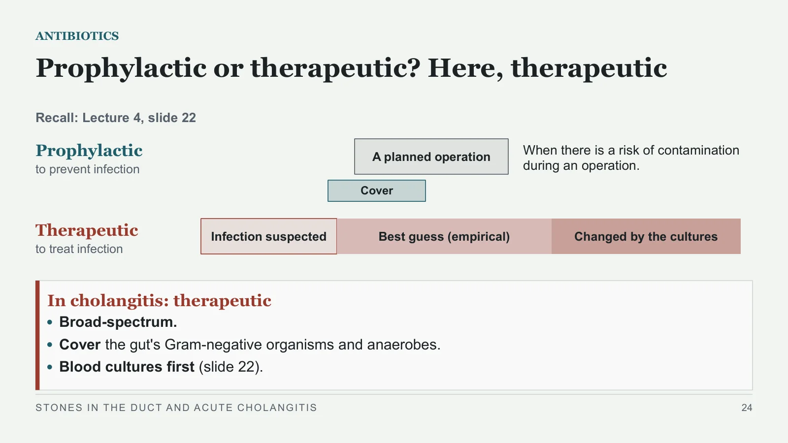
Prophylactic or therapeutic? Here, therapeutic
ANTIBIOTICS
Recall: Lecture 4, slide 22
Prophylactic to prevent infection
Therapeutic to treat infection
A planned operation
Cover
When there is a risk of contamination during an operation.
Infection suspected
Best guess (empirical)
Changed by the cultures
In cholangitis: therapeutic
Broad-spectrum.
Cover the gut's Gram-negative organisms and anaerobes.
Blood cultures first (slide 22).
STONES IN THE DUCT AND ACUTE CHOLANGITIS
24
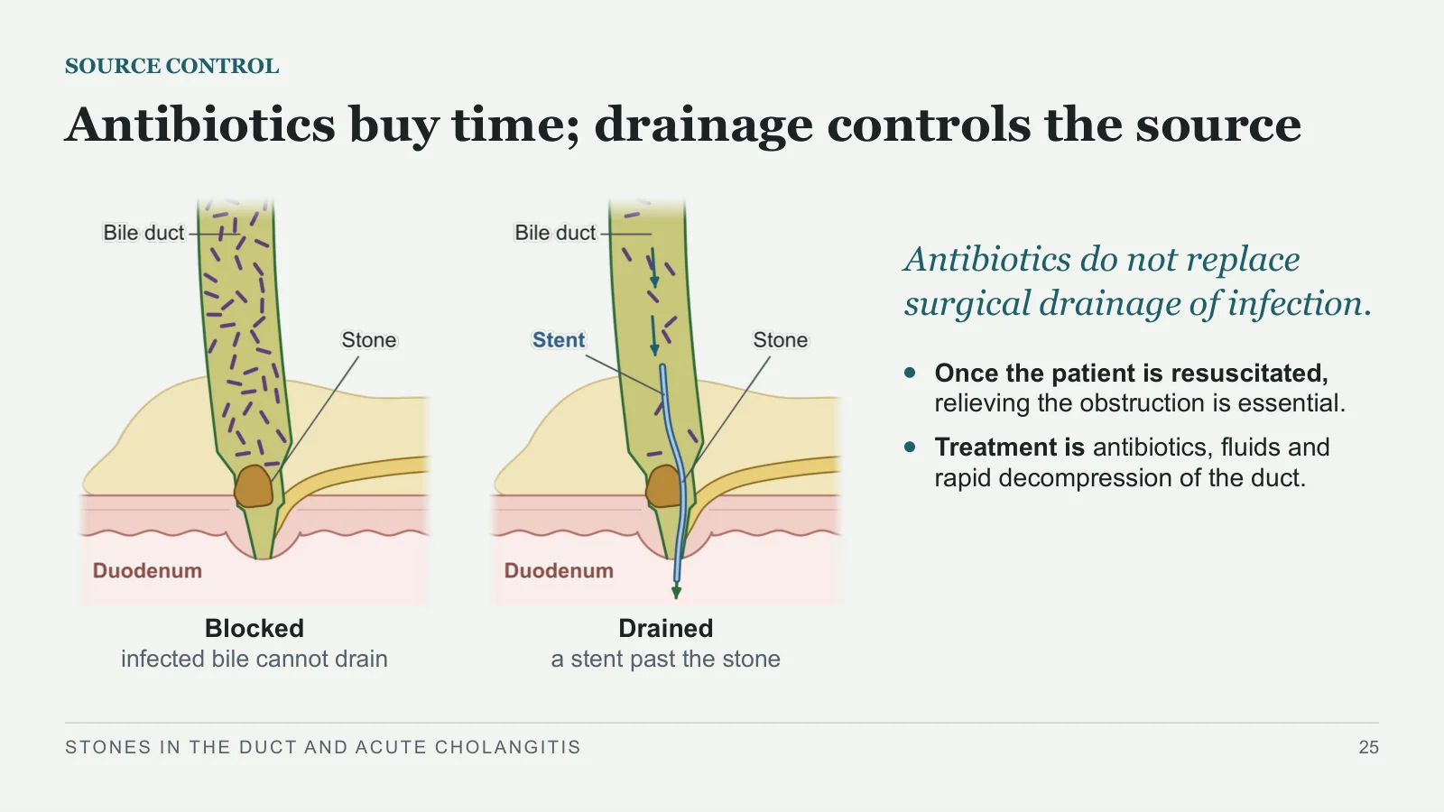
Antibiotics buy time; drainage controls the source
SOURCE CONTROL
Blocked
infected bile cannot drain
Drained
a stent past the stone
Antibiotics do not replace surgical drainage of infection.
Once the patient is resuscitated, relieving the obstruction is essential.
Treatment is antibiotics, fluids and rapid decompression of the duct.
STONES IN THE DUCT AND ACUTE CHOLANGITIS
25
Draining the duct
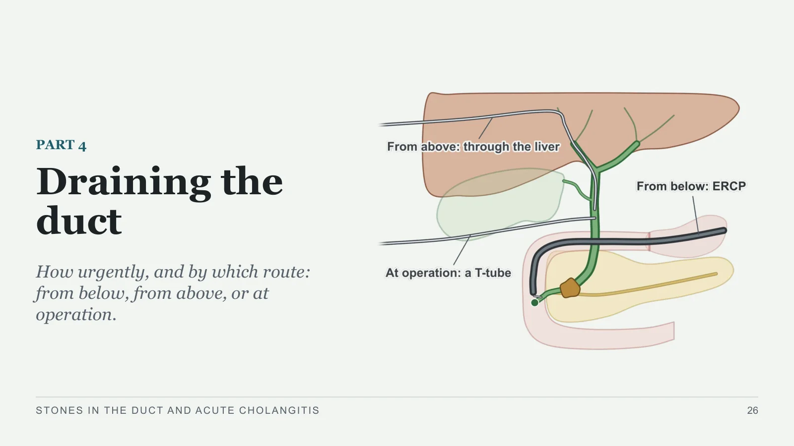
Draining the duct
PART 4
How urgently, and by which route: from below, from above, or at operation.
STONES IN THE DUCT AND ACUTE CHOLANGITIS
26
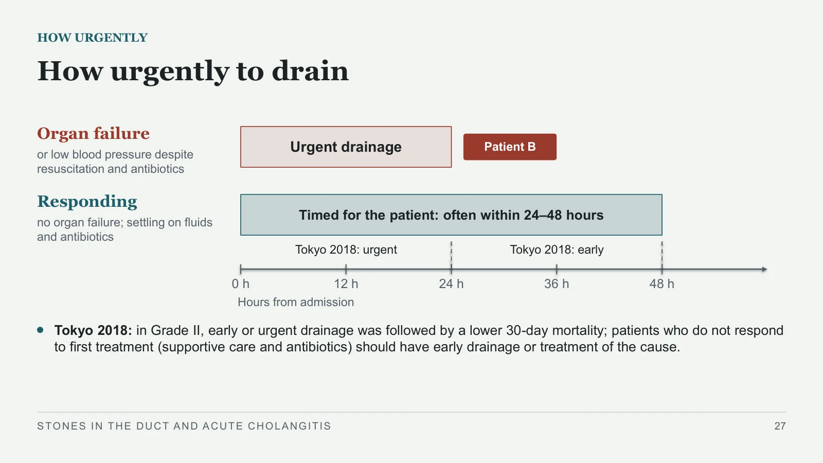
How urgently to drain
HOW URGENTLY
Organ failure
or low blood pressure despite resuscitation and antibiotics
Responding
no organ failure; settling on fluids and antibiotics
Urgent drainage
Patient B
Timed for the patient: often within 24–48 hours
0 h
12 h
24 h
36 h
48 h
Hours from admission
Tokyo 2018: urgent
Tokyo 2018: early
Tokyo 2018: in Grade II, early or urgent drainage was followed by a lower 30-day mortality; patients who do not respond to first treatment (supportive care and antibiotics) should have early drainage or treatment of the cause.
STONES IN THE DUCT AND ACUTE CHOLANGITIS
27
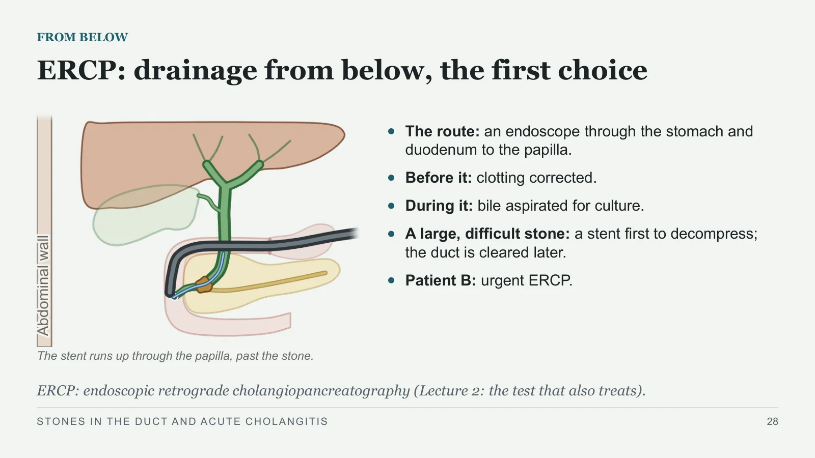
ERCP: drainage from below, the first choice
FROM BELOW
The route: an endoscope through the stomach and duodenum to the papilla.
Before it: clotting corrected.
During it: bile aspirated for culture.
A large, difficult stone: a stent first to decompress; the duct is cleared later.
Patient B: urgent ERCP.
The stent runs up through the papilla, past the stone.
ERCP: endoscopic retrograde cholangiopancreatography (Lecture 2: the test that also treats).
STONES IN THE DUCT AND ACUTE CHOLANGITIS
28
At the papilla: cut, clear, or stent
FROM BELOW
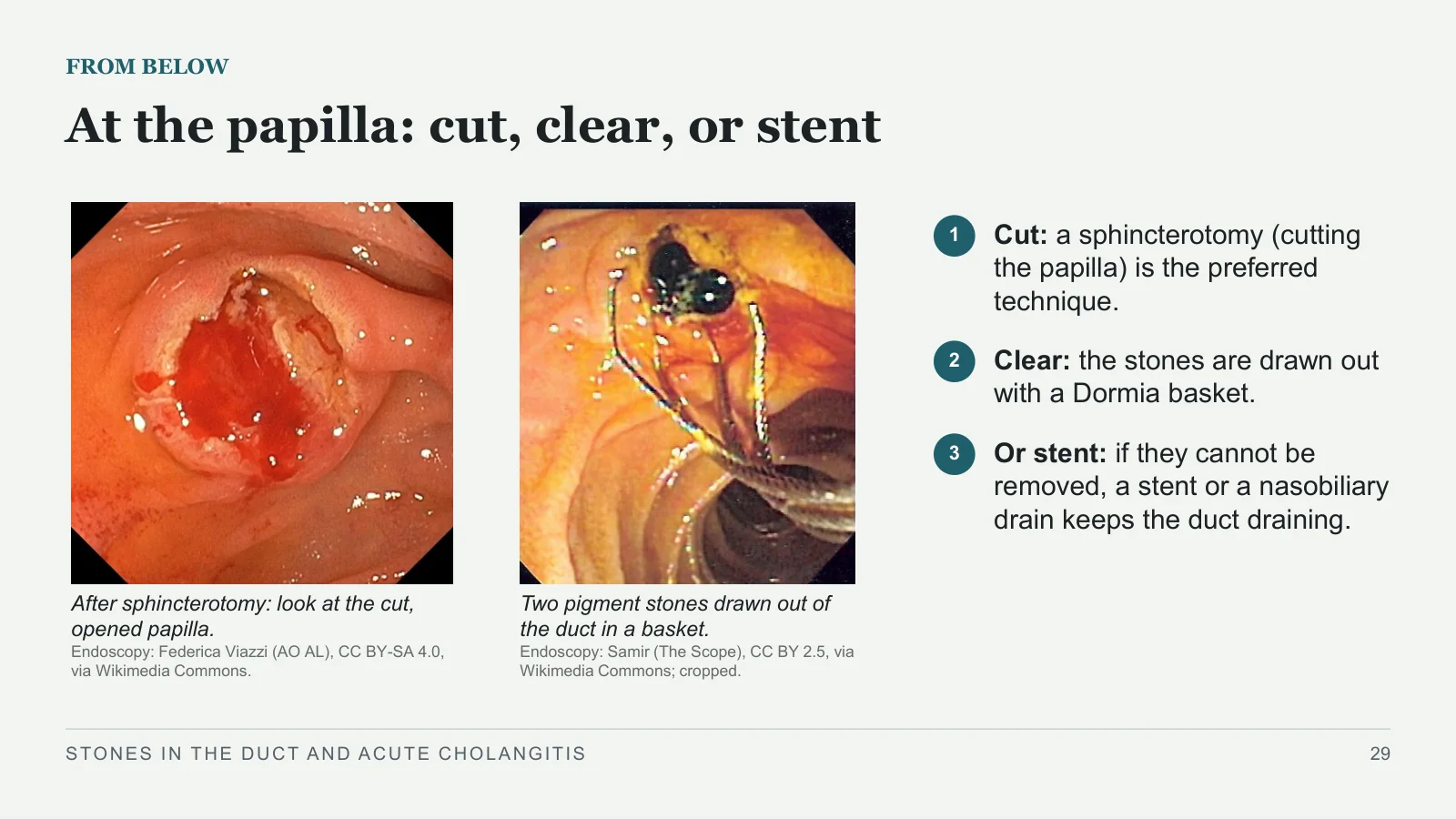
After sphincterotomy: look at the cut, opened papilla.
Endoscopy: Federica Viazzi (AO AL), CC BY-SA 4.0, via Wikimedia Commons.
Two pigment stones drawn out of the duct in a basket.
Endoscopy: Samir (The Scope), CC BY 2.5, via Wikimedia Commons; cropped.
1
Cut: a sphincterotomy (cutting the papilla) is the preferred technique.
2
Clear: the stones are drawn out with a Dormia basket.
3
Or stent: if they cannot be removed, a stent or a nasobiliary drain keeps the duct draining.
STONES IN THE DUCT AND ACUTE CHOLANGITIS
29
When ERCP fails or cannot reach: drain from above
FROM ABOVE
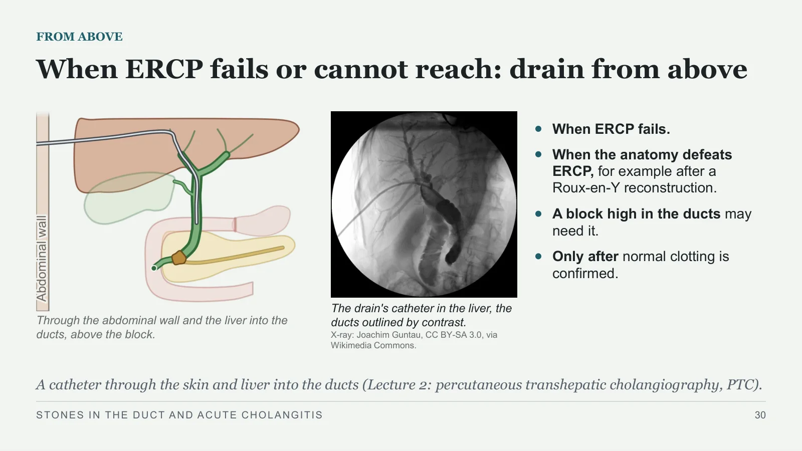
Through the abdominal wall and the liver into the ducts, above the block.
The drain's catheter in the liver, the ducts outlined by contrast.
X-ray: Joachim Guntau, CC BY-SA 3.0, via Wikimedia Commons.
When ERCP fails.
When the anatomy defeats ERCP, for example after a Roux‑en‑Y reconstruction.
A block high in the ducts may need it.
Only after normal clotting is confirmed.
A catheter through the skin and liver into the ducts (Lecture 2: percutaneous transhepatic cholangiography, PTC).
STONES IN THE DUCT AND ACUTE CHOLANGITIS
30
Surgery when the others are unavailable or have failed
AT OPERATION
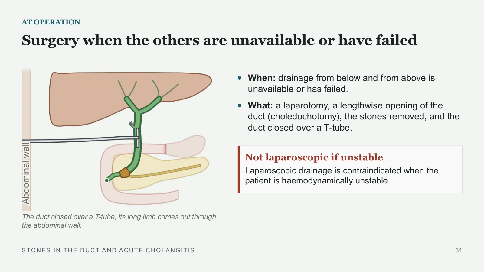
The duct closed over a T-tube; its long limb comes out through the abdominal wall.
When: drainage from below and from above is unavailable or has failed.
What: a laparotomy, a lengthwise opening of the duct (choledochotomy), the stones removed, and the duct closed over a T-tube.
Not laparoscopic if unstable
Laparoscopic drainage is contraindicated when the patient is haemodynamically unstable.
STONES IN THE DUCT AND ACUTE CHOLANGITIS
31
Three ways to drain the duct: when to use each
THREE ROUTES
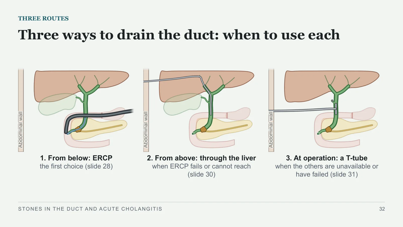
1. From below: ERCP
the first choice (slide 28)
2. From above: through the liver
when ERCP fails or cannot reach (slide 30)
3. At operation: a T-tube
when the others are unavailable or have failed (slide 31)
STONES IN THE DUCT AND ACUTE CHOLANGITIS
32
Predicting and proving
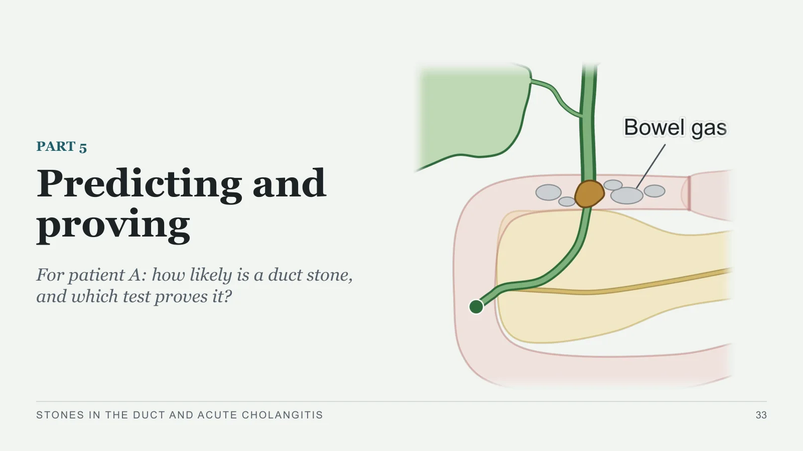
Predicting and proving
PART 5
For patient A: how likely is a duct stone, and which test proves it?
STONES IN THE DUCT AND ACUTE CHOLANGITIS
33
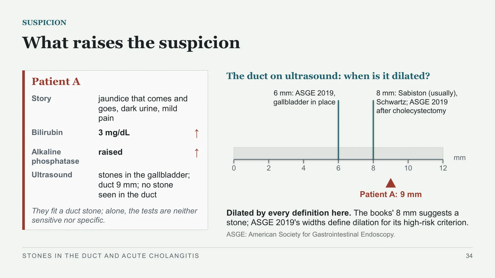
What raises the suspicion
SUSPICION
Patient A
Story
jaundice that comes and goes, dark urine, mild pain
Bilirubin
3 mg/dL
↑
Alkaline phosphatase
raised
↑
Ultrasound
stones in the gallbladder; duct 9 mm; no stone seen in the duct
They fit a duct stone; alone, the tests are neither sensitive nor specific.
The duct on ultrasound: when is it dilated?
0
2
4
6
8
10
12
mm
6 mm: ASGE 2019, gallbladder in place
8 mm: Sabiston (usually), Schwartz; ASGE 2019 after cholecystectomy
Patient A: 9 mm
Dilated by every definition here. The books' 8 mm suggests a stone; ASGE 2019's widths define dilation for its high-risk criterion.
ASGE: American Society for Gastrointestinal Endoscopy.
STONES IN THE DUCT AND ACUTE CHOLANGITIS
34
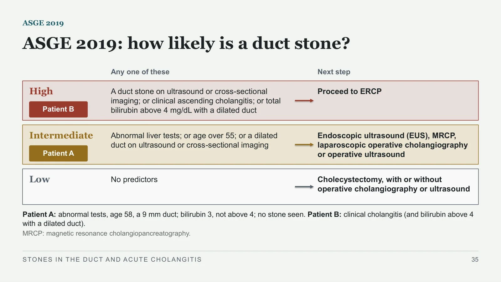
ASGE 2019: how likely is a duct stone?
ASGE 2019
Any one of these
Next step
High
Patient B
A duct stone on ultrasound or cross-sectional imaging; or clinical ascending cholangitis; or total bilirubin above 4 mg/dL with a dilated duct
Proceed to ERCP
Intermediate
Patient A
Abnormal liver tests; or age over 55; or a dilated duct on ultrasound or cross-sectional imaging
Endoscopic ultrasound (EUS), MRCP, laparoscopic operative cholangiography or operative ultrasound
Low
No predictors
Cholecystectomy, with or without operative cholangiography or ultrasound
Patient A: abnormal tests, age 58, a 9 mm duct; bilirubin 3, not above 4; no stone seen. Patient B: clinical cholangitis (and bilirubin above 4 with a dilated duct).
MRCP: magnetic resonance cholangiopancreatography.
STONES IN THE DUCT AND ACUTE CHOLANGITIS
35
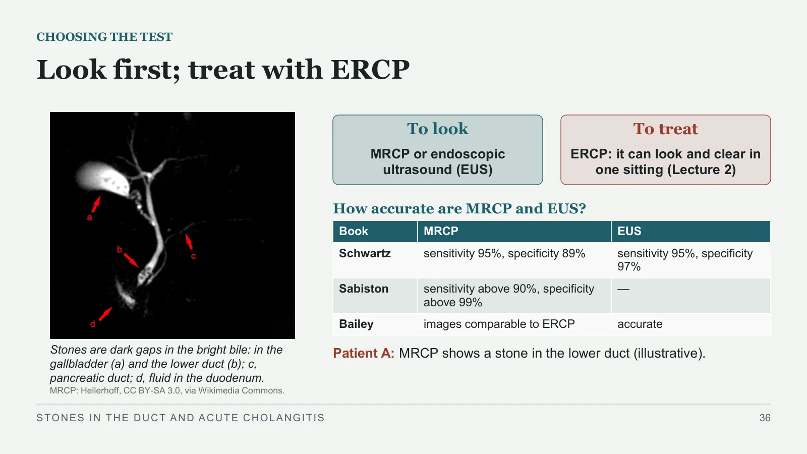
Look first; treat with ERCP
CHOOSING THE TEST
Stones are dark gaps in the bright bile: in the gallbladder (a) and the lower duct (b); c, pancreatic duct; d, fluid in the duodenum.
MRCP: Hellerhoff, CC BY-SA 3.0, via Wikimedia Commons.
To look
MRCP or endoscopic ultrasound (EUS)
To treat
ERCP: it can look and clear in one sitting (Lecture 2)
How accurate are MRCP and EUS?
Patient A: MRCP shows a stone in the lower duct (illustrative).
STONES IN THE DUCT AND ACUTE CHOLANGITIS
36
| Book | MRCP | EUS |
| Schwartz | sensitivity 95%, specificity 89% | sensitivity 95%, specificity 97% |
| Sabiston | sensitivity above 90%, specificity above 99% | — |
| Bailey | images comparable to ERCP | accurate |
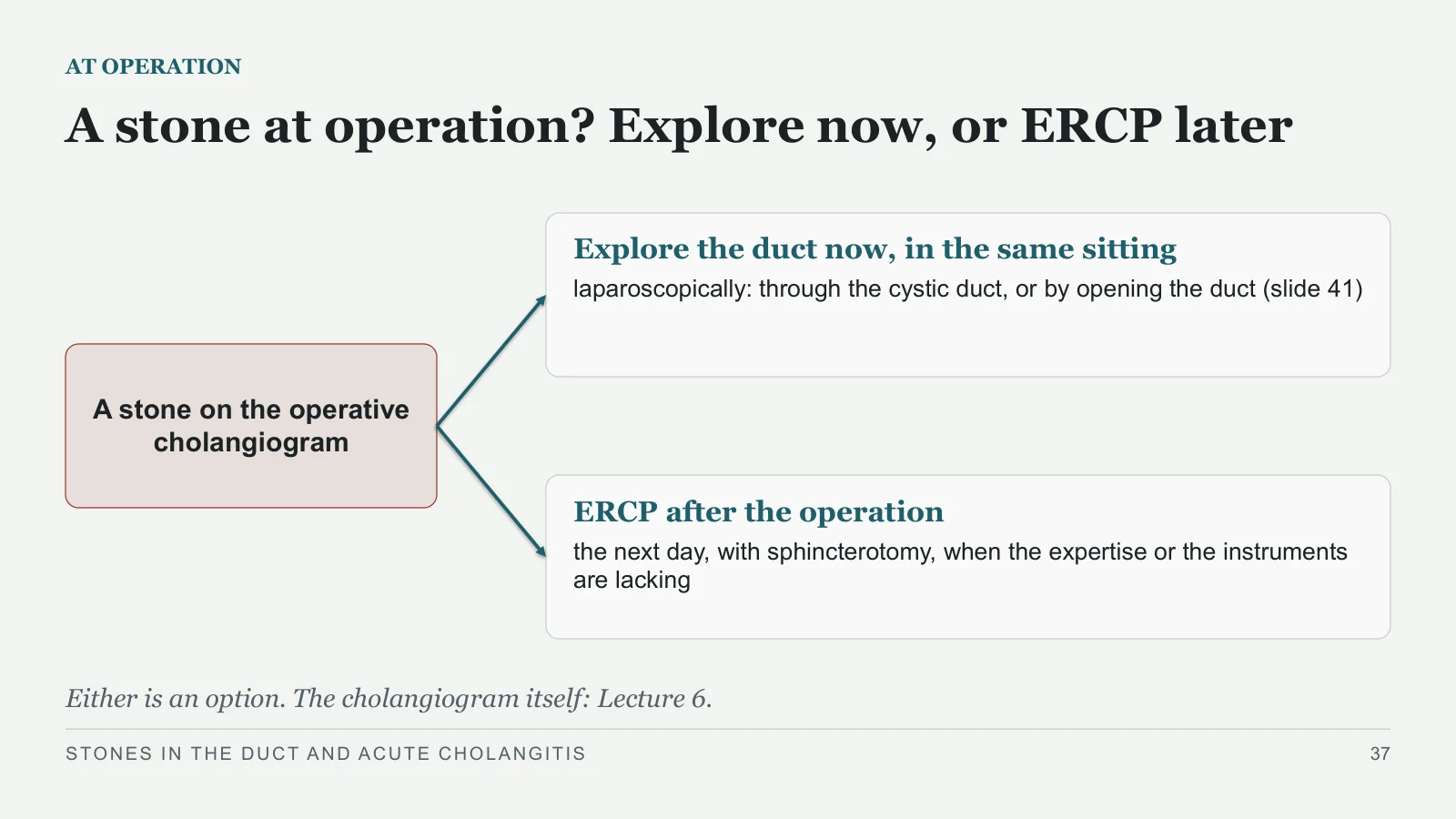
A stone at operation? Explore now, or ERCP later
AT OPERATION
A stone on the operative cholangiogram
Explore the duct now, in the same sitting
laparoscopically: through the cystic duct, or by opening the duct (slide 41)
ERCP after the operation
the next day, with sphincterotomy, when the expertise or the instruments are lacking
Either is an option. The cholangiogram itself: Lecture 6.
STONES IN THE DUCT AND ACUTE CHOLANGITIS
37
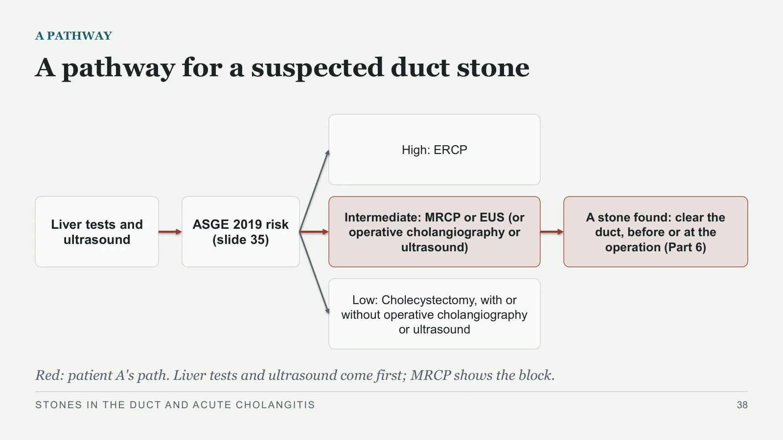
A pathway for a suspected duct stone
A PATHWAY
Liver tests and ultrasound
ASGE 2019 risk (slide 35)
High: ERCP
Intermediate: MRCP or EUS (or operative cholangiography or ultrasound)
Low: Cholecystectomy, with or without operative cholangiography or ultrasound
A stone found: clear the duct, before or at the operation (Part 6)
Red: patient A's path. Liver tests and ultrasound come first; MRCP shows the block.
STONES IN THE DUCT AND ACUTE CHOLANGITIS
38
Clearing the duct
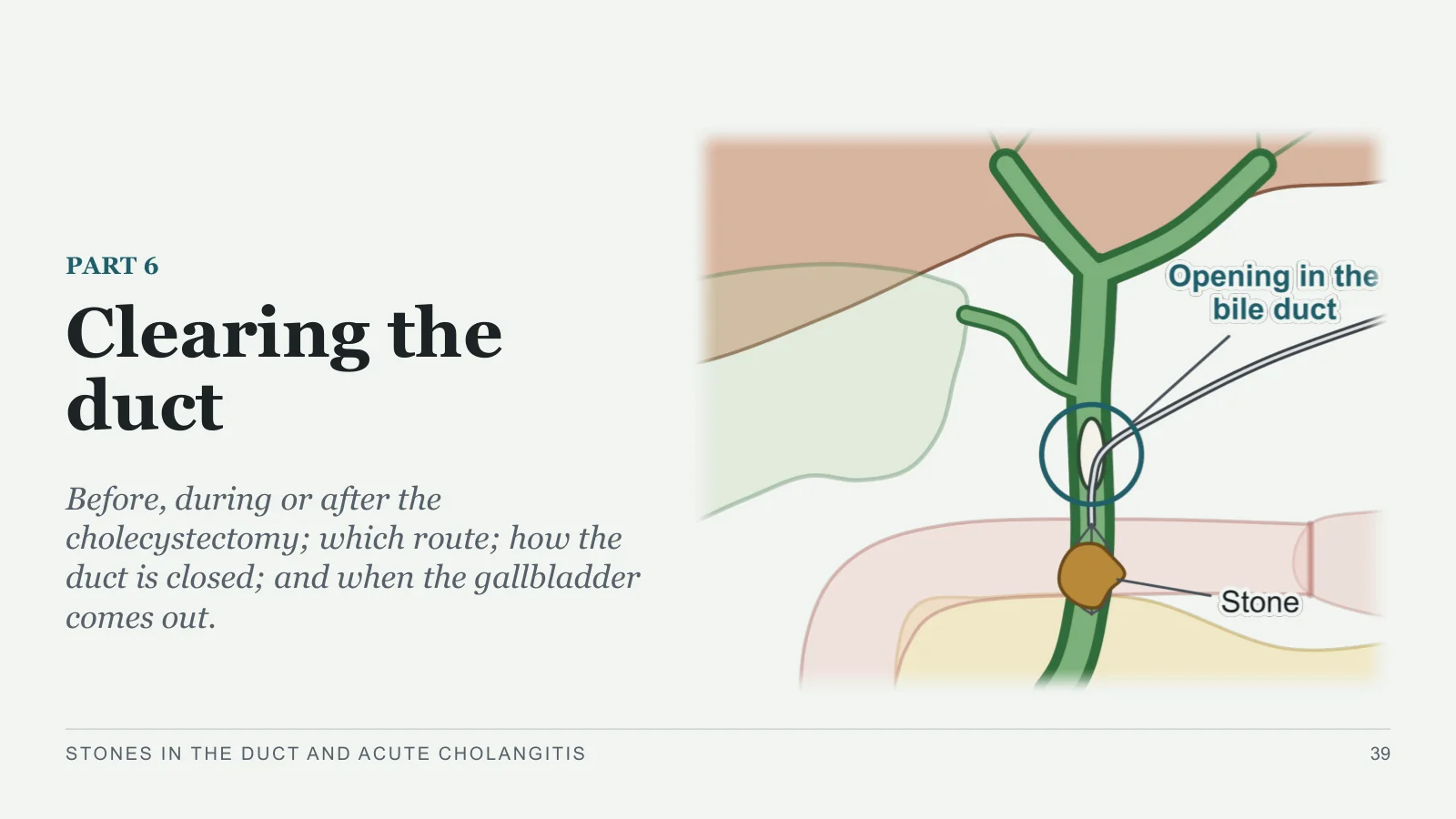
Clearing the duct
PART 6
Before, during or after the cholecystectomy; which route; how the duct is closed; and when the gallbladder comes out.
STONES IN THE DUCT AND ACUTE CHOLANGITIS
39
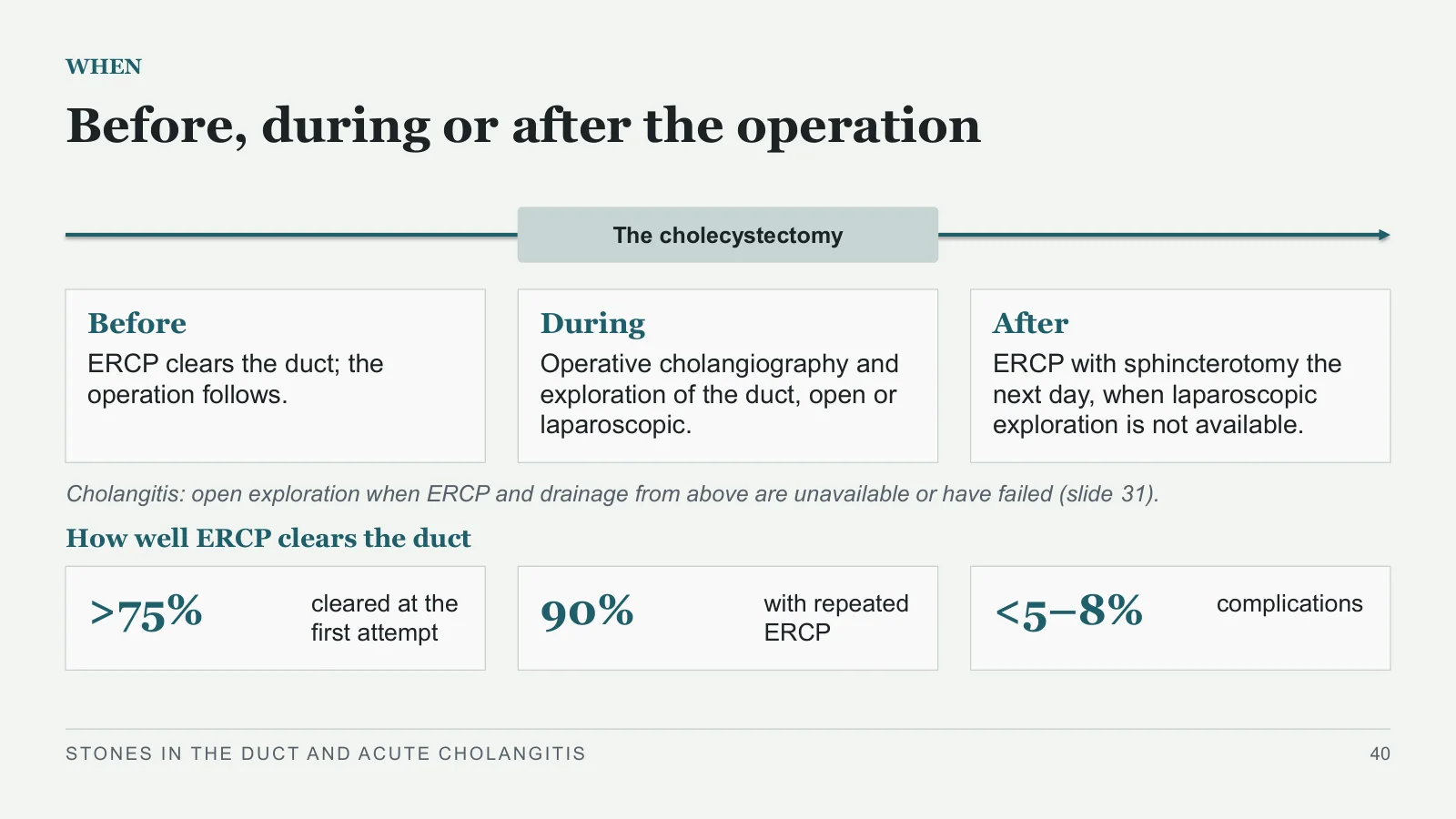
Before, during or after the operation
WHEN
The cholecystectomy
Before
ERCP clears the duct; the operation follows.
During
Operative cholangiography and exploration of the duct, open or laparoscopic.
After
ERCP with sphincterotomy the next day, when laparoscopic exploration is not available.
Cholangitis: open exploration when ERCP and drainage from above are unavailable or have failed (slide 31).
How well ERCP clears the duct
>75%
cleared at the first attempt
90%
with repeated ERCP
<5–8%
complications
STONES IN THE DUCT AND ACUTE CHOLANGITIS
40
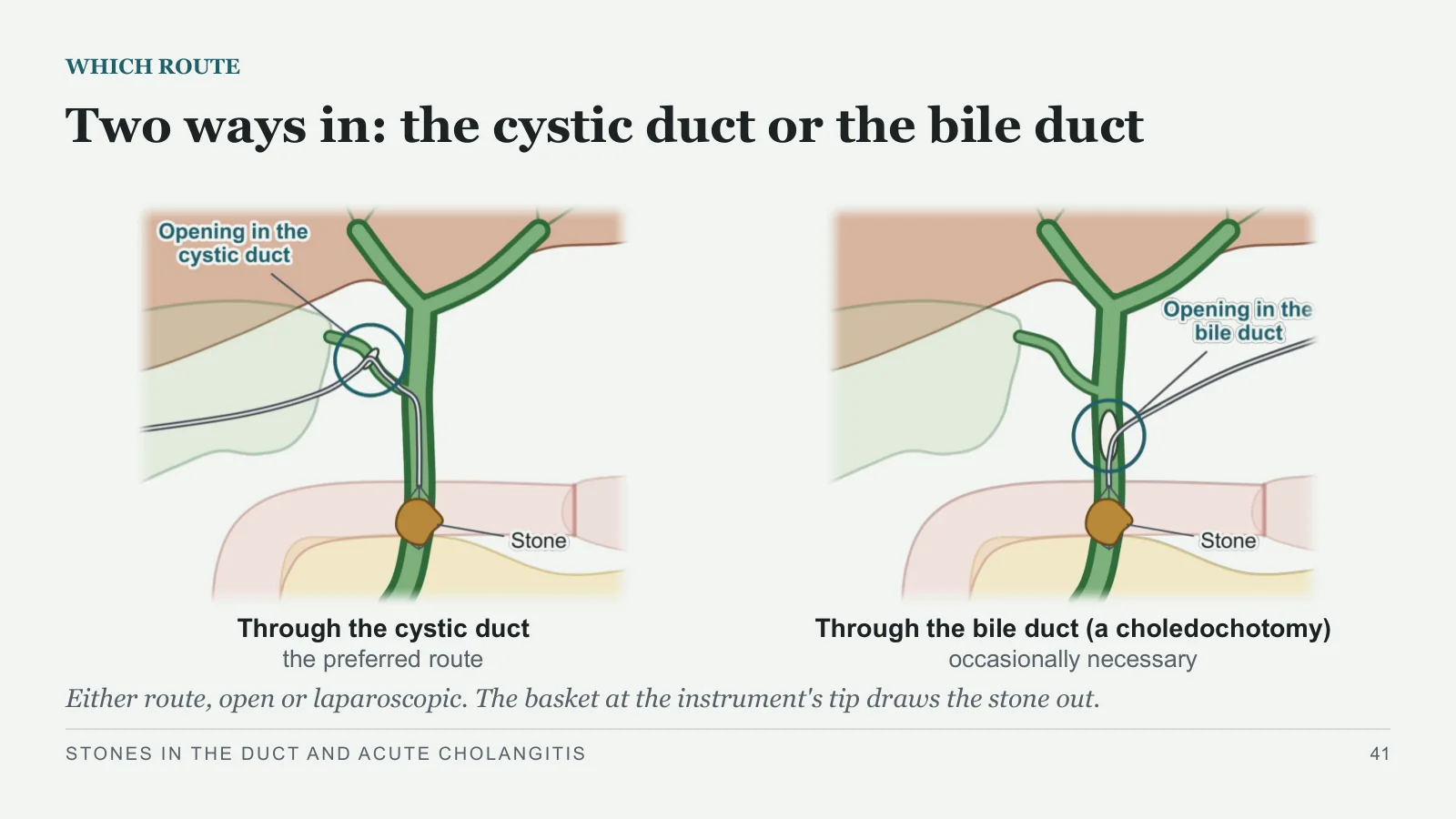
Two ways in: the cystic duct or the bile duct
WHICH ROUTE
Through the cystic duct
the preferred route
Through the bile duct (a choledochotomy)
occasionally necessary
Either route, open or laparoscopic. The basket at the instrument's tip draws the stone out.
STONES IN THE DUCT AND ACUTE CHOLANGITIS
41
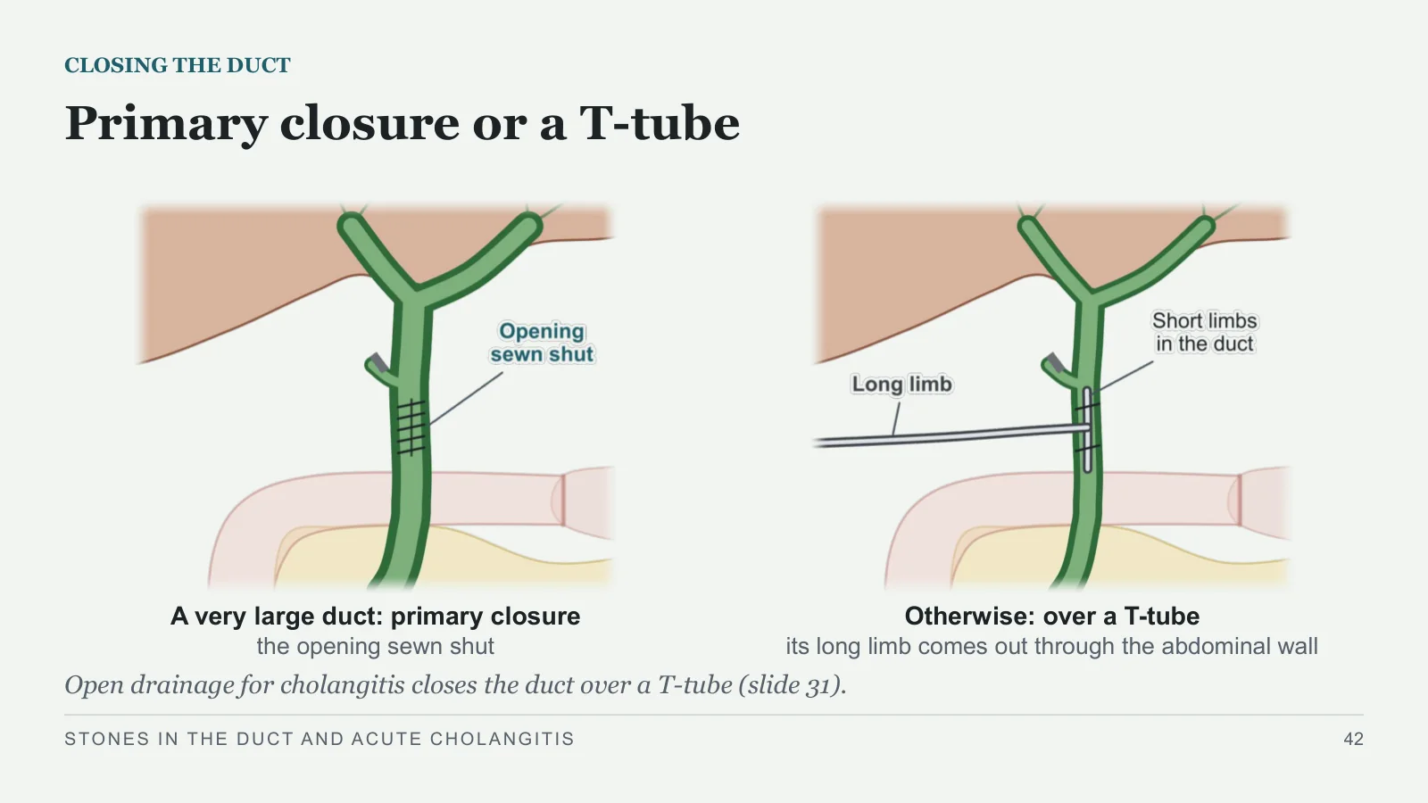
Primary closure or a T-tube
CLOSING THE DUCT
A very large duct: primary closure
the opening sewn shut
Otherwise: over a T-tube
its long limb comes out through the abdominal wall
Open drainage for cholangitis closes the duct over a T-tube (slide 31).
STONES IN THE DUCT AND ACUTE CHOLANGITIS
42
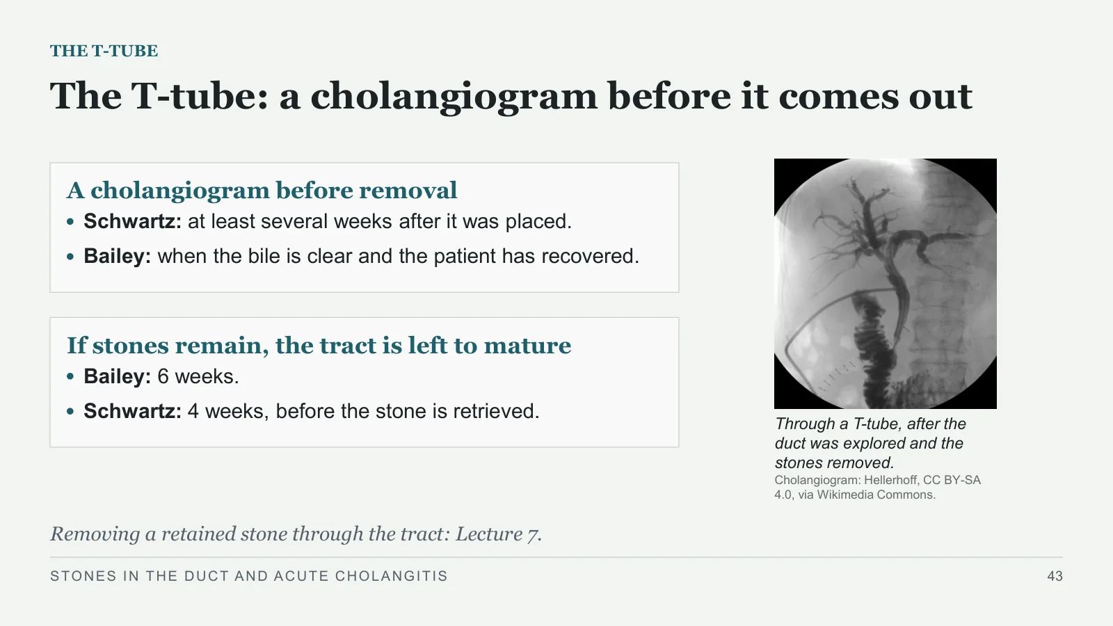
The T-tube: a cholangiogram before it comes out
THE T-TUBE
A cholangiogram before removal
Schwartz: at least several weeks after it was placed.
Bailey: when the bile is clear and the patient has recovered.
If stones remain, the tract is left to mature
Bailey: 6 weeks.
Schwartz: 4 weeks, before the stone is retrieved.
Through a T-tube, after the duct was explored and the stones removed.
Cholangiogram: Hellerhoff, CC BY-SA 4.0, via Wikimedia Commons.
Removing a retained stone through the tract: Lecture 7.
STONES IN THE DUCT AND ACUTE CHOLANGITIS
43
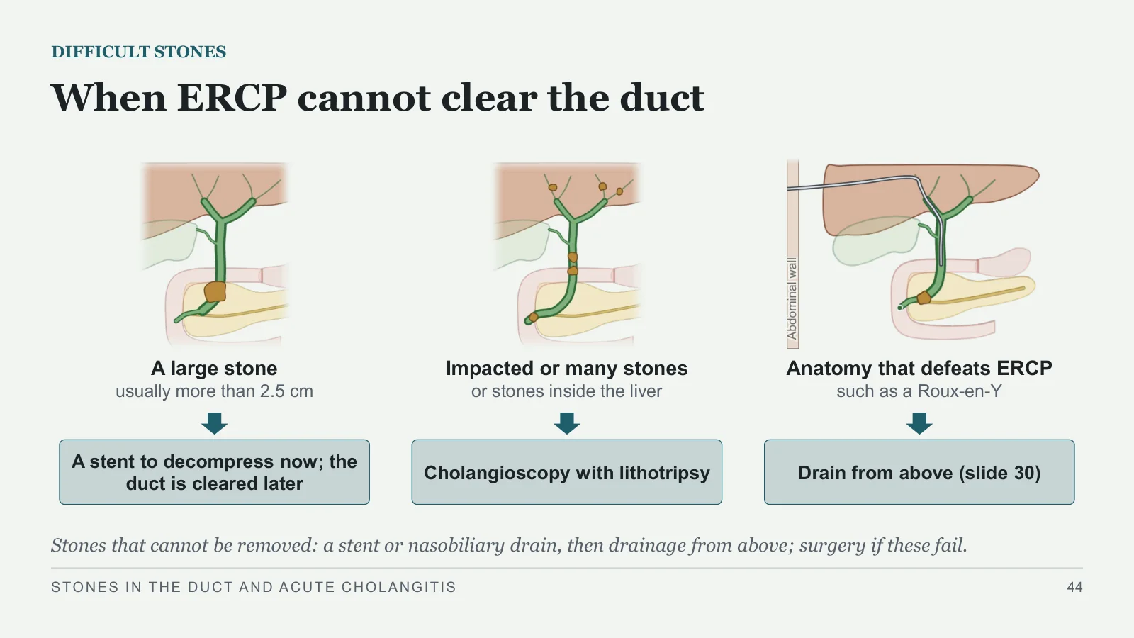
When ERCP cannot clear the duct
DIFFICULT STONES
A large stone
usually more than 2.5 cm
A stent to decompress now; the duct is cleared later
Impacted or many stones
or stones inside the liver
Cholangioscopy with lithotripsy
Anatomy that defeats ERCP
such as a Roux-en-Y
Drain from above (slide 30)
Stones that cannot be removed: a stent or nasobiliary drain, then drainage from above; surgery if these fail.
STONES IN THE DUCT AND ACUTE CHOLANGITIS
44
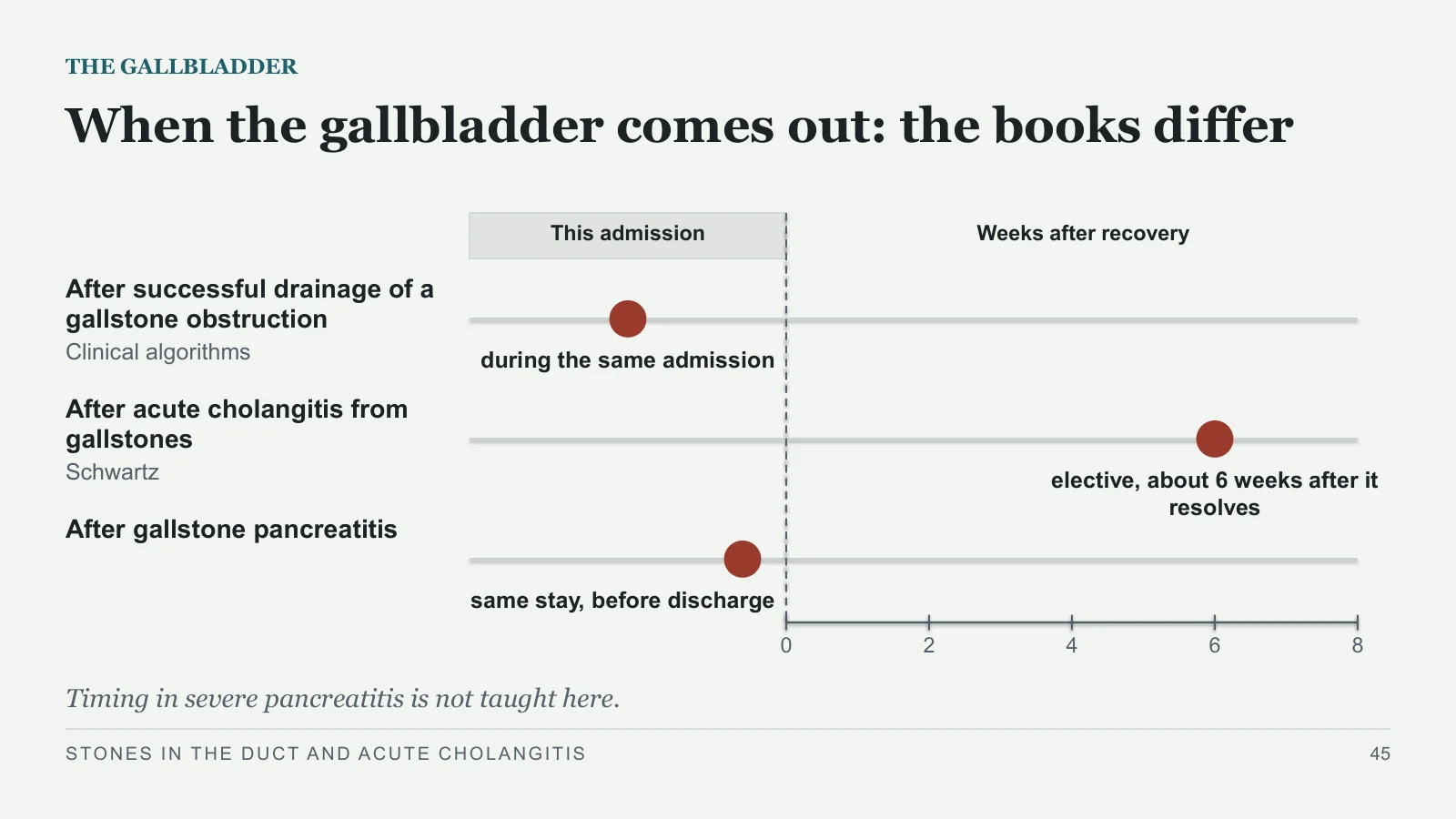
When the gallbladder comes out: the books differ
THE GALLBLADDER
This admission
Weeks after recovery
After successful drainage of a gallstone obstruction
Clinical algorithms
during the same admission
After acute cholangitis from gallstones
Schwartz
elective, about 6 weeks after it resolves
After gallstone pancreatitis
same stay, before discharge
0
2
4
6
8
Timing in severe pancreatitis is not taught here.
STONES IN THE DUCT AND ACUTE CHOLANGITIS
45
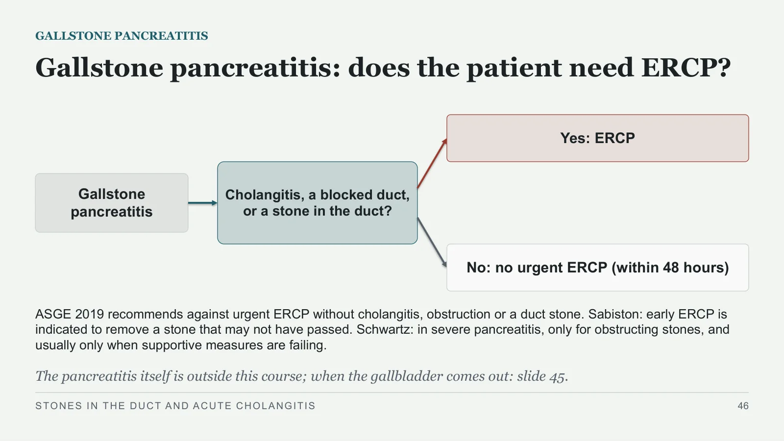
Gallstone pancreatitis: does the patient need ERCP?
GALLSTONE PANCREATITIS
Gallstone pancreatitis
Cholangitis, a blocked duct, or a stone in the duct?
Yes: ERCP
No: no urgent ERCP (within 48 hours)
ASGE 2019 recommends against urgent ERCP without cholangitis, obstruction or a duct stone. Sabiston: early ERCP is indicated to remove a stone that may not have passed. Schwartz: in severe pancreatitis, only for obstructing stones, and usually only when supportive measures are failing.
The pancreatitis itself is outside this course; when the gallbladder comes out: slide 45.
STONES IN THE DUCT AND ACUTE CHOLANGITIS
46
Recap and references
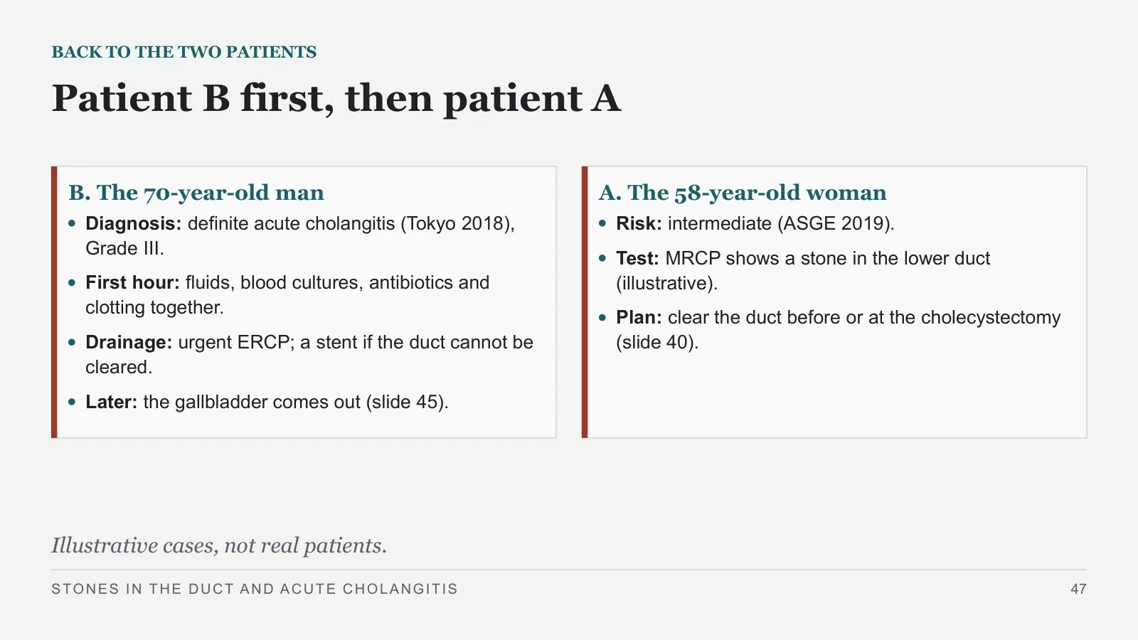
Patient B first, then patient A
BACK TO THE TWO PATIENTS
B. The 70-year-old man
Diagnosis: definite acute cholangitis (Tokyo 2018), Grade III.
First hour: fluids, blood cultures, antibiotics and clotting together.
Drainage: urgent ERCP; a stent if the duct cannot be cleared.
Later: the gallbladder comes out (slide 45).
A. The 58-year-old woman
Risk: intermediate (ASGE 2019).
Test: MRCP shows a stone in the lower duct (illustrative).
Plan: clear the duct before or at the cholecystectomy (slide 40).
Illustrative cases, not real patients.
STONES IN THE DUCT AND ACUTE CHOLANGITIS
47
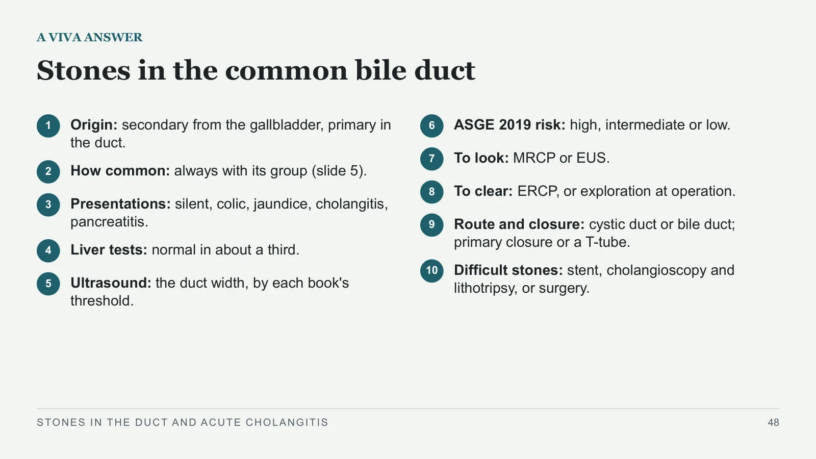
Stones in the common bile duct
A VIVA ANSWER
1
Origin: secondary from the gallbladder, primary in the duct.
2
How common: always with its group (slide 5).
3
Presentations: silent, colic, jaundice, cholangitis, pancreatitis.
4
Liver tests: normal in about a third.
5
Ultrasound: the duct width, by each book's threshold.
6
ASGE 2019 risk: high, intermediate or low.
7
To look: MRCP or EUS.
8
To clear: ERCP, or exploration at operation.
9
Route and closure: cystic duct or bile duct; primary closure or a T-tube.
10
Difficult stones: stent, cholangioscopy and lithotripsy, or surgery.
STONES IN THE DUCT AND ACUTE CHOLANGITIS
48
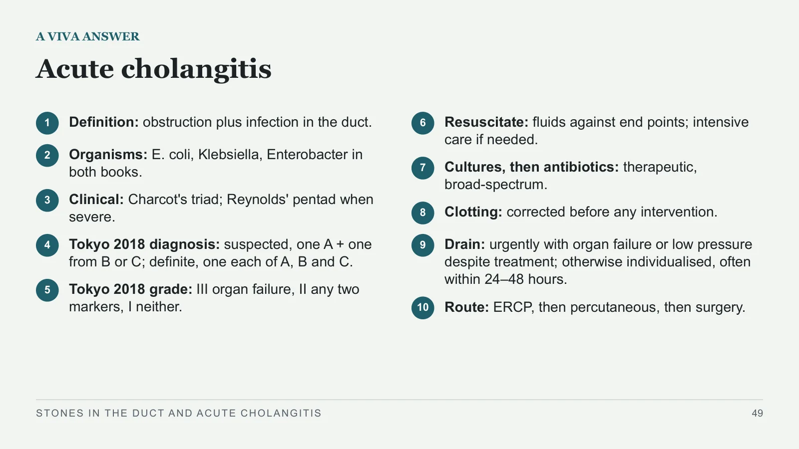
Acute cholangitis
A VIVA ANSWER
1
Definition: obstruction plus infection in the duct.
2
Organisms: E. coli, Klebsiella, Enterobacter in both books.
3
Clinical: Charcot's triad; Reynolds' pentad when severe.
4
Tokyo 2018 diagnosis: suspected, one A + one from B or C; definite, one each of A, B and C.
5
Tokyo 2018 grade: III organ failure, II any two markers, I neither.
6
Resuscitate: fluids against end points; intensive care if needed.
7
Cultures, then antibiotics: therapeutic, broad‑spectrum.
8
Clotting: corrected before any intervention.
9
Drain: urgently with organ failure or low pressure despite treatment; otherwise individualised, often within 24–48 hours.
10
Route: ERCP, then percutaneous, then surgery.
STONES IN THE DUCT AND ACUTE CHOLANGITIS
49
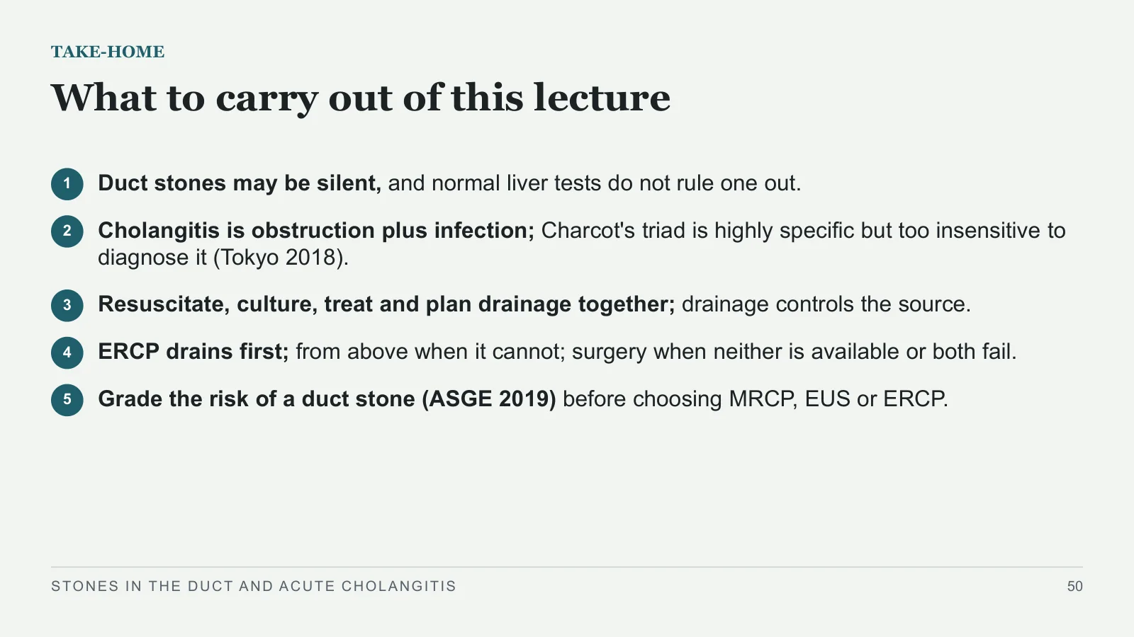
What to carry out of this lecture
TAKE-HOME
1
Duct stones may be silent, and normal liver tests do not rule one out.
2
Cholangitis is obstruction plus infection; Charcot's triad is highly specific but too insensitive to diagnose it (Tokyo 2018).
3
Resuscitate, culture, treat and plan drainage together; drainage controls the source.
4
ERCP drains first; from above when it cannot; surgery when neither is available or both fail.
5
Grade the risk of a duct stone (ASGE 2019) before choosing MRCP, EUS or ERCP.
STONES IN THE DUCT AND ACUTE CHOLANGITIS
50
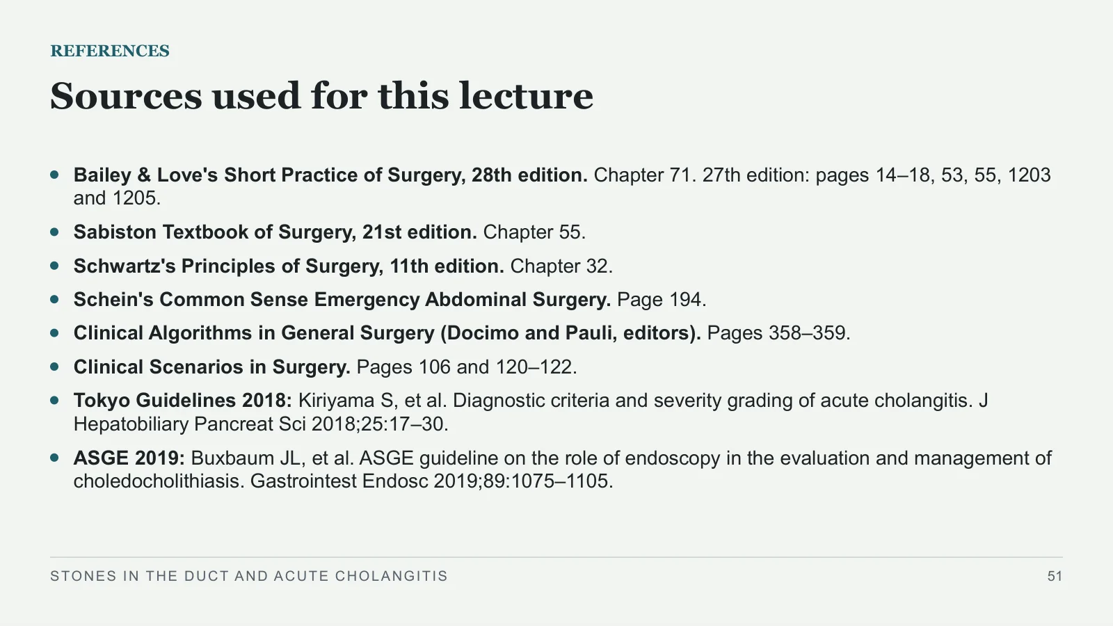
Sources used for this lecture
REFERENCES
Bailey & Love's Short Practice of Surgery, 28th edition. Chapter 71. 27th edition: pages 14–18, 53, 55, 1203 and 1205.
Sabiston Textbook of Surgery, 21st edition. Chapter 55.
Schwartz's Principles of Surgery, 11th edition. Chapter 32.
Schein's Common Sense Emergency Abdominal Surgery. Page 194.
Clinical Algorithms in General Surgery (Docimo and Pauli, editors). Pages 358–359.
Clinical Scenarios in Surgery. Pages 106 and 120–122.
Tokyo Guidelines 2018: Kiriyama S, et al. Diagnostic criteria and severity grading of acute cholangitis. J Hepatobiliary Pancreat Sci 2018;25:17–30.
ASGE 2019: Buxbaum JL, et al. ASGE guideline on the role of endoscopy in the evaluation and management of choledocholithiasis. Gastrointest Endosc 2019;89:1075–1105.
STONES IN THE DUCT AND ACUTE CHOLANGITIS
51
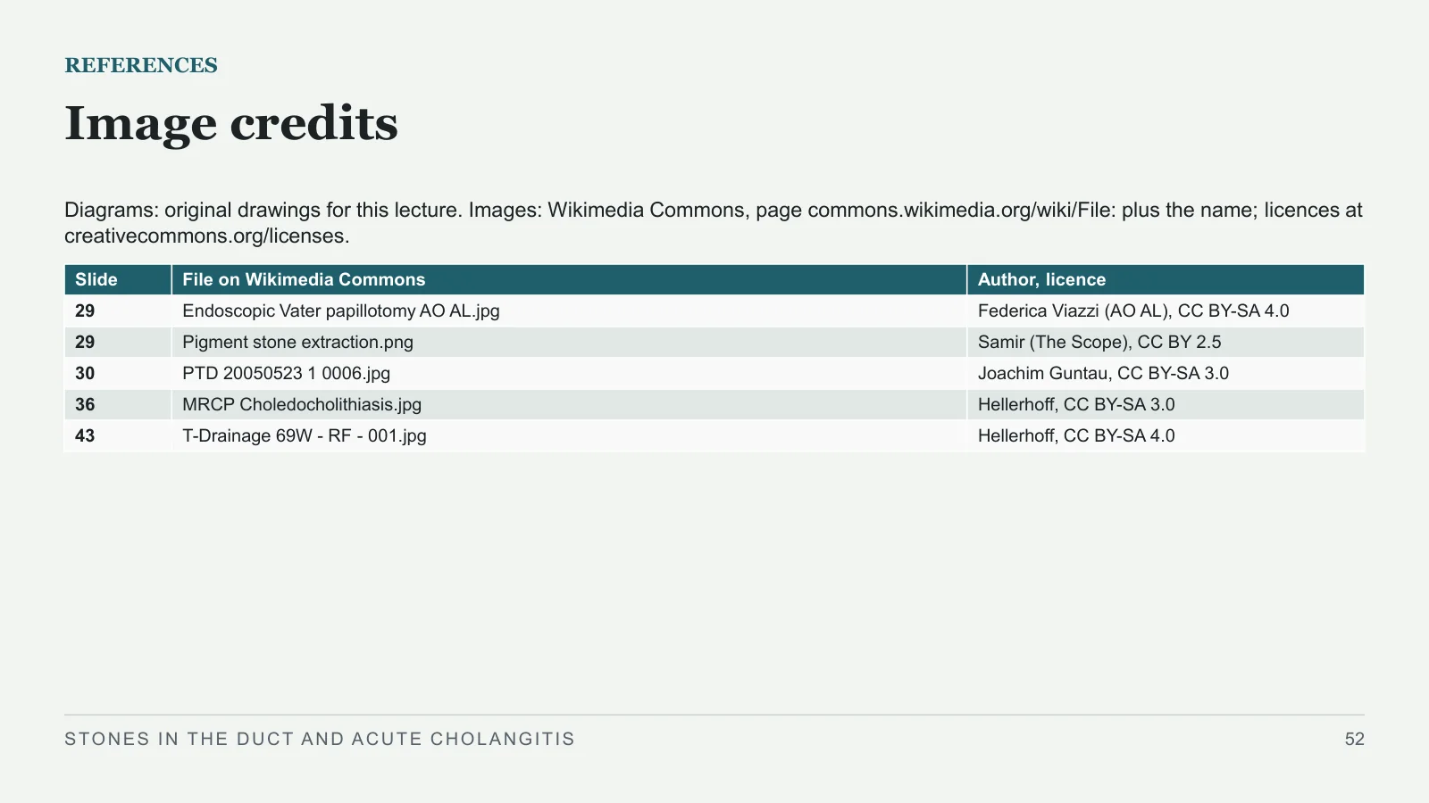
Image credits
REFERENCES
Diagrams: original drawings for this lecture. Images: Wikimedia Commons, page commons.wikimedia.org/wiki/File: plus the name; licences at creativecommons.org/licenses.
STONES IN THE DUCT AND ACUTE CHOLANGITIS
52
| Slide | File on Wikimedia Commons | Author, licence |
| 29 | Endoscopic Vater papillotomy AO AL.jpg | Federica Viazzi (AO AL), CC BY-SA 4.0 |
| 29 | Pigment stone extraction.png | Samir (The Scope), CC BY 2.5 |
| 30 | PTD 20050523 1 0006.jpg | Joachim Guntau, CC BY-SA 3.0 |
| 36 | MRCP Choledocholithiasis.jpg | Hellerhoff, CC BY-SA 3.0 |
| 43 | T-Drainage 69W - RF - 001.jpg | Hellerhoff, CC BY-SA 4.0 |
Sources and image credits
Original lecture diagrams and source/image credits are preserved from the PowerPoint. Each photograph or scan below links to its source and applicable licence. Changes are described in the slide captions; adapted ShareAlike images retain that licence.
Images
- Slide 29: Endoscopic Vater papillotomy AO AL.jpg · Federica Viazzi (AO AL), CC BY-SA 4.0. Slide caption: Endoscopy: Federica Viazzi (AO AL), CC BY-SA 4.0, via Wikimedia Commons. Endoscopy: Samir (The Scope), CC BY 2.5, via Wikimedia Commons; cropped. Adaptations of this image retain the same Creative Commons ShareAlike licence.
- Licence — Endoscopic Vater papillotomy AO AL.jpg · CC BY-SA 4.0
- Slide 29: Pigment stone extraction.png · Samir (The Scope), CC BY 2.5. Source attribution: Samir (The Scope), English Wikipedia. Slide caption: Endoscopy: Federica Viazzi (AO AL), CC BY-SA 4.0, via Wikimedia Commons. Endoscopy: Samir (The Scope), CC BY 2.5, via Wikimedia Commons; cropped.
- Licence — Pigment stone extraction.png · CC BY 2.5
- Slide 30: PTD 20050523 1 0006.jpg · Joachim Guntau, CC BY-SA 3.0. Slide caption: X-ray: Joachim Guntau, CC BY-SA 3.0, via Wikimedia Commons. Adaptations of this image retain the same Creative Commons ShareAlike licence. The Commons page documents CC BY-SA 3.0 through its GFDL licensing update.
- Licence — PTD 20050523 1 0006.jpg · CC BY-SA 3.0
- Slide 36: MRCP Choledocholithiasis.jpg · Hellerhoff, CC BY-SA 3.0. Slide caption: MRCP: Hellerhoff, CC BY-SA 3.0, via Wikimedia Commons. Adaptations of this image retain the same Creative Commons ShareAlike licence.
- Licence — MRCP Choledocholithiasis.jpg · CC BY-SA 3.0
- Slide 43: T-Drainage 69W - RF - 001.jpg · Hellerhoff, CC BY-SA 4.0. Slide caption: Cholangiogram: Hellerhoff, CC BY-SA 4.0, via Wikimedia Commons. Adaptations of this image retain the same Creative Commons ShareAlike licence.
- Licence — T-Drainage 69W - RF - 001.jpg · CC BY-SA 4.0


Sources
- [1] REFERENCES
- [2] Sources used for this lecture
- [3] Bailey & Love's Short Practice of Surgery, 28th edition. Chapter 71. 27th edition: pages 14–18, 53, 55, 1203 and 1205.
- [4] Sabiston Textbook of Surgery, 21st edition. Chapter 55.
- [5] Schwartz's Principles of Surgery, 11th edition. Chapter 32.
- [6] Schein's Common Sense Emergency Abdominal Surgery. Page 194.
- [7] Clinical Algorithms in General Surgery (Docimo and Pauli, editors). Pages 358–359.
- [8] Clinical Scenarios in Surgery. Pages 106 and 120–122.
- [9] Tokyo Guidelines 2018: Kiriyama S, et al. Diagnostic criteria and severity grading of acute cholangitis. J Hepatobiliary Pancreat Sci 2018;25:17–30.
- [10] ASGE 2019: Buxbaum JL, et al. ASGE guideline on the role of endoscopy in the evaluation and management of choledocholithiasis. Gastrointest Endosc 2019;89:1075–1105.